
In vivo screen using chemical inducers of dimerization

a dimerization and chemical inducer technology, applied in the field of enzyme evolution and drug screening in vivo, can solve the problems of limited to existing reactions, difficult protein engineering, and naive notions of how enzymes work

Inactive Publication Date: 2010-10-14
THE TRUSTEES OF COLUMBIA UNIV IN THE CITY OF NEW YORK
View PDF1 Cites 2 Cited by
  • Summary
  • Abstract
  • Description
  • Claims
  • Application Information

AI Technical Summary

Benefits of technology

The invention provides a compound that can be used to screen proteins for their ability to catalyze bond cleavage or formation. This compound has a specific formula and can be used to activate or inhibit a cellular readout when the proteins it is designed to bind to dimerize. The method involves expressing a pair of fusion proteins in a cell and then adding the compound to see if it changes the cellular readout. This invention can help identify new proteins with catalytic activity and aid in the development of new treatments for diseases that require the formation or cleavage of bonds.

Problems solved by technology

(Kaiser 1984; Knowles 1987) Despite some successes, (Wharton 1985; Wilks 1988; Hilvert 1985, 1989, 1994; Imperiali 1994; Johnson 1993) protein engineering has proven to be difficult and has suggested that notions of how enzymes work may still be naive.
In vivo complementation of essential enzymes, such as chorismate mutase and triosephosphate isomerase, offers a direct selection for catalysis but is limited to existing reactions.
However, as with proteins, it is difficult to design screens for non-protein catalysts.
FK506, however, is not an ideal chemical handle.
However, it has not heretofore been suggested to use small molecule induced protein dimerization to screen for catalysis in vivo., and specifically, it has not been suggested to use an enzyme cleavable moiety to link two molecules to dimerize proteins.

Method used


Image

  • In vivo screen using chemical inducers of dimerization
  • In vivo screen using chemical inducers of dimerization
  • In vivo screen using chemical inducers of dimerization

Examples


example 1

[0185]We have shown that Dex-Mtx can dimerize a LexA-DHFR and a B42-rGR protein chimera in vivo (Table I). (Lin, 2000) Dex-Mtx was assayed using both plate and liquid assays at extracellular concentrations of 1-100 μM. No activation was observed at concentrations ≦0.1 μM. 100 μM is the limit of Dex-Mtx solubility. Control experiments established that lacZ transcription is dependent on Dex-Mtx. There are only background levels of lacZ transcription when Dex-Mtx is omitted, LexA-DHFR is replaced with LexA, or B42-GR is replaced with B42. Likewise, a 10-fold excess of Mtx competes out Dex-Mtx-dependent LacZ transcription. Interestingly, of the 10 protein chimera combinations tested, Dex-Mtx could only activate lacZ transcription in the context of the LexA-eDHFR and the B42-(Gly6)-rGR chimeras (Table 1). None of the 9 other protein combinations tested worked. This result is consistent with our view that the Dex-Mtx systems (and other dimerization systems) could be further improved both ...

example 2

Cephalosporin Hydrolysis by the 908R Cephalosporinase in the Yeast Three-Hybrid System

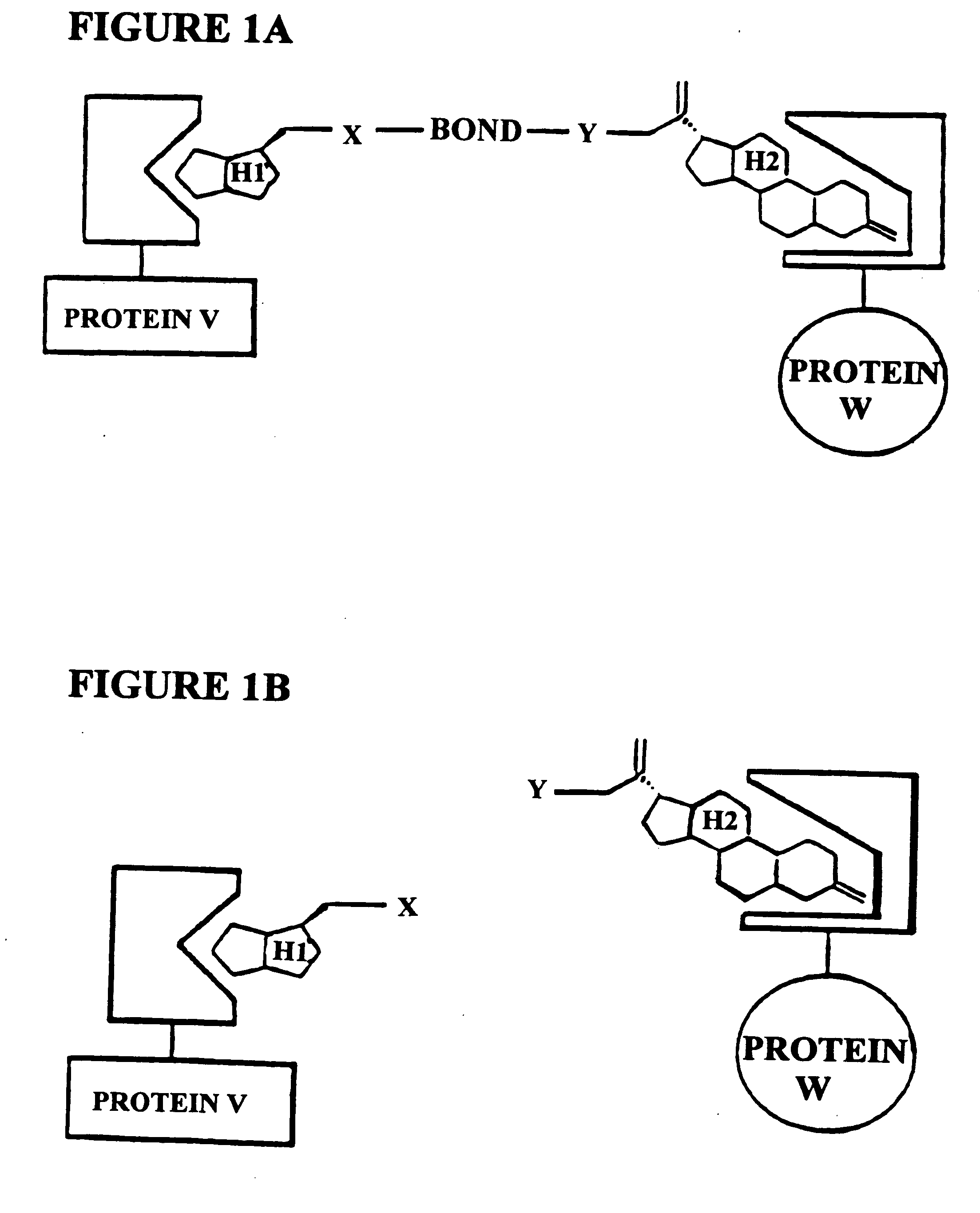
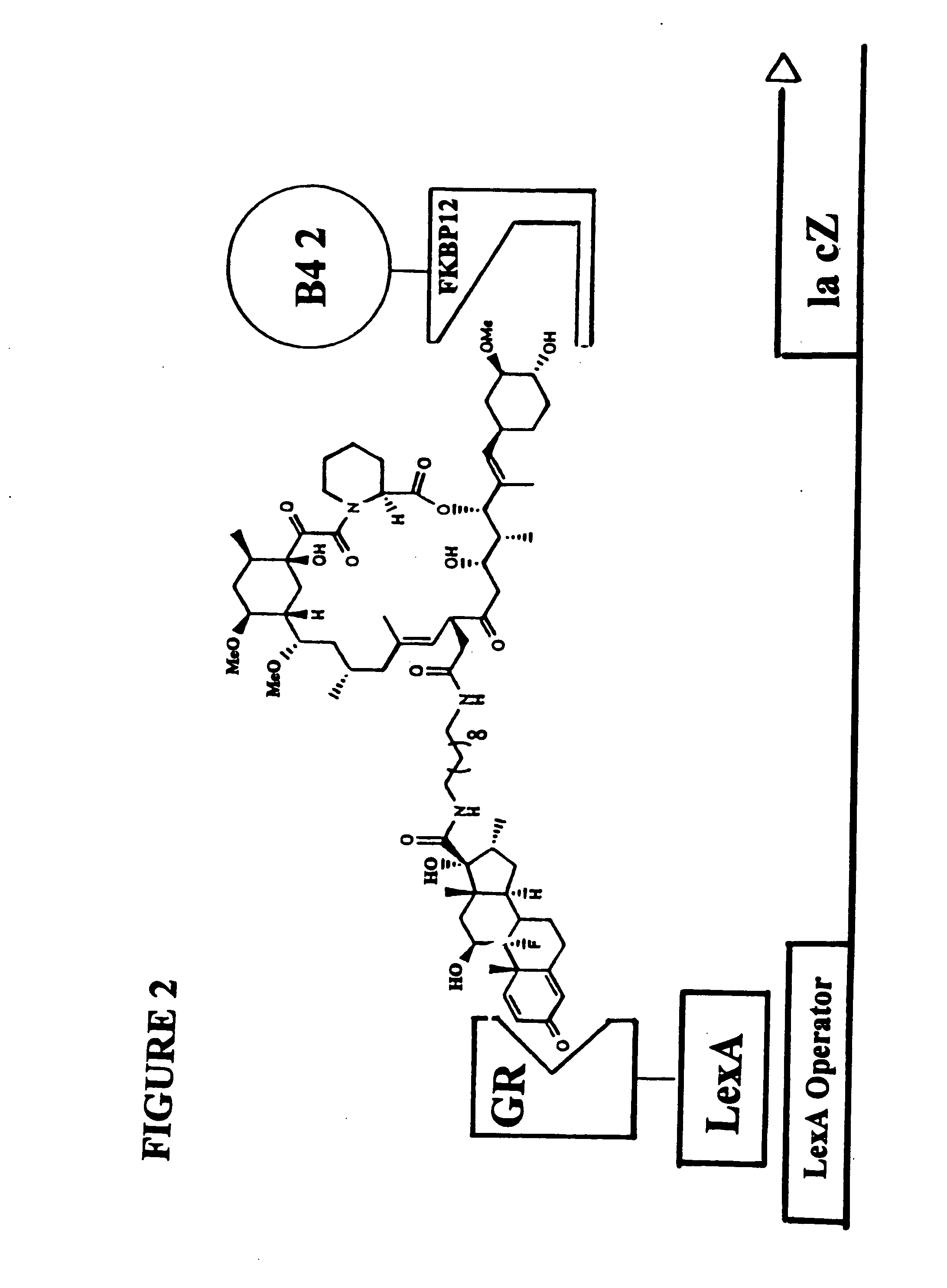
[0186]The subject invention is exemplified using the components of the yeast three-hybrid system (Licitra, represented in FIG. 2, see also U.S. Pat. No. 5,928,868). In this system DEX-FK506 (exemplifying H1-H2) mediates dimerization of the protein fusions LexA-GR (representing reporter V—H1 receptor) and B42-FKBP12 (representing reporter W—H2 receptor) thus activating transcription of a lacZ reporter gene. The chemical handles H1 and H2 and the protein dimerization assay, however, all can be varied.

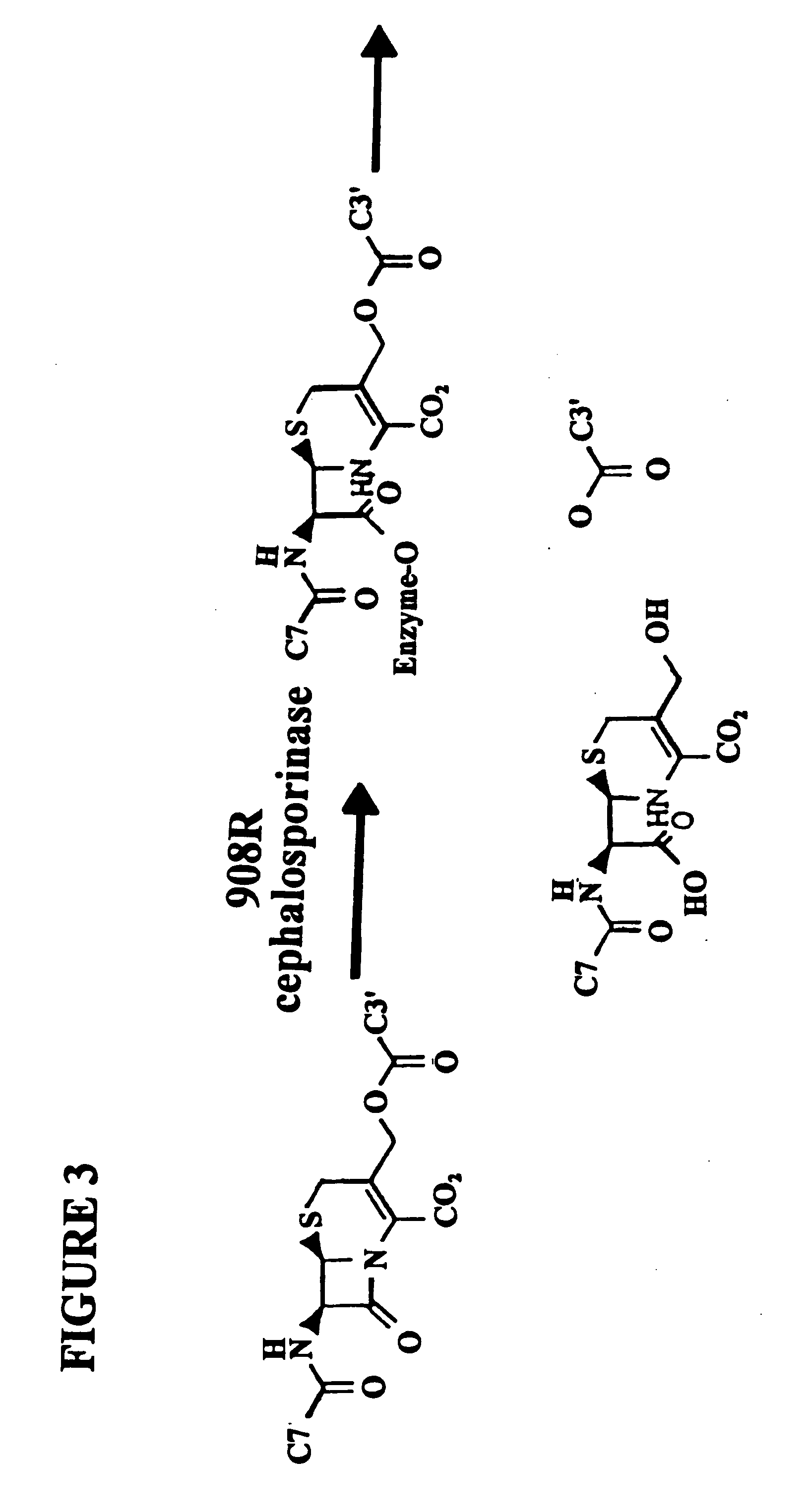
[0187]In the subject invention, however, the yeast three-hybrid system is altered by inserting a BOND, B, as well as any required spacers X and Y, so as to form a small molecule having the structure H1-X—B—Y—H2. While there is ample precedent for small-molecule mediated protein dimerization, what remains is to show these assays can be used to select for catalysts. Cephalosporin hydrolysis by a cephalospo...

example 3

[0202]CIDs can be used to screen cDNA libraries based on biochemical function. This glycosidase example is used to determine the best method for expressing the cDNA clones and to optimize the screening process.

Proof of Principle—β-Galactosidase Activity Assays

[0203]Table III explains the components of each strain. Each strain was constructed from the parent yeast strain FY250 and also contains the pMW106 plasmid, which has the LacZ reporter gene that is turned on only in when the LexA DNA binding domain and the B42 activation are brought in to the vicinity of each other. We use several different strains because we use DHFR from two different species, mDHFR is from murine, while eDHFR is from E. coli. We are asl oable to switch the small moleculebinding domains. For example, the strain containing LexA-eDHFR with B42-rGH2 is a different strain and behaves differently from the strain containing LexA-rGR2 with B42-eDHFR. We also put in short 6 amino acid linkers between the two domains ...


PUM

PropertyMeasurementUnit
structure-activity relationshipsaaaaaaaaaa
acid and base sensitiveaaaaaaaaaa
affinityaaaaaaaaaa

Abstract

A method for identifying a molecule that binds a known target in a cell from a pool of candidate molecules, comprising: (a) covalently bonding each molecule in the pool of candidate molecules to a methotrexate moiety to form a screening molecule; (b) introducing the screening molecule into a cell which expresses a first fusion-protein comprising a binding domain capable of binding methotrexate, a second fusion protein comprising the known target, and a reporter gene wherein expression of the reporter gene is conditioned on the proximity of the first fusion protein to the second fusion protein; (c) permitting the screening molecule to bind to the first fusion protein and to the second fusion protein so as to activate the expression of the reporter gene; (d) selecting which cell expresses the reporter gene; and (e) identifying the small molecule that binds the known target.

Description

[0001]This application is a divisional of U.S. Ser. No. 09 / 768,479, filed Jan. 24, 2001, which is a continuation-in-part of U.S. Ser. No. 09 / 490,320, filed Jan. 24, 2000, now abandoned, the contents of all of which are hereby incorporated by reference.[0002]This invention has been made with government support under National Science Foundation grants CHE-9626981, CHE-9977402, and CHE-9984928. Accordingly, the U.S. Government has certain rights in the invention.[0003]Throughout this application, various publications are referenced by author or author and date. Full citations for these publications may be found listed alphabetically at the end of the specification immediately preceding the claims. The disclosures of these publications in their entireties are hereby incorporated by reference into this application in order to more fully describe the state of the art as known to those skilled therein as of the date of the invention described and claimed herein.FIELD OF INVENTION[0004]The ...

Claims


Application Information

Patent Timeline
Patent Type & Authority Applications(United States)
IPC IPC(8): C12Q1/68A61K47/48A61K49/00C07J41/00C07J43/00C07K5/078C07K19/00C12N15/62G01N33/542
CPCA61K47/48023A61K49/0006C07J41/0066G01N33/542C07K5/06139C07K19/00C12N15/1055C07J43/003A61K47/54
Inventor CORNISH, VIRGINIA W.
Owner THE TRUSTEES OF COLUMBIA UNIV IN THE CITY OF NEW YORK
Learn More