Method for Micromanipulation of Cells
a cell and micromanipulation technology, applied in the field of cell micromanipulation, can solve the problems of many errors in the initial target area selection of the regenerating axon, little is known about the targeting of specific cells, and the targeting may not proceed smoothly
- Summary
- Abstract
- Description
- Claims
- Application Information
AI Technical Summary
Benefits of technology
Problems solved by technology
Method used
Image
Examples
example 1
Materials and Methods
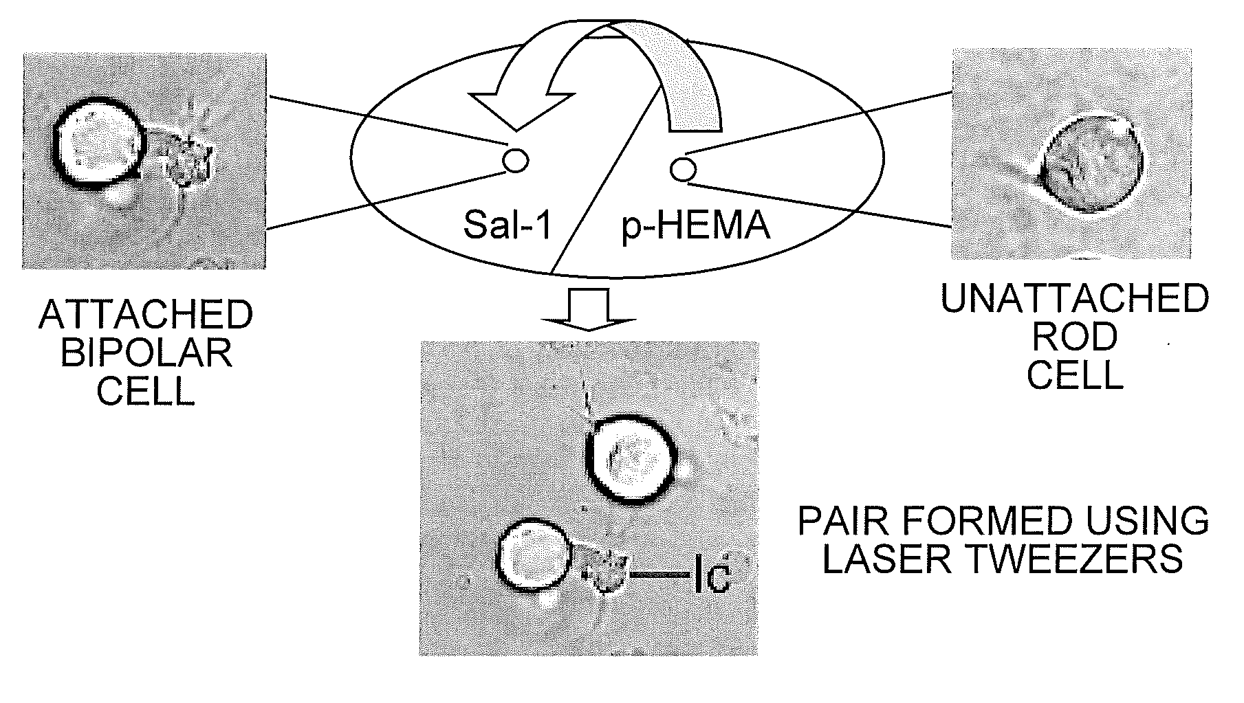
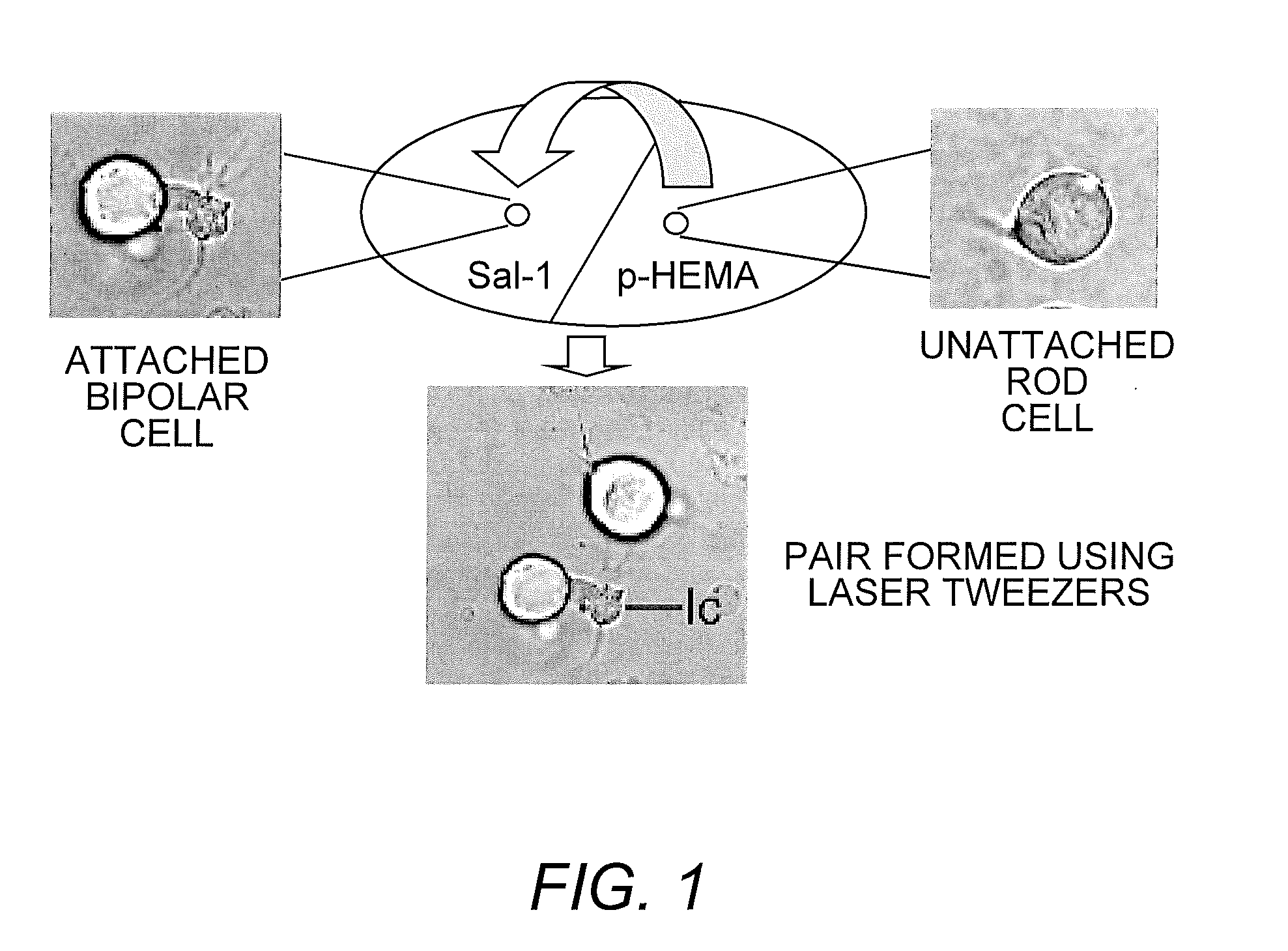
[0023]Preparation of Culture Dishes for Optical Tweezers. Trapping forces of optical tweezers are generated from the momentum of light (Ashkin (1991) supra; Ashkin, et al. (1986) supra). Although these forces easily trap cells in suspension, they are not able to move cells that adhere to a surface. In initial experiments to manipulate retinal neurons with optical tweezers (Townes-Anderson, et al. (1998) supra), a thin layer of SYLGARD was used to reduce cell adhesion to the culture dish. For the current studies, poly-HEMA (poly-2-hydroxyethylmethacrylate), a nontoxic compound with cell repellent properties (Folkman & Moscona (1978) supra), was used.
[0024]Acid-cleaned, #1 glass coverglass (VWR Scientific Inc., Media, Pa.) was prepared so that one half was coated first with poly-HEMA (20 mg / ml 95% ethanol, Sigma Chemical Co. St Louis Mo.). Allowing a few drops of poly-HEMA solution to flow down the surface of the coverglass held at a steep incline ensured a thin, ...
example 2
Interaction Between Photoreceptors and Neurons
[0034]To examine the effect of cell type on photoreceptor targeting, pairs of rod-bipolar (second-order), rod-multipolar (third-order), cone-bipolar, and cone-multipolar cells were created. Optical tweezing was done within hours after retinal dissociation and cell plating and thus before any modification in cell shape. Although the neurons sustain some loss of cell processes during isolation, cell types remain distinct. Only cells positively identified were used to create cell pairs. Cell identification was augmented by immunocytochemistry: anti-rod opsin immunolabel distinguished rod from cone cells; anti-Goα antibody labeled most ON bipolar cells.
[0035]In culture, photoreceptors initially create actin-filled filopodia which emanate from all points on the cell's circumference (Mandell (1993) supra). Lamellipodia appear as well, frequently formed from existing synaptic pedicles (Nachman-Clewner & Townes-Anderson (1996) supra). Actin- and...
example 3
Interactions Between Photoreceptors and Bipolar Cell Subtypes
[0039]It was contemplated that although layer-specific markers associated with specific cell classes may determine targeting, cell subtypes may also influence the outcome of cell class pairings. To investigate the effects of cell subtype, photoreceptor-bipolar interactions were examined because these second order neurons are divided into two basic categories, the ON and OFF cells. This division depends on the response to light: ON cells are active in the light; OFF cells are active in the dark. The functional differences are due in part to the differential presence of metabotropic and tonotropic glutamate receptors on ON and OFF cells respectively. In adult tiger salamander, ON and OFF cells are approximately equal in number (Maple, et al. (2005) Vision Res. 45:697-705). Since, for both cone and rod cells, about half the bipolar cells were attractive targets, it is possible that either the ON or the OFF cells were the pref...
PUM
| Property | Measurement | Unit |
|---|---|---|
| diameter | aaaaa | aaaaa |
| diameter | aaaaa | aaaaa |
| wavelength | aaaaa | aaaaa |
Abstract
Description
Claims
Application Information
 - R&D
- Intellectual Property
- Life Sciences
- Materials
- Tech Scout
- Unparalleled Data Quality
- Higher Quality Content
- 60% Fewer Hallucinations
Browse by: Latest US Patents, China's latest patents, Technical Efficacy Thesaurus, Application Domain, Technology Topic, Popular Technical Reports.
© 2025 PatSnap. All rights reserved.Legal|Privacy policy|Modern Slavery Act Transparency Statement|Sitemap|About US| Contact US: help@patsnap.com


