Methods to identify and enrich for populations of ovarian cancer stem cells and somatic ovarian stem cells and uses thereof

a technology which is applied in the field of methods to identify and enrich populations of ovarian cancer stem cells and somatic ovarian stem cells, can solve the problems of rarely a cure, and achieve the effects of reducing inhibiting the self-renewal capacity of an ovarian cancer stem cell, and reducing the proliferation

Inactive Publication Date: 2010-10-28
THE GENERAL HOSPITAL CORP
View PDF6 Cites 107 Cited by
  • Summary
  • Abstract
  • Description
  • Claims
  • Application Information

AI Technical Summary

Benefits of technology

[0050]In some embodiments, the present invention further provides a method to identify agents that reduce the self-renewal capacity of an ovarian cancer stem cell, comprising contacting an ovarian cancer stem cell as identified according the methods as disclosed herein with an agent, and measuring the proliferation of the ovarian cancer cell, whereby an agent that decreases the proliferation as compared to a reference agent or absence of an agent identifies an agent that inhibits the self-renewal capacity of the ovarian cancer stem cell. In some embodiments, the method can further comprise contacting a somatic ovarian stem cell identified according to the methods as disclosed herein with an agent, and measuring the proliferation of the somatic ovarian cell, and comparing the rate of proliferation of the somatic ovarian stem cell in the presence of the agent with the rate of proliferation of an ovarian cancer stem cell in the presence of the same agent, whereby if the agent decreases the rate of proliferation of the ovarian cancer stem cells as compared to the rate of proliferation of somatic ovarian stem cells identifies an agent that inhibits the self-renewal capacity of the ovarian cancer stem cell.
[0051]In another embodiment, the present invention further provides a method for identifying agents which kill or decrease the rate of proliferation of ovarian cancer stem cells, the method comprising: (a) culturing a population of ovarian cancer stem cells as identified according to the methods as disclosed herein, (b) culturing a population of somatic ovarian stem cells as identified according to the methods as disclosed herein, (c) adding to the media of the population of ovarian cancer stem cells one or more agents and adding to the media of the population of somatic ovarian stem cells one or more of the same agents; (d) measuring the rate of proliferation of the population of ovarian cancer stem cells and measuring the rate of proliferation of the population of somatic ovarian stem cells; (e) comparing the rate of proliferation of the population of ovarian cancer stem cells with rate of proliferation of the population of somatic ovarian stem cells, wherein an agent which decreases the rate of proliferation of the ovarian cancer stem cells as compared to the rate of proliferation of somatic ovarian stem cells identifies an agent that kills or decreases the rate of proliferation of an ovarian cancer stem cell. In particular embodiments, the population of somatic ovarian stem cell used in such an assay to identify agents that selectively inhibit the ovarian cancer stem cells are coelomic ovarian stem cell population. In some embodiments, agents used in the methods to identify agents that kill or decrease the rate of proliferation of an ovarian cancer stem cell, such an agent is a nucleic acid, nucleic acid analogue, small molecule, protein, peptiomimetic, antibody, peptide, aptamer, ribozyme, and variants and fragments thereof. Further, proliferation assays useful in identifying agents that kill or decrease the rate of proliferation of an ovarian cancer stem cell are the methyliazoletetrazolium (MTT) proliferation assay or the colony forming unit (CFU) assay, as disclosed herein.
[0052]In further aspect, the present invention provides a method of treating a subject affected with cancer, the method comprising assessing the presence of ovarian cancer stem cells in a biological sample obtained from the subject, wherein a clinician reviews the results and if the results indicate the presence of ovarian cancer stem cells in the biological sample, the clinician directs the subject to be treated with an effective amount of a pharmaceutical composition comprising MIS or a homologue or variant or...

Problems solved by technology

Chemotherapy alone can yield several l...

Method used


Image

  • Methods to identify and enrich for populations of ovarian cancer stem cells and somatic ovarian stem cells and uses thereof
  • Methods to identify and enrich for populations of ovarian cancer stem cells and somatic ovarian stem cells and uses thereof
  • Methods to identify and enrich for populations of ovarian cancer stem cells and somatic ovarian stem cells and uses thereof

Examples


example 1

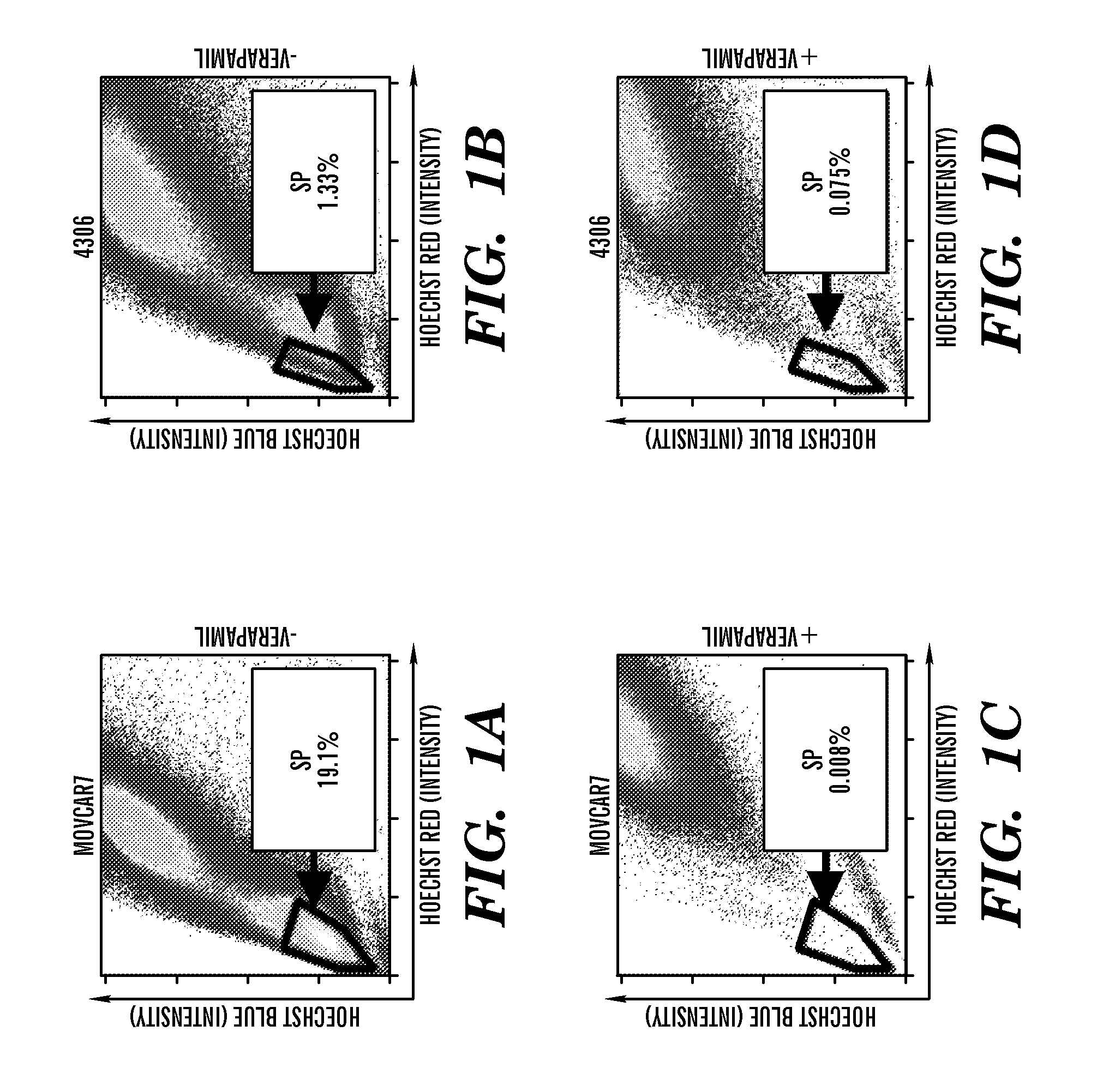
[0263]Identification of SPs in Mouse Ovarian Cancer Cell Lines. To determine whether mouse ovarian cancer cell lines contain candidate cancer stem cells, Hoechst 33342 was used to sort for the SP phenotype. The serous adenocarcinoma-recapitulating MOVCAR 7 and 8 cell lines were developed by using the MIS type II receptor (MISRII) promoter to drive the SV40 T antigen (19). The endometrioid carcinoma-recapitulating 4306 cell line was developed from conditional LSL-K-rasG12D / + / PtenloxP / loxP mice in which the ovarian surface epithelium was infected with adenovirus expressing Cre recombinase (20). Flow cytometry demonstrated a very high percentage of HoechstLow SP cells in the MOVCAR 7 and 4306 cell lines (FIGS. 1A and B), whereas SP was not detected in MOVCAR 8. Verapamil, a BCRP1 inhibitor (18), effectively eliminated the SP in both MOVCAR 7 and 4306 cells (FIGS. 1C and D). The average first sort percentage of SP cells was 6.28% (n=6) for MOVCAR 7 and 1.83% (n=4) for 4306 cells, which ...

example 2

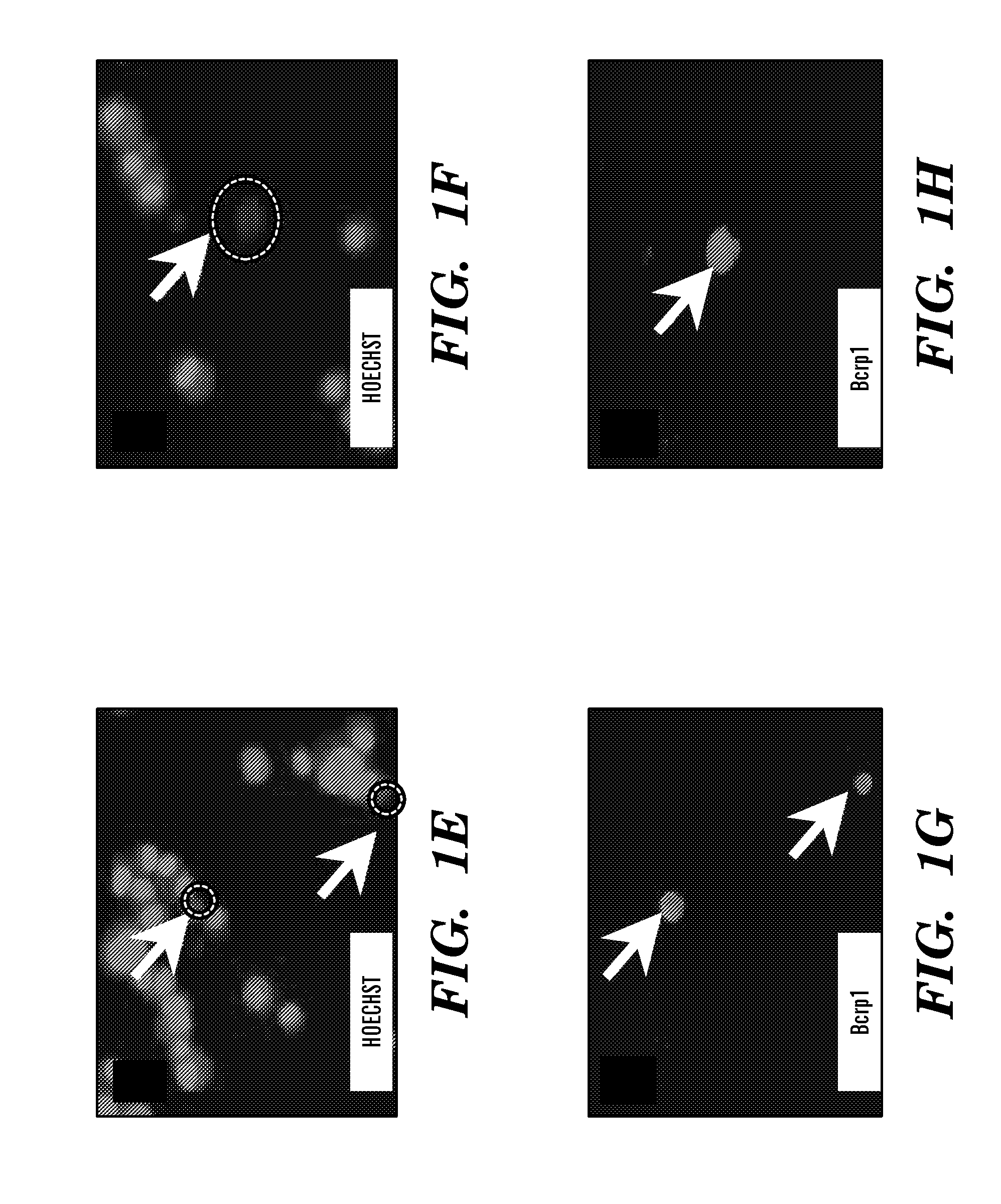
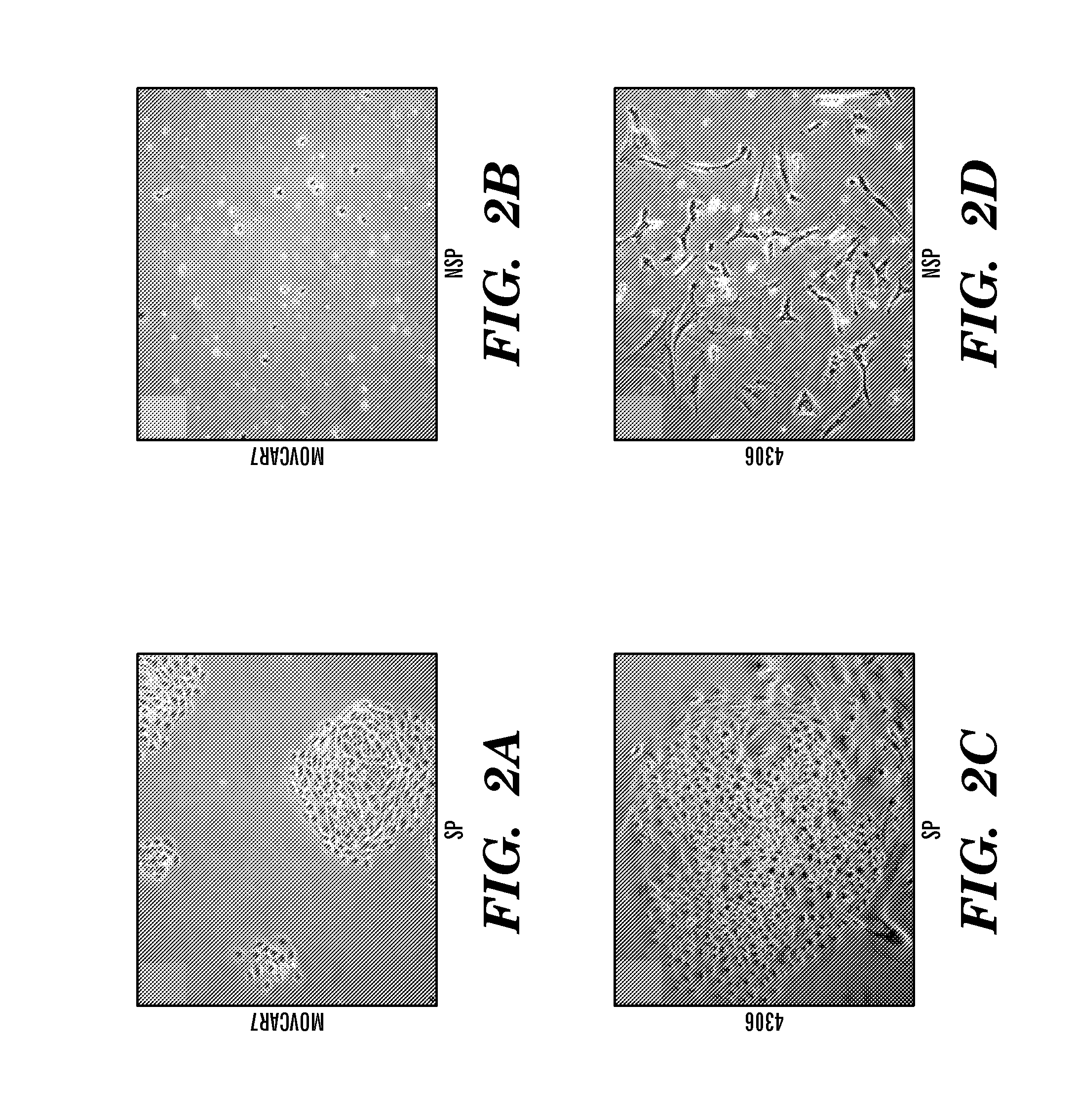
[0264]Ex Vivo Growth of SP and NSP Cells. Growth characteristics of the SP and NSP cells were consistent with previous findings for cancer stem cells (21). MOVCAR 7 and 4306 cells were sorted by flow cytometry and equal numbers of SP and NSP cells cultured. SP cells from both cell lines formed characteristic compact circular colonies with a cobblestone appearance and survived numerous passages (FIGS. 2 A and C; n=9). NSP cells from both cell lines were sparse and failed to proliferate beyond 1-2 weeks (FIGS. 2 B and D; n=9). These differences were not a consequence of prolonged Hoechst retention in the NSP cells because propidium iodide was used to gate out all nonviable cells. Serial sorting and reanalysis (total passages=3) of SP cells demonstrated enrichment of the SP and the presence of NSP cells (FIG. 2 E-J), suggesting asymmetric division occurred during culture (FIG. 2 F, G, I, and J). Thus, SP cells are able to self-renew, be enriched, and produce NSP cells when recovered an...

example 3

[0265]SP Cells Are in G1 Cell Cycle Arrest and Resistant to Doxorubicin in Vitro. By definition, SP cells should express high levels of BCRP1 and thus be able to efflux the lipophilic dye Hoechst 33342 and some lipophilic anticancer drugs, including those used in the treatment of ovarian cancer (18, 22). The lipophilic anticancer drug doxorubicin is a substrate of the BCRP1 transporter, whereas the lipophilic microtubule inhibitor paclitaxel is not (23). To investigate the functional significance of the Bcrp1 transporter found in MOVCAR 7 SP cells, we tested their response to doxorubicin and paclitaxel, as compared with that observed in the NSP, by methylthiazoletetrazolium (MTT) proliferation assays (FIG. 3). MOVCAR 7 SP cells were only inhibited by 30% after treatment with doxorubicin, whereas the NSP cells showed 81% inhibition (FIG. 3A). In contrast to doxorubicin, MOVCAR 7 SP and NSP cells were almost equally inhibited by the Bcrp1-resistant paclitaxel (FIG. 3B; SP=85% inhibiti...


PUM

PropertyMeasurementUnit
Lipophilicityaaaaaaaaaa

Abstract

The present invention relates to compositions and methods for treating, characterizing and diagnosing ovarian cancer. In particular, the present invention provides methods for treating and/or preventing ovarian cancer in a subject by administering to the subject an effective amount of Mullerian Inhibiting substance and/or an effective amount of an agent that inhibits BCRP1. The present invention further provides methods to identify and/or enrich for populations of ovarian cancer stem cells and populations of somatic ovarian stem cells, in particular, enrichment for populations of coelomic somatic ovarian stem cells, subcoelomic/stromal somatic ovarian stem cells and periphilar medullary somatic ovarian stem cells. The present invention also provides somatic ovarian stem cell markers and ovarian cancer stem cell markers, as well as methods to identify agents which selectively inhibit the proliferation of ovarian cancer stem cells as compared to somatic ovarian stem cells.

Description

CROSS REFERENCED APPLICATIONS[0001]This application claims the benefit under 35 U.S.C. 119(e) of U.S. Provisional Application Ser. No. 60 / 959,866 filed on Jul. 17, 2007, the contents of which is incorporated herein by reference.GOVERNMENT SUPPORT[0002]The present application was made with Government support under Grant Numbers CA17393 and HD32112 awarded by the National Institutes for Health (NIH). The Government of the United States has certain rights in the invention.FIELD OF THE INVENTION[0003]The present invention relates generally to methods and compositions to treat cancer, and more particularly to treating and / or preventing ovarian cancer, in particular where the ovarian cancer comprises ovarian cancer stem cells. The present invention further provides methods to identify and / or enrich for populations of ovarian cancer stem cells and populations of somatic ovarian stem cells, in particular, enrichment for populations of coelomic somatic ovarian stem cells, subcoelomic / stromal...

Claims


Application Information

Patent Timeline
IPC IPC(8): C12Q1/04C12N5/09C12Q1/02C12Q1/68C12N5/095
CPCA61K31/00A61K31/277A61K31/7088A61K45/06C12N5/0695C12N2501/998G01N2800/54C12N2503/02G01N33/5011G01N33/5091G01N33/57449G01N2800/44C12N2503/00C12N5/0672
Inventor DONAHOE, PATRICIA K.SZOTEK, PAUL P.MACLAUGHLIN, DAVID T.PREFFER, FREDERICPIERETTI-VANMARCKE, RAFAELDOMBKOWSKI, DAVID MICHAEL
Owner THE GENERAL HOSPITAL CORP
Why Eureka
Try Eureka