
Methods and kits for the inhibition of hyperplasia in vascular fistulas and grafts

Inactive Publication Date: 2002-05-14
PHARMASONICS
View PDF8 Cites 56 Cited by
  • Summary
  • Abstract
  • Description
  • Claims
  • Application Information

AI Technical Summary

Benefits of technology

Treatment according to the present invention is effected by exposing the anastomotic junction to vibrational energy at a mechanical index and for a time sufficient to inhibit hyperplasia of smooth muscle cells within the neointimal layer of the blood vessel. Surprisingly, it has been found that the strength of vibrational energy (as measured by the mechanical index) and the duration of the treatment (as measured by elapsed treatment time, duty cycle, and pulse repetition frequency (PRF)) can be selected to provide highly effective hyperplasia inhibition in the neointimal layer without significant damage to surrounding tissues or structures within the blood vessel(s) and / or the anastomotic junction itself. In particular, by exposing an anastomotic junction at risk of neointimal hyperplasia to a vibrational energy having a mechanical index in the range from 0.1 to 50, preferably from 0.2 to 10, and more preferably from 0.5 to 5, for a treatment time in the range from 10 seconds to 1000 seconds, preferably from 30 seconds to 500 seconds, and more preferably from 60 seconds to 300 seconds, the proliferation of vascular smooth muscle cells in the neointimal layer of the artery can be reduced by at least 2% (in comparison with untreated controls) after seven days, often at least 4% , and sometimes 6% or greater. The resulting reduction in hyperplasia mass after 28 days will typically be at least 10%, usually at least 20%, and preferably at least 30%. Such inhibitions can be achieved without significant necrosis of the smooth muscle cells.
The methods of the present invention will preferably be performed under conditions which cause little or no cavitation within the smooth muscle cells and other cells within or near the treatment region. While the initiation of cavitation will be governed to a large extent by the power and mechanical index of the vibrational energy, the presence of cavitation nucleii, such as gas microbubbles, can also contribute to cavitation. Thus, the methods of the present invention will preferable be performed in the absence of introduced microbubbles and / or other cavitation nucleii. Moreover, the treatment conditions of the present invention will result in little or no inhibition of migration of smooth muscle cells into the neointimal layer. Instead, the migration will be generally normal, but the migrated cells will have a quiescent phenotype rather than the proliferative phenotype associated with the formation of neointimal hyperplasia. In their proliferative phenotype, vascular smooth muscle cells not only divide rapidly but also excrete extracellular matrix which accounts for most of the volume of the neointimal layer responsible for hyperplasia. Quiescent smooth muscle cells divide less rapidly, do not secrete significant amounts of extracellular matrix, and promote healing and reformation of an intact endothelial layer over the neointimal layer. Additionally, the duty cycles and pulse repetition frequencies of the treatment will be selected to limit the heating within the neointimal layer to a temperature rise below 10.degree. C., preferably below 5.degree. C., and more preferably below 2.degree. C. Such limited temperature rise further assures the viability and normalcy of the treated cells to enhance healing and re-endothelialization of the neointimal layer in a rapid manner.
The duration of treatment is defined as the actual time during which vibrational energy is being applied to the arterial wall. Duration will thus be a function of the total elapsed treatment time, i.e., the difference in seconds between the initiation and termination of treatment; burst length, i.e., the length of time for a single burst of vibrational energy; and pulse repetition frequency (PRF). Usually, the vibrational energy will be applied in short bursts of high intensity (power) interspersed in relatively long periods of no excitation or energy output. An advantage of the spacing of short energy bursts is that heat may be dissipated and operating temperature reduced.

Problems solved by technology

Hyperplasia can occur as the result of surgical creation of the anastomotic junction, and it has been found that the surgical attachment can damage cells lining the involved blood vessels in a manner which results in an injury response characterized by secretion of extracellular matrix and excessive proliferation of the smooth muscle cells lining the blood vessels which together form the neointimal layer lining the blood vessel wall adjacent the anastomotic junction.
In the latter case, intimal hyperplasia has been a particular problem at the anastomotic junction formed between the graft and the vein.

Method used


Image

  • Methods and kits for the inhibition of hyperplasia in vascular fistulas and grafts
  • Methods and kits for the inhibition of hyperplasia in vascular fistulas and grafts
  • Methods and kits for the inhibition of hyperplasia in vascular fistulas and grafts

Examples


Embodiment Construction

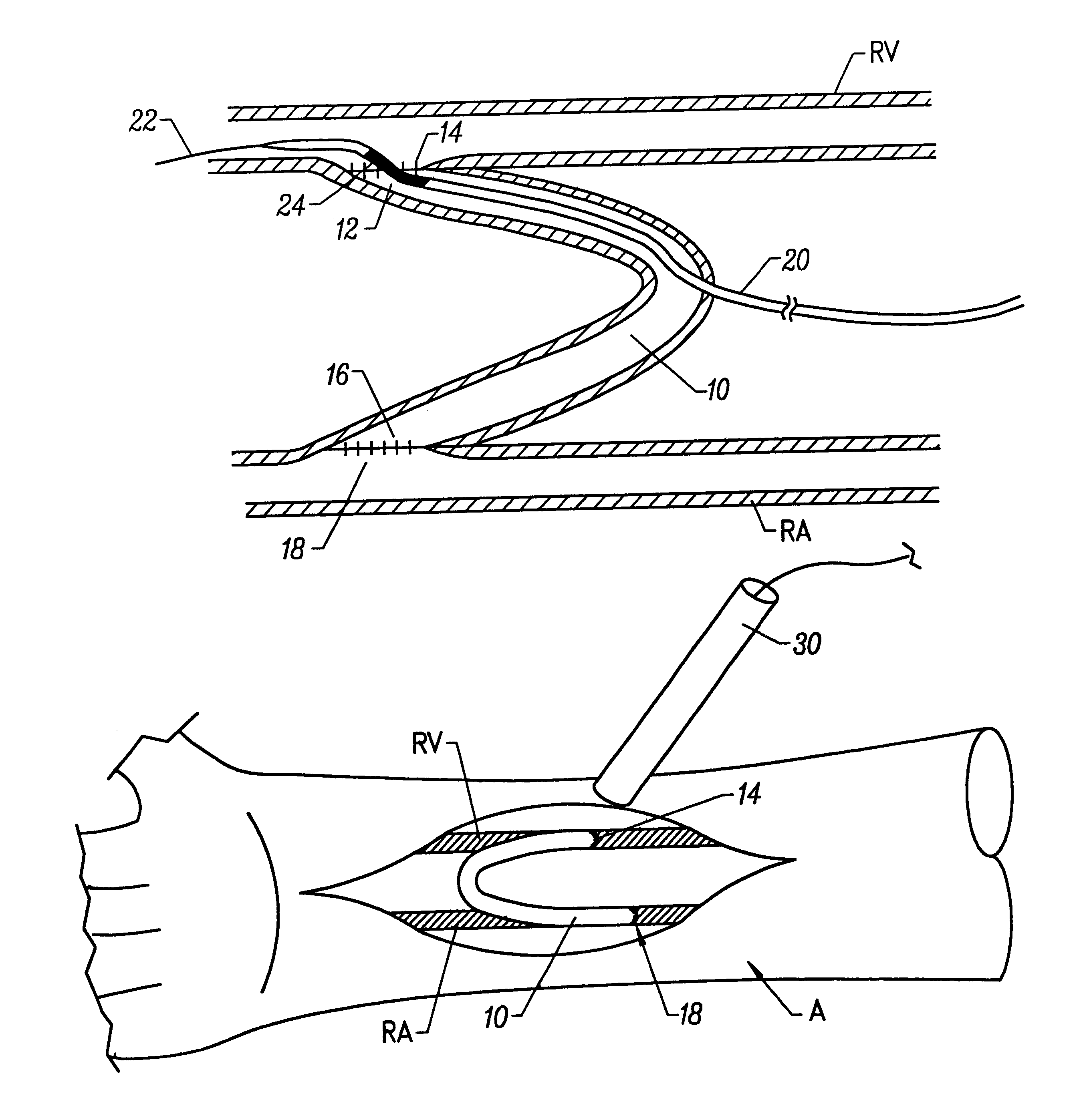
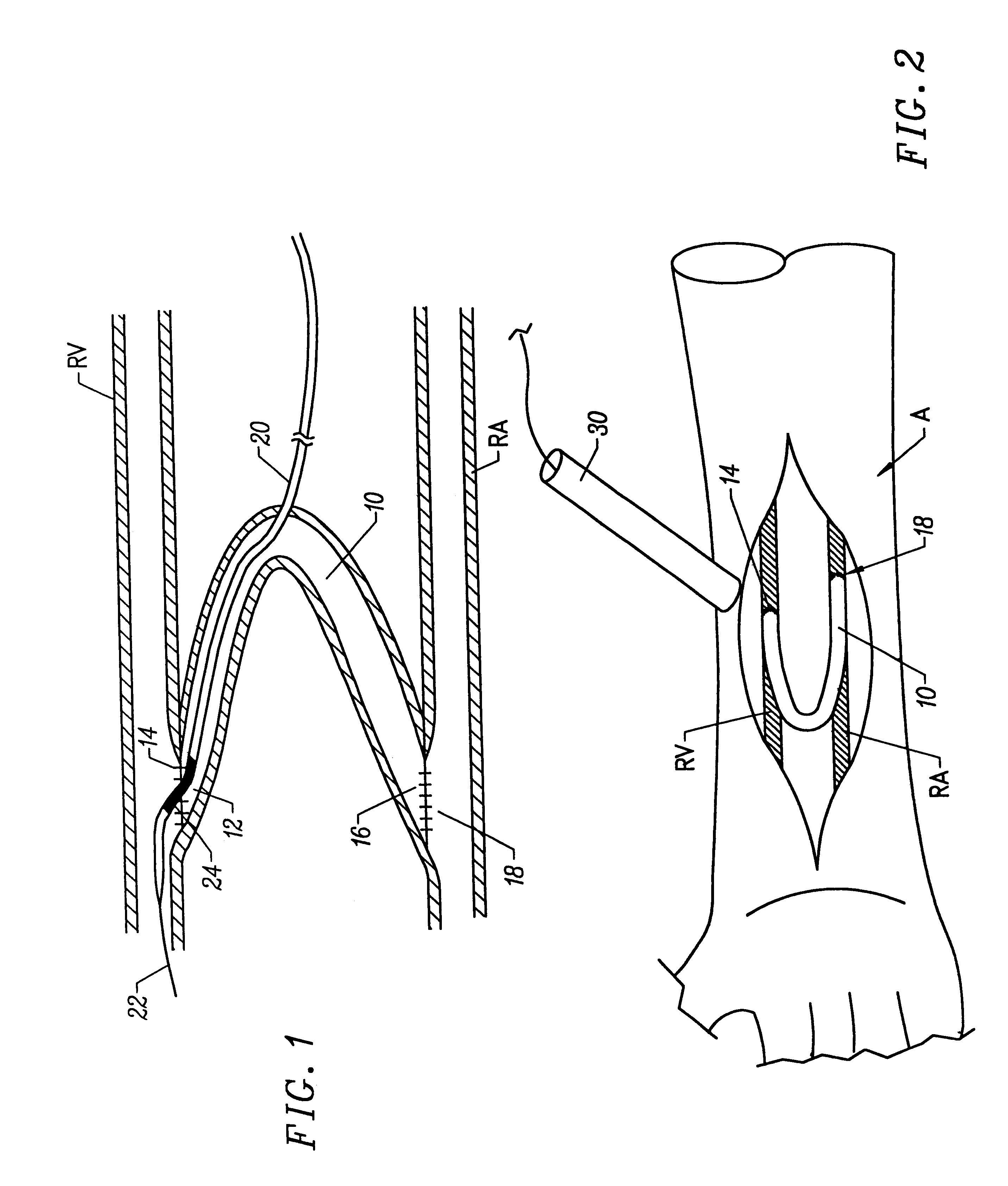
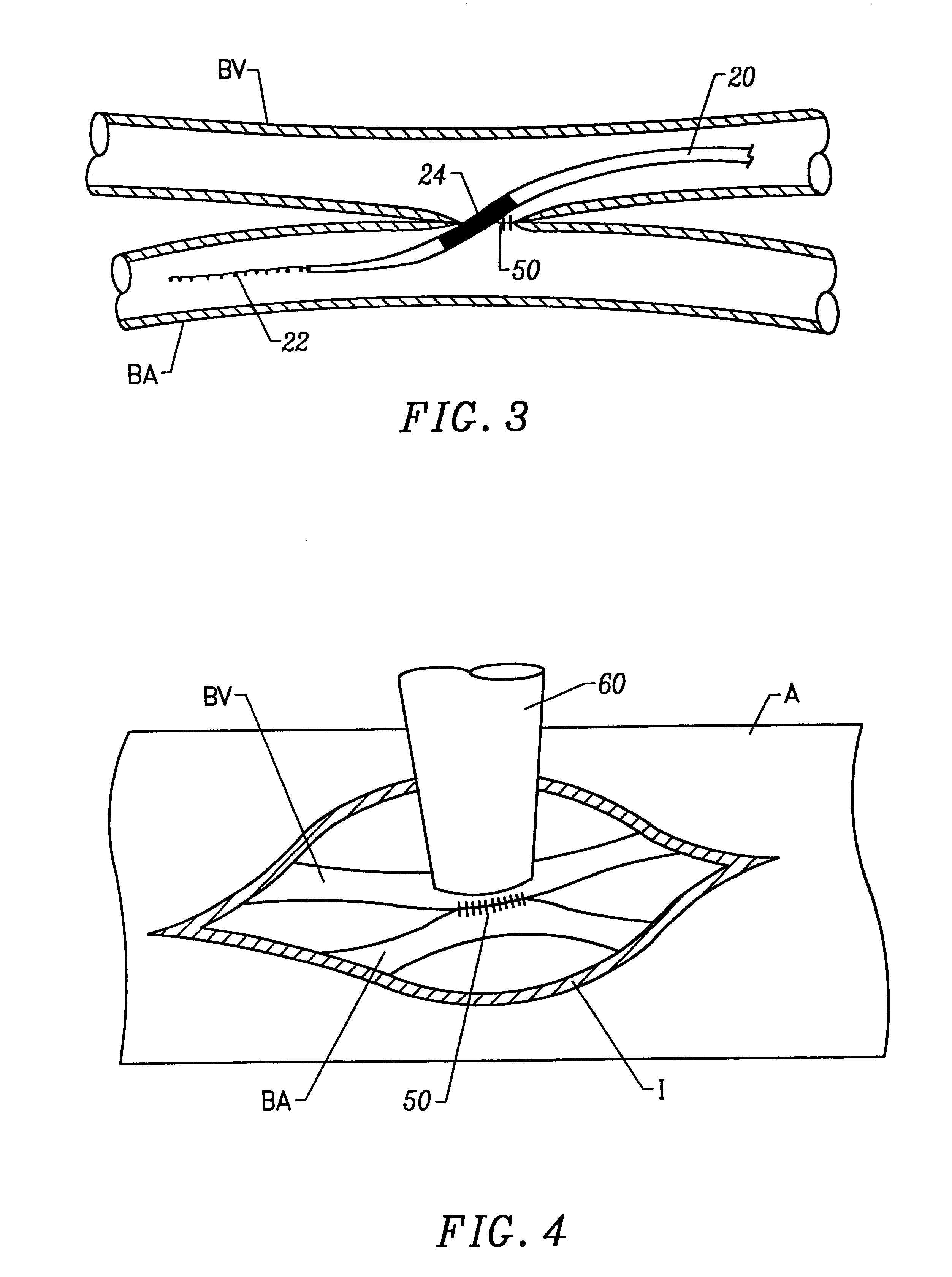
Anastomotic junctions between blood vessels and / or a blood vessel and a biological or synthetic graft are subject to occlusion due to hyperplasia and other injury responses, as discussed above. The present invention relies on exposure of such anastomotic junctions to vibrational energy at a mechanical index, and for a time sufficient to inhibit hyperplasia and thus reduce the risk of occlusion of the anastomotic junction and failure of the graft or fistula which has been created. The vibrational energy can be directed at the anastomotic junction in a variety of ways and at a variety of different times. When surgical access is available, e.g., at the time of fistula creation or graft implantation, it will often be desirable to apply the vibrational energy directly to an exterior surface of the junction using a probe or other suitable acoustic generator. It will be appreciated that the probe can be engaged directly against the surface of the blood vessels or blood vessel and graft. Wh...


PUM


Abstract

A method for inhibiting hyperplasia at an anastomotic junction of a vascular fistula or vascular graft comprises exposing the anastomotic junction to vibrational energy at a mechanical index and for a time sufficient to inhibit hyperplasia. Kits are described comprising the catheter or a probe suitable for performing the methods together with instructions for use setting forth the methods. The methods and kits are particularly suitable for treating anastomotic junctions formed to provide vascular access for hemodialysis, hemofiltration, and the like. The methods and kits are also suitable for treating other vascular grafts, such as those formed during cardiac bypass graft surgery, vascular repair, and the like.

Description

1. Field of the InventionThe present invention relates generally to medical methods, systems, and kits. More particularly, the present invention relates to methods for treating vascular fistulas and grafts to inhibit hyperplasia and subsequent occlusion thereof.Vascular fistulas and grafts are used in a wide variety of circumstances when it is desired to reconfigure or re-establish vascular circulation in a patient. For example, both fistulas and grafts are used to create access sites for blood withdrawal and return in patient's undergoing periodic kidney dialysis, hemofiltration, or other extracorporeal blood treatments. Usually, either a native artery and vein are connected together via a side-to-side anastomosis or a saphenous vein or synthetic graft is placed between an artery and a vein and attached at each end via an end-to-side anastomosis. Both natural grafts (usually a vein harvested from the patient being treated) and synthetic grafts are used in open and / or minimally inva...

Claims


Application Information

Patent Timeline
IPC IPC(8): A61F2/02A61F2/06
CPCA61F2/06A61F2/02
Inventor MCKENZIE, JOHN R.ZUK, ROBERT F.BRISKEN, AXEL F.CORL, PAUL D.
Owner PHARMASONICS
Learn More