
ErbB Surface Receptor Complexes as Biomarkers

a surface receptor and complex technology, applied in the field of biomarkers, can solve the problems of impracticality of use, individual receptor expression level alone is not always a reliable indicator, etc., and achieve the effect of increasing cost and labor

Inactive Publication Date: 2010-11-18
LAB OF AMERICA HLDG
View PDF5 Cites 17 Cited by
  • Summary
  • Abstract
  • Description
  • Claims
  • Application Information

AI Technical Summary

Benefits of technology

The invention is about a method for determining the status of a disease or healthful condition in a patient by measuring the amounts of specific receptors in cell surface membranes. This is particularly useful for diseases or conditions that are associated with the aberrant expression of these receptors. The method can be performed using fixed or frozen samples, and can also predict the effectiveness of drugs that target these receptors. The invention provides a way to better understand the disease status of a patient and to improve the diagnosis and treatment of cancer.

Problems solved by technology

However, individual receptor expression level alone is not always a reliable indicator of a disease status or condition, e.g. Chow et al, Clin.
Despite the important role that receptor dimerization plays in cellular and disease processes, receptor dimer expression has not been employed as a biomarker, in part due to the inconvenience and lack of sensitivity of current measurement technologies and the inability or impracticality of using such technologies to carry out measurements on patient samples, which may be formalin fixed and / or in too small a quantity for analysis, e.g. Price et al, Methods in Molecular Biology, 218: 255-267 (2003); Stagljar, Science STKE 2003, pe56 (2003); Koll et al, International patent publication WO 2004 / 008099; Golemis, editor, Protein-Protein Interactions (Cold Spring Harbor Laboratory Press, New York, 2002); Sorkin et al, Curr. Biol., 10: 1395-1398 (2000); McVey et al, J. Biol. Chem., 17: 14092-14099 (2001); Salim et al, J. Biol. Chem., 277: 15482-15485 (2002); Angers et al, Annu Rev. Pharmacol. Toxicol., 42: 409-435 (2002); Szollosi et al, Reviews in Molecular Biotechnology, 82: 251-266 (2002); Matko et al, Meth. in Enzymol., 278: 444-462 (1997); Reed-Gitomer, U.S. Pat. No. 5,192,660.

Method used


Image

  • ErbB Surface Receptor Complexes as Biomarkers
  • ErbB Surface Receptor Complexes as Biomarkers
  • ErbB Surface Receptor Complexes as Biomarkers

Examples


example 1

Analysis of Cell Lysates for Her-2 Heterodimerization and Receptor Phosphorylation

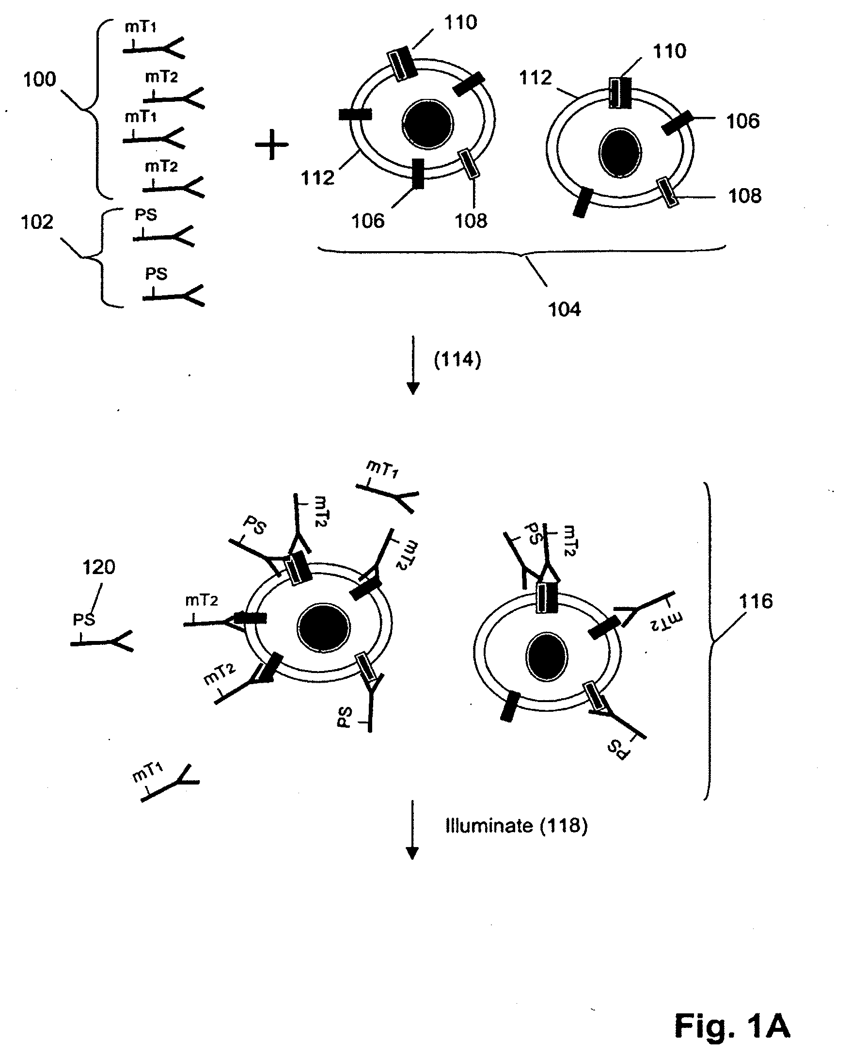
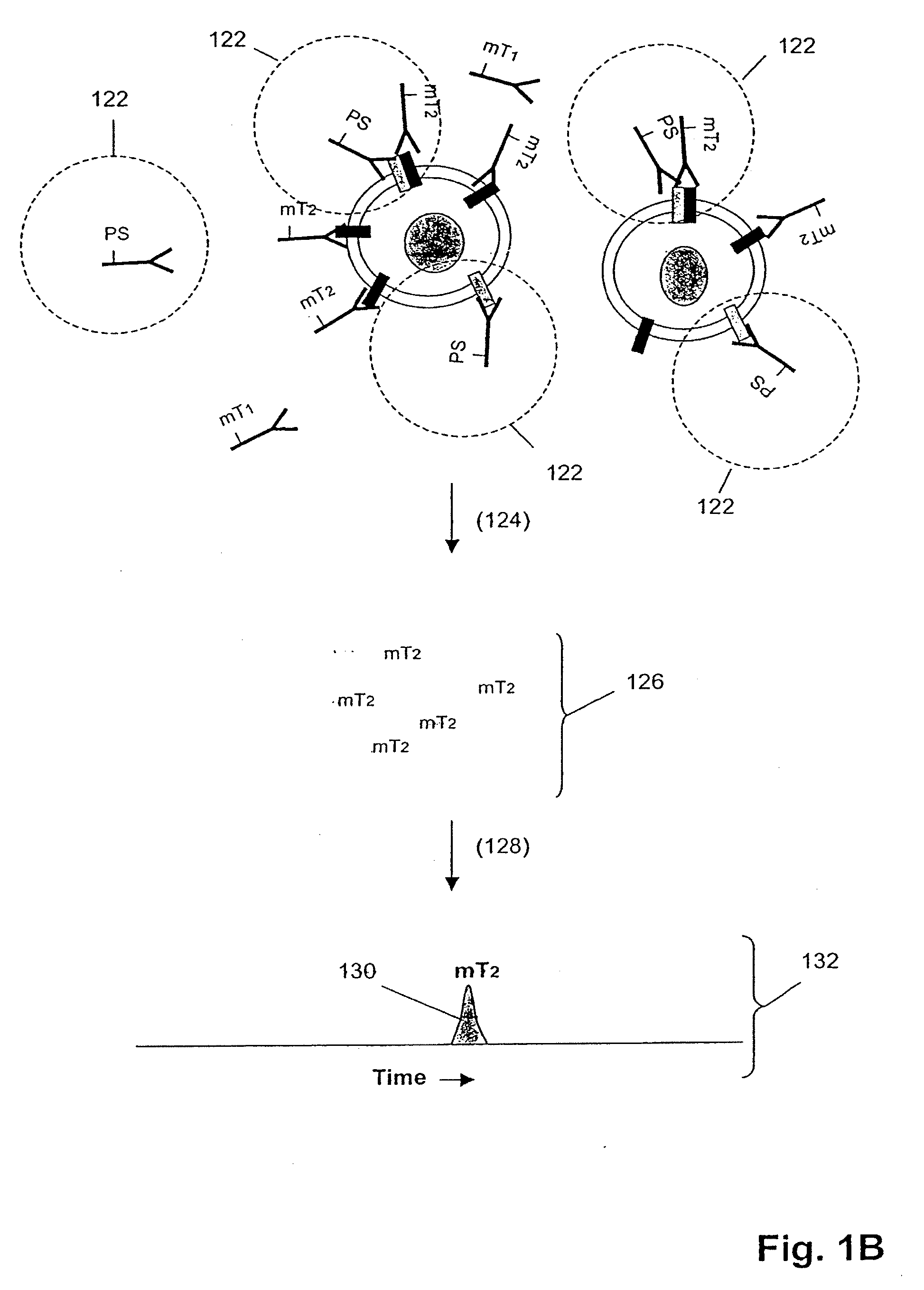
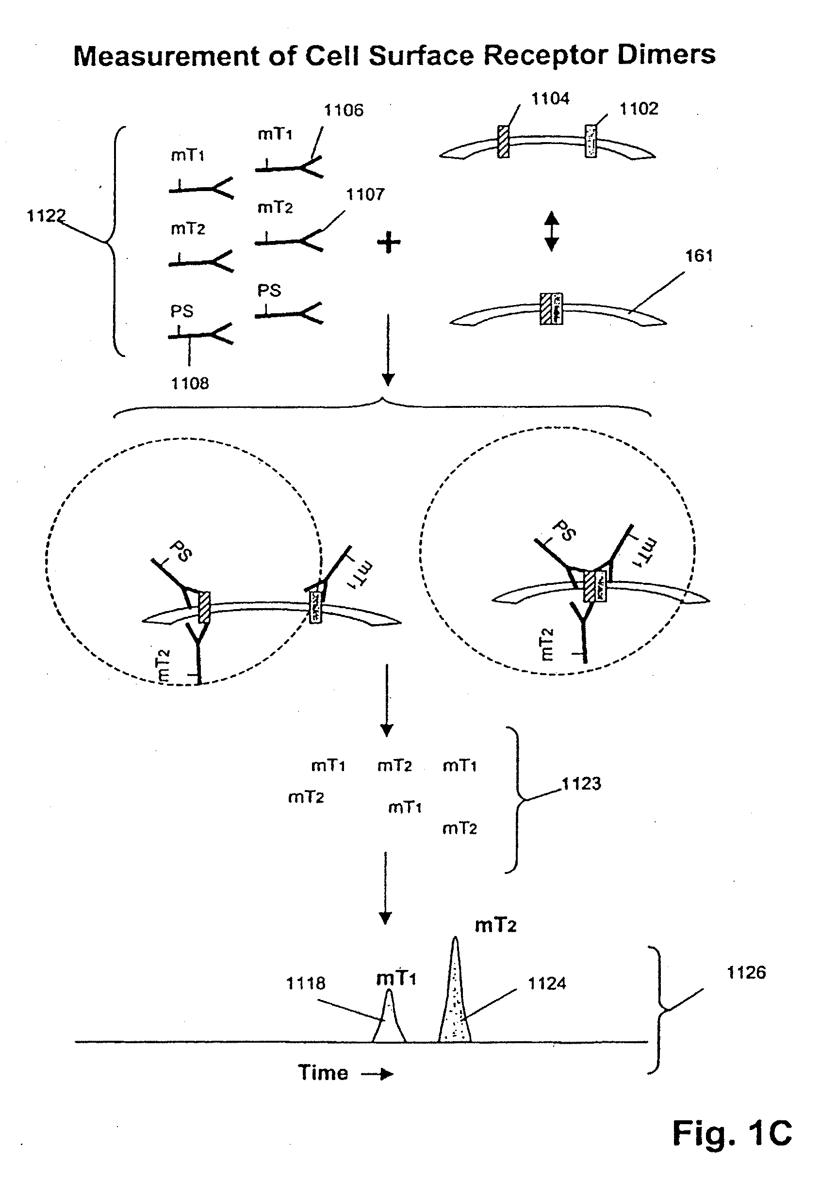
[0135]In this example, Her1-Her2 and Her2-Her3 heterodimers and phosphorylation states are measured in cell lysates from several cell lines after treatment with various concentrations of epidermal growth factor (EGF) and heregulin (HRG). Measurements are made using three binding compounds and a cleaving probe as described below.

Sample Preparation:

[0136]1. Serum-starve breast cancer cell line culture overnight before use.[0137]2. Stimulate cell lines with EGF and / or HRG in culture media for 10 minutes at 37° C. Exemplary doses of EGF / HRG are 0, 0.032, 0.16, 0.8, 4, 20, 100 nM for all cell lines (e.g. MCF-7, T47D, SKBR-3) except BT20 for which the maximal dose is increased to 500 nM because saturation is not achieved with 100 nM EGF.[0138]3. Aspirate culture media, transfer onto ice, and add lysis buffer to lyse cells in situ.[0139]4. Scrape and transfer lysate to microfuge tube. Incubate on ice for 30 m...

example 2

Analysis of Tissue Lysates for Her2 Heterodimerization and Receptor Phosphorylation

[0163]In this example, Her1-Her2 and Her2-Her3 heterodimers and phosphorylation states are measured in tissue lysates from human breast cancer specimens.

Sample Preparation:

[0164]1. Snap frozen tissues are mechanically disrupted at the frozen state by cutting.[0165]2. Transfer tissues to microfuge tube and add 3× tissue volumes of lysis buffer (from appendix I) followed by vortexing to disperse tissues in buffer.[0166]3. Incubate on ice for 30 min with intermittent vortexing to mix.[0167]4. Centrifuge at 14,000 rpm, 4° C., for 20 min.[0168]5. Collect supernatants as lysates and determine total protein concentration with BCA assay (Pierce) using a small aliquot.[0169]6. Aliquot the rest for storage at −80° C. until use.

Assay Design:

[0170]1. The total assay volume is 40 ul.[0171]2. The lysates are tested in serial titration series of 40, 20, 10, 5, 2.5, 1.25, 0.63, 0.31 ug total-equivalents and the volum...

example 3

Analysis of Cell Lysates for Her1 or Her2 Homodimerization and Receptor Phosphorylation

[0202]Sample preparation was carried out essentially as described in Example 2. Her1 homodimerization was induced by treating the cell lines with EGF or TGFα. For homodimerization of Her2 which does not have a ligand, unstimulated SKBR-3 or MDA-MD-453 cells that overexpress Her2 are compared to unstimulated MCF-7 cells that express a low level of Her2.

[0203]Assay design: A monoclonal antibody specific to the receptor is separately conjugated with either a molecular tag or biotin (that is then linked to a photosensitizer via an avidin bridge), so that the cleaving probe and a binding compound compete to bind to the same epitope in this example. Another binding compound is used that consists of a second anibody recognizing an overlapping epitope on the receptor, so that a ratiometric signal can be generated as a measure of homodimerization. The signal derived from the second antibody also provides a...


PUM

PropertyMeasurementUnit
molecular weightaaaaaaaaaa
thickaaaaaaaaaa
thicknessaaaaaaaaaa

Abstract

The invention is directed to a new class of biomarker in patient samples comprising dimers of ErbB cell surface membrane receptors. In one aspect, the invention includes a method of determining the status of a disease or healthful condition by correlating such condition to amounts of one or more dimers of ErbB cell surface membrane receptors measured directly in a patient sample, in particular a fixed tissue sample. In another aspect, the invention includes a method of determining a status of a cancer in a specimen from an individual by correlating measurements of amounts of one or more dimers of ErbB cell surface membrane receptors in cells of the specimen to such status, including presence or absence of a pre-cancerous state, presence or absence of a cancerous state, prognosis of a cancer, or responsiveness to treatment. Preferably, methods of the invention are implemented by using sets of binding compounds having releasable molecular tags that are specific for multiple components of one or more types of receptor dimers. After binding, molecular tags are released and separated from the assay mixture for analysis.

Description

[0001]This is a continuation of U.S. patent application Ser. No. 11 / 041,073, filed Jan. 21, 2005, which is a continuation of application Ser. No. 10 / 831,417, filed on Mar. 30, 2004, which is a continuation-in-part of application Ser. No. 10 / 623,057 filed Jul. 17, 2003, and a benefit of priority is further claimed to U.S. Provisional Application No. 60 / 459,888, filed Apr. 1, 2003, U.S. Provisional Application No. 60 / 494,482, filed Aug. 11, 2003, U.S. Provisional Application No. 60 / 508,034, filed Oct. 1, 2003, U.S. Provisional Application No. 60 / 512,941, filed Oct. 20, 2003, and U.S. Provisional Application No. 60 / 523,258, filed Nov. 18 2003, all of the above applications are incorporated by reference in their entireties herein.FIELD OF THE INVENTION [0002]The present invention relates generally to biomarkers, and more particularly, to the use of ErbB cell surface receptor complexes, such as dimmers and oligomers, as biomarkers.BACKGROUND OF THE INVENTION[0003]A biomarker is a charact...

Claims


Application Information

Patent Timeline
Patent Type & Authority Applications(United States)
IPC IPC(8): G01N33/574
CPCG01N33/6893G01N33/56905
Inventor CHAN-HUI, PO-YINGDUA, RAJIVMUKHERJEE, ALIPIDAPARTHI, SAILAJASALIMI-MOOSAVI, HOSSEINSHI, YININGSINGH, SHARAT
Owner LAB OF AMERICA HLDG
Learn More