Method for quantifying caries

a caries and quantitative technology, applied in the field of dental imaging, can solve the problems of permanent tooth damage and even tooth loss, exposure to x-ray radiation, risk of damaging weakened teeth, spreading infection with tactile methods, etc., and achieve the effect of efficient workflow in caries identification and monitoring

Active Publication Date: 2010-12-23
CARESTREAM DENTAL TECH TOPCO LTD
View PDF32 Cites 88 Cited by
  • Summary
  • Abstract
  • Description
  • Claims
  • Application Information

AI Technical Summary

Benefits of technology

[0023]An advantage of the present invention is that carious lesions in tooth images are extracted and quantified without user intervention, thus providing an efficient workflow in caries identification and monitoring.
[0024]These objects are given only by way of illustrative example, and such objects may be exemplary of one or more embodiments of the invention. Other desirable objectives and advantages inherently achieved by the disclosed invention may occur or become apparent to those skilled in the art. The invention is defined by the appended claims.
[0025]According to one aspect of the invention, there is provided a method for quantifying caries, executed at least in part on data processing hardware such as computer hardware, the method comprising steps of generating a digital image of a tooth, the image comprising actual intensity values for a region of pixels corresponding to the tooth, gum, and background; extracting a lesion area from sound tooth regions by identifying tooth regions, extracting suspicious lesion areas, and removing false positives; identifying an adjacent sound region that is adjacent to the extracted lesion area; reconstructing intensity values for tooth tissue within the lesion area according to values in the adjacent sound region; and quantifying the condition of the caries using the reconstructed intensity values and intensity values from the lesion area.

Problems solved by technology

If not properly and promptly treated, caries could lead to permanent tooth damage and even to loss of teeth.
There are also hazards associated with conventional detection techniques, including the risk of damaging weakened teeth and spreading infection with tactile methods as well as exposure to x-ray radiation.
By the time a caries condition is evident under visual and tactile examination, the disease is generally in an advanced stage, requiring a filling and, if not timely treated, possibly leading to tooth loss.
One recognized drawback with existing techniques that employ fluorescence imaging relates to image contrast.
The image provided by fluorescence generation techniques such as QLF can be difficult to assess due to relatively poor contrast between healthy and infected areas.
As noted in the '2356 Stookey et al. disclosure, spectral and intensity changes for incipient caries can be very slight, making it difficult to differentiate non-diseased tooth surface irregularities from incipient caries.
In such cases, detection accuracy using fluorescence techniques may not show marked improvement over conventional methods.
Because of this shortcoming, the use of fluorescence effects appears to have some practical limits that prevent accurate diagnosis of incipient caries.
As a result, a caries condition may continue undetected until it is more serious, requiring a filling, for example.
As noted earlier, conventional techniques generally fail to detect caries at a stage at which the condition can be reversed.
More advanced caries, however, grows increasingly more difficult to treat, most often requiring some type of filling or other type of intervention.
In many cases, as is acknowledged in the '2356 Stookey et al. disclosure, this level of detection has been found to be difficult to achieve using existing fluorescence imaging techniques, such as QLF.
As a result, early caries can continue undetected, so that by the time positive detection is obtained, the opportunity for reversal using low-cost preventive measures can be lost.
Manual extraction of lesion regions from the image presents two problems.
Firstly, the extraction process is slow, requiring the user to make many mouse clicks or to draw lines on the images to indicate the boundary of a lesion region.
Secondly, manual extraction requires considerable caries diagnostic experience on the part of the user and is generally subjective.
In addition, fluorescence-only images display incipient caries at relatively low contrast, further adding difficulty to the manual lesion extraction process.
Therefore, in the disclosed methods, only compromised caries quantification results are achieved at best.

Method used


Image

  • Method for quantifying caries
  • Method for quantifying caries
  • Method for quantifying caries

Examples


Embodiment Construction

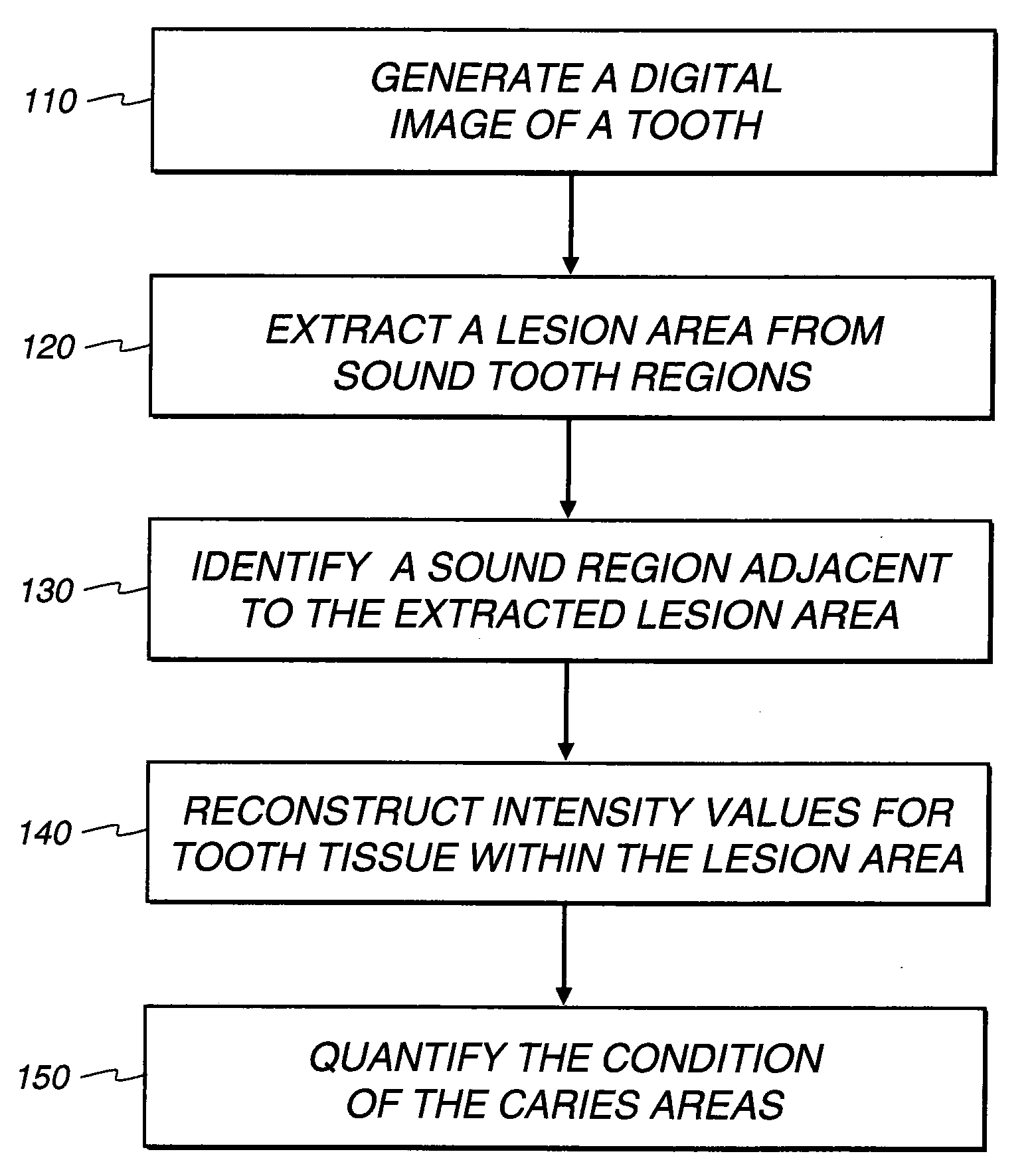
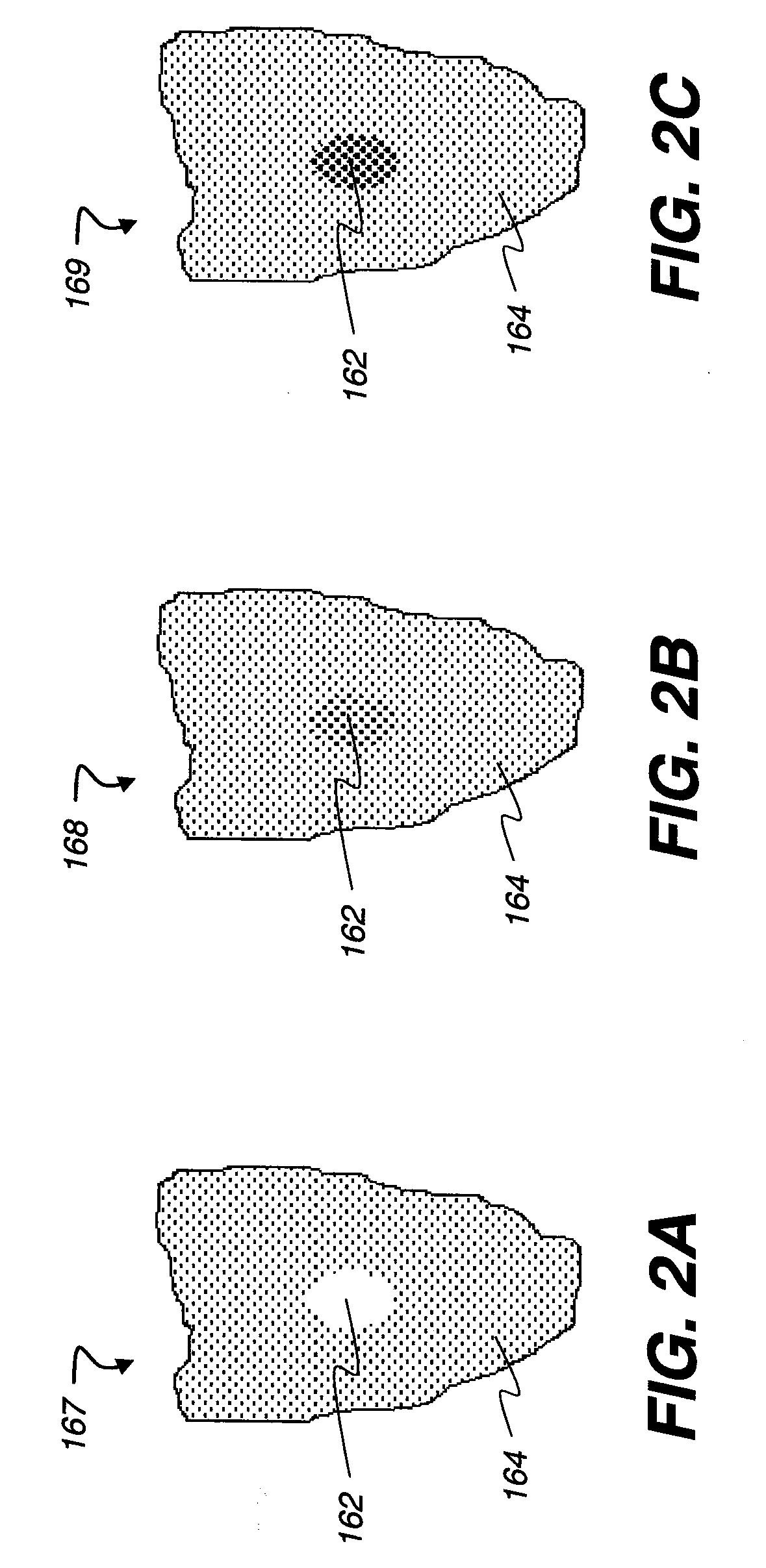
[0047]The following is a detailed description of the preferred embodiments of the invention, reference being made to the drawings in which the same reference numerals identify the same elements of structure in each of the several figures.

[0048]Reference is made to PCT / CN2009 / 000078, filed on Jan. 20, 2009, entitled METHOD FOR DETECTION OF CARIES, by Wei Wang et al.

[0049]Reference is made to U.S. Patent Application Publication No. 2008 / 0056551, published Mar. 6, 2008, entitled METHOD FOR DETECTION OF CARIES, by Wong et al.

[0050]Reference is made to U.S. Patent Application Publication No. 2008 / 0063998, published Mar. 13, 2008, entitled APPARATUS FOR CARIES DETECTION, by Liang et al.

[0051]Reference is made to U.S. Patent Application Publication No. 2008 / 0170764, published Jul. 17, 2008, entitled SYSTEM FOR EARLY DETECTION OF DENTAL CARIES, by Bums et al.

[0052]Reference is made to U.S. Patent Publication No. 2007 / 0099148, published on May 3, 2007, entitled METHOD AND APPARATUS FOR DETEC...


PUM


Abstract

A method for quantifying caries, executed at least in part on data processing hardware, the method comprising generating a digital image of a tooth, the image comprising intensity values for a region of pixels corresponding to the tooth, gum, and background; extracting a lesion area from sound tooth regions by identifying tooth regions, extracting suspicious lesion areas, and removing false positives; identifying an adjacent sound region that is adjacent to the extracted lesion area; reconstructing intensity values for tooth tissue within the lesion area according to values in the adjacent sound region; and quantifying the condition of the caries using the reconstructed intensity values and intensity values from the lesion area.

Description

FIELD OF THE INVENTION[0001]The invention relates generally to the field of dental imaging, and in particular to a method and an apparatus for early detection of caries. More specifically, the invention relates to a method and an apparatus for quantifying caries in tooth images captured using fluorescence and scattering of light.BACKGROUND OF THE INVENTION[0002]While there have been improvements in detection, treatment and prevention techniques, dental caries remains a prevalent condition affecting people of all age groups. If not properly and promptly treated, caries could lead to permanent tooth damage and even to loss of teeth.[0003]Traditional methods for caries detection include visual examination and tactile probing with a sharp dental explorer device, often assisted by radiographic (x-ray) imaging. Detection using these methods can be somewhat subjective, varying in accuracy due to many factors, including practitioner expertise, location of the infected site, extent of infect...

Claims


Application Information

Patent Timeline
Patent Type & Authority Applications(United States)
IPC IPC(8): G06K9/00
CPCA61B5/0088G06T7/00G06T7/0012G06T2207/30036G06T2207/10064G06T2207/20212G06T2207/10024A61B5/4547A61B5/06A61B5/064A61B10/00A61C19/04A61C19/06
Inventor PAN, LIANGLIANGYAN, JIAYONGWANG, WEISHI, LIXINGWONG, VICTOR C.
Owner CARESTREAM DENTAL TECH TOPCO LTD
Why Eureka
Try Eureka