
Methods and reagents for the early detection of melanoma

Inactive Publication Date: 2011-01-13
VERIDEX LCC
View PDF9 Cites 3 Cited by
  • Summary
  • Abstract
  • Description
  • Claims
  • Application Information

AI Technical Summary

Benefits of technology

[0004]The present invention provides a method of identifying a melanoma by: obtaining a tissue sample; assaying and measuring the expression levels in the sample for genes encoding mRNA corresponding to SILV (me20m) (SEQ. ID NO: 1-3), and tyorsinase (TYR) (SEQ. ID NO: 4). TYR was used as a mormalization control that confirms the presence of melanocytes in the tested sample. The invention fur

Problems solved by technology

Cutaneous malignant melanoma is a serious health are problem with at least 62,000 new, invasive melanoma cases diagnosed in people expected in 2008 in the United States, among which 8,200 will die of this disease.

Method used


Image

  • Methods and reagents for the early detection of melanoma
  • Methods and reagents for the early detection of melanoma

Examples


example 1

Patient Clinical and Pathological Characteristics

[0043]204 FFPE skin biopsy tissue specimens were selected from patients with primary melanocytic skin lesions diagnosed at Georgetown University Hospital. The patient series included specimens with 102 unequivocal features of invasive melanoma or benign nevi and 102 specimens with various degrees of cellular atypia. These atypical specimens were initially classified as suspicious / atypical and subsequently resolved by expert dermatopathologists as atypical nevi or malignant melanoma. Two patient samples were excluded because of insufficient RNA yield (less than 350 ng) after a sample preparation step. An additional nine RNA samples were excluded due to the failure of the PCR control. The final sample set eligible for analysis consisted of 193 biopsy tissues (95% of the original sample set) representing 47 melanomas, 48 benign nevi and 98 atypical / suspicious, including 48 atypical nevi and 50 melanomas as assigned by dermatopathologists...

example 2

Tissue Preparation

[0045]Two hundred four tissue samples were collected from individuals diagnosed with primary melanocytic skin lesions. All samples were collected using excisional, punch, or shave biopsy depending on lesion size, depth, and physician judgment and embedded in FFPE blocks.

[0046]Total RNA was isolated from FFPE blocks using a standard High Pure RNA paraffin Kit from Roche (catalogue # 3270289) with the following modifications. Paraffin embedded tissue samples were sectioned according to the size of the embedded tumor (2-5 mm or smaller=9×10 μm, 6-8 mm or greater=6×10 μm). Sections were de-paraffinized according to the manufacturer's instructions. The isolated RNA was stored in RNase free water at −80° C. until used.

[0047]The distribution by biopsy type and extracted RNA yield are presented in Tables 2a and 2b, respectively. Median RNA yields, corresponding to a total average of 10.5, 4.5 and 6 slides were equivalent among all three types of biopsies. No bias in assay ...

example 3

Single One-Step qRTPCR Assays Using RNA-Specific Primers and Cutoff Establishment

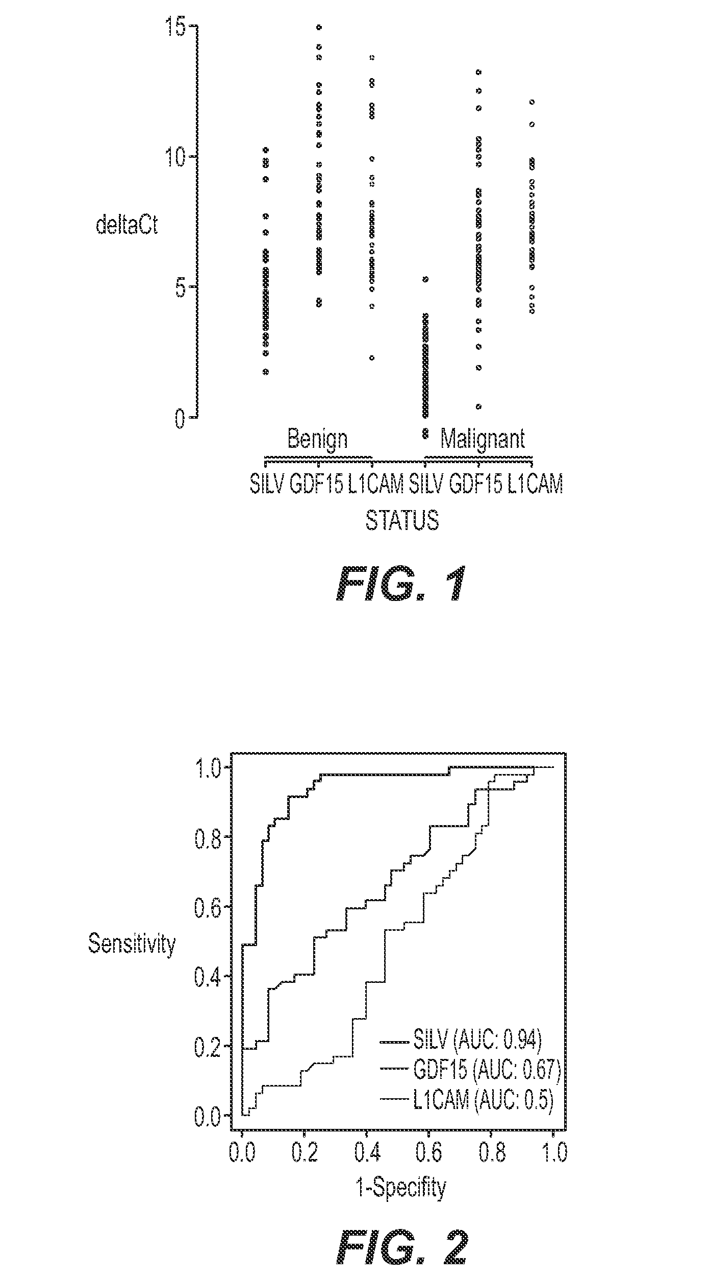
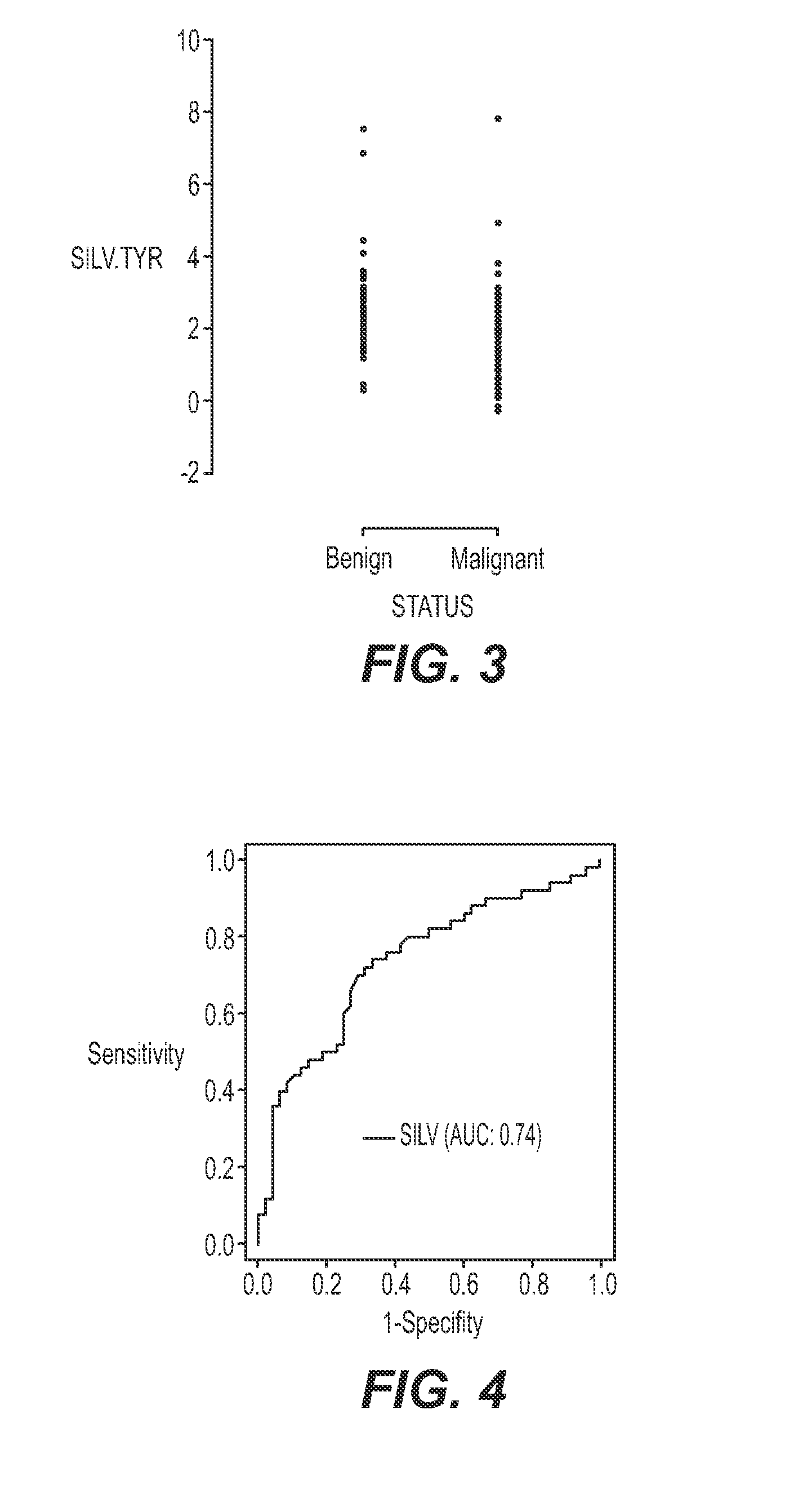
[0048]Evaluation of expression of selected genes was carried out with one-step RT-PCR with RNA from melanoma, benign nevi, and atypical / suspicious FFPE tissue. The specimens included two series of samples: 1) unequivocal, or clear-cut, melanoma and benign nevi cases and 2.) samples with various degrees of atypia. Tyrosinase (“TYR”) was used as a housekeeping gene to control for the input quantity and quality of RNA in the reactions. DNase treatment was not used. Instead, primers or probes were designed to span an intron so they would not report on genomic DNA. All primer / probe sets were pre-screened on a set of 20 total RNA specimens isolated from 10 melanoma and 10 benign nevi FFPE tissues from a commercial vendor (Oncomatrix). The best performing primer-probe set was selected for each of the four markers. The sequences are listed in Table 3 below.

[0049]The gene expression markers GDF-15, SILV, and L1C...


PUM

PropertyMeasurementUnit
Massaaaaaaaaaa
Massaaaaaaaaaa
Massaaaaaaaaaa

Abstract

An assay for identifying early stage malignant melanocyte in biopsy tissues is provided by determining whether differential expression of a particular gene indicative of melanoma exceed a cut-off value.

Description

CROSS-REFERENCE TO RELATED APPLICATIONS[0001]This application is a non-provisional of U.S. Provisional Patent Application No. 61 / 223,894, filed Jul. 8, 2009, which is hereby incorporated by reference in its entirety.BACKGROUND OF THE INVENTION[0002]Cutaneous malignant melanoma is a serious health are problem with at least 62,000 new, invasive melanoma cases diagnosed in people expected in 2008 in the United States, among which 8,200 will die of this disease. The incidence of melanoma continues to increase faster than that of any other malignancy. Also, it is one of the most common cancers in young adults and, thus, is the number two type of cancer in terms of average years of life lost.[0003]Certain proteins have been shown to be associated with melanoma and its metastases. These proteins or their activities have been used in immunohistochemistry to identify metastases and include L1CAM (Thies et al. (2002); Fogel et al. (2003)); and S-100 (Diego et al. (2003)). High-density microar...

Claims


Application Information

Patent Timeline
IPC IPC(8): C40B30/04C12Q1/68
CPCC12Q1/6886C12Q2600/158C12Q2600/112C12Q1/6837C12Q1/6844
Inventor TALANTOV, DMITRIPALMA, JOHN F.VENER, TATIANAJATKOE, TIMWANG, HAIYINGWANG, YIXIN
Owner VERIDEX LCC
Features
  • Generate Ideas
  • Intellectual Property
  • Life Sciences
  • Materials
  • Tech Scout
  • Unparalleled Data Quality
  • Higher Quality Content
  • 60% Fewer Hallucinations
Learn More