Laser-perforated intra-parenchymal micro-probe

a micro-probe and laser-perforated technology, applied in the field of medical and neuroscience instruments for research, clinical diagnostics and therapy, can solve the problems of frequent clogging of cannulae, inability to collect and deliver large particles,

Inactive Publication Date: 2011-03-24
LENOX LASER CORP
View PDF3 Cites 1 Cited by
  • Summary
  • Abstract
  • Description
  • Claims
  • Application Information

AI Technical Summary

Benefits of technology

[0004]The Laser-Perforated Intra-Parenchymal Micro-Probe (“LAPP”) comprises a fluid manifold body having inlet and outlet ports connected respectively to the interior volume of a nested, coaxial dual-lumen cannula or microtube, in which the inlet port feeds the inner cannula, and the outlet port drains the annular volume (external to the inner cannula and internal to the outer cannula), such that the tip of the outer cannula is sealed, and the only fluid access between fluid inside the microtube assembly and the external biological tissue in which the microtube is inserted is provided by an array of laser-perforated apertures having a uniform size selected to enable extraction of molecules or fluid-borne material, but excluding any material of size greater than that of the apertures. Conversely, the aperture size also allows delivery of size-limited fluid-borne material. Connection of the inlet and outlet ports to independently programmable fluid pumps allows operation of the micro-probe according to a variety of protocols, enabling sampling (extraction) or delivery of fluid-borne material with net zero or non-zero fluid volume extracted or delivered, along with positive sampling or delivery of the fluid-borne molecules or material.

Problems solved by technology

This method was widely used in the sixties and seventies; it then became clear that the technique has the serious problems of frequent clogging of the cannulae by tissue or by clotting of fluids and damage to tissue by fluid build-up around the open-end cannula-tips.
Yet, the very innovation that gave birth to this technique, the use of the microdialysis membrane, led to another problem: the inability to collect and deliver large particles, including such critical biological substances as proteins; these particles and molecules are very large and cannot pass through the membrane.

Method used


Image

  • Laser-perforated intra-parenchymal micro-probe
  • Laser-perforated intra-parenchymal micro-probe
  • Laser-perforated intra-parenchymal micro-probe

Examples


Embodiment Construction

Intra-Parenchymal Micro-Probe Structure

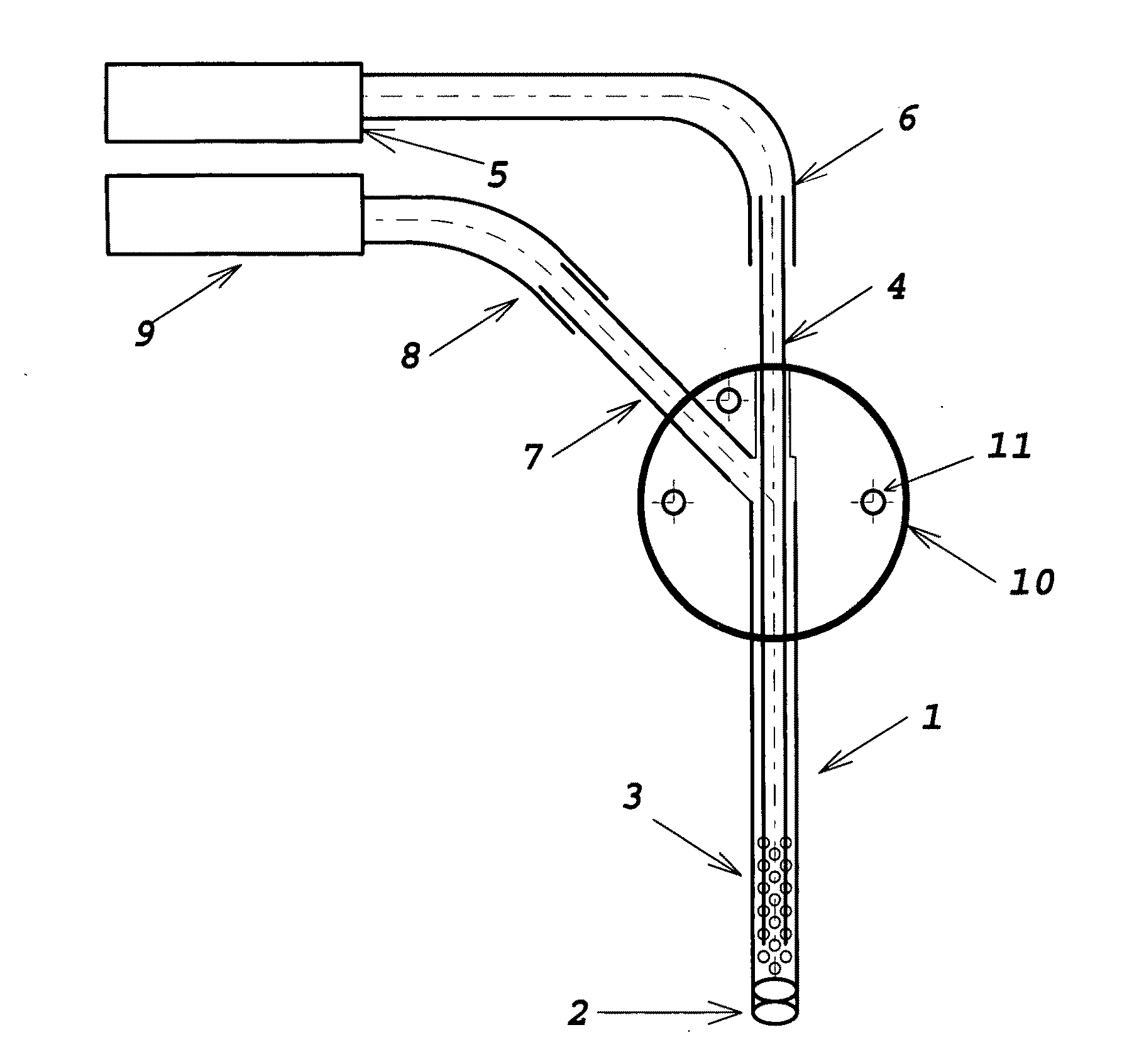
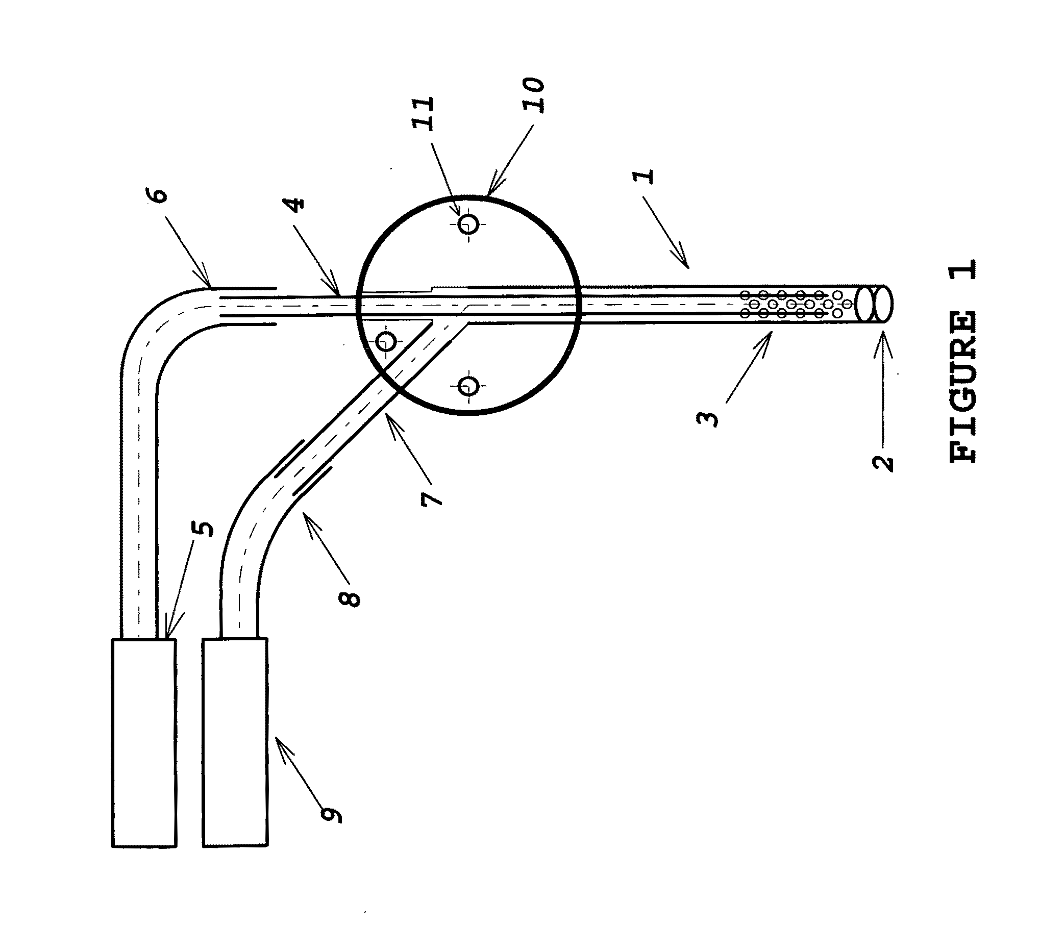
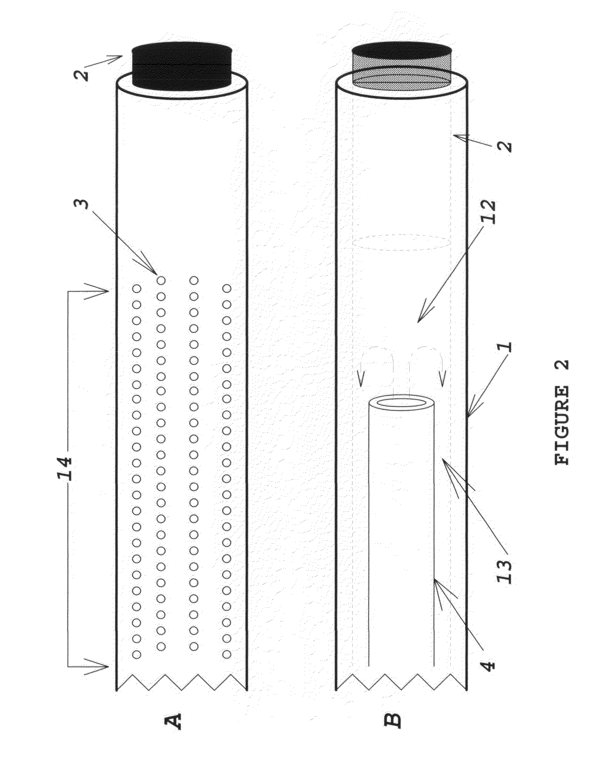
[0012]The device comprises multiple sections of thin-wall tubing, retained in intersecting bores in a multi-port manifold body. The manifold and tubing provide access for fluid extraction or delivery. The manifold body may be fabricated from stainless steel, titanium, ceramic, glass, acetyl, or some other biocompatible material. The tubing must also be a biocompatible material, not necessarily the same as that of the manifold body. Appropriate material selection allows fabrication of probes which are compatible with MRI and other diagnostic procedures. The laser-perforated design has the ability to size-selectively exclude materials from extracted or delivered fluid; it may also minimize tissue damage at the sampling or delivery site by distributing the fluid volume interface over multiple small orifices covering a much larger area than a plain needle tip.

[0013]FIG. 1 illustrates a preferred embodiment of the invention. The functional portion o...


PUM


Abstract

Apparatus and methods in which very small volumes of biological fluid-borne material, particularly large molecules such as proteins, may be selectively extracted from or delivered to interstitial fluid (in vivo or in vitro) by means of intra-parenchymal micro-probes inserted in the brain. The primary use of the micro-probe is in neuroscience research, clinical diagnostics or treatment of epilepsy and other neurological conditions; it may also be applied to other organs and biological systems. Eventual human clinical applications may include neurosurgical monitoring, functional tracking of devices or materials introduced in a surgical procedure, or cerebro-spinal fluid sampling.

Description

FIELD OF THE INVENTION[0001]{The field of the invention is medical and neuroscience instrumentation for research, clinical diagnostics and therapy, particularly for size-selective molecular sampling and delivery of fluid-borne agents to and from interstitial fluid in the brain, or in cerebro-spinal fluid in the spinal column.}BACKGROUND[0002]A micro-probe capable of sampling and delivery of relatively large particles, such as protein molecules, cells and microorganisms with minimal fluid transfer or trauma to selected sites in the brain may be of great utility in neuroscience research, clinical diagnostics or treatment of epilepsy and other neurological conditions.[0003]Relevant prior art includes the use of the push-pull cannula, which comprises two adjacent, open-end cannulae with one cannula carrying the “pushed fluid” downward, whereas the other one carrying the “pulled fluid” upward, creating an open molecule-exchange zone at the tip of the two cannulas. This method was widely ...

Claims


Application Information

Patent Timeline
Patent Type & Authority Applications(United States)
IPC IPC(8): A61B10/02A61M1/00A61N1/36
CPCA61N1/36017A61B10/02
Inventor LUDVIG, NANDORBLUMENFELD, WALTERMEDVECZKY, GEZA
Owner LENOX LASER CORP
Why Eureka
Try Eureka