

Antibody library display by yeast cell plasma membrane
a technology of yeast cell plasma membrane and antibody library, which is applied in the field of antibody library display by yeast cell plasma membrane, can solve the problems of not only laborious process, but also phage display technology, and rely on the screening of antibody fragments rather than full-length antibodies
- Summary
- Abstract
- Description
- Claims
- Application Information
AI Technical Summary
Benefits of technology
Problems solved by technology
Method used
Image


Examples
example 1
6.1 Example 1
Antibody Display on Yeast Plasma Membrane
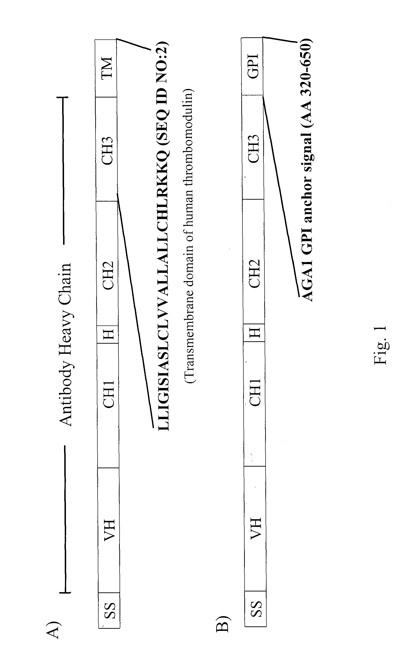
[0301]The following sections describe the generation and characterization of antibody fusion polypeptides that are efficiently displayed on the extracellular surface of the yeast plasma membrane.
6.1.1 Construction of Light Chain Display Vector
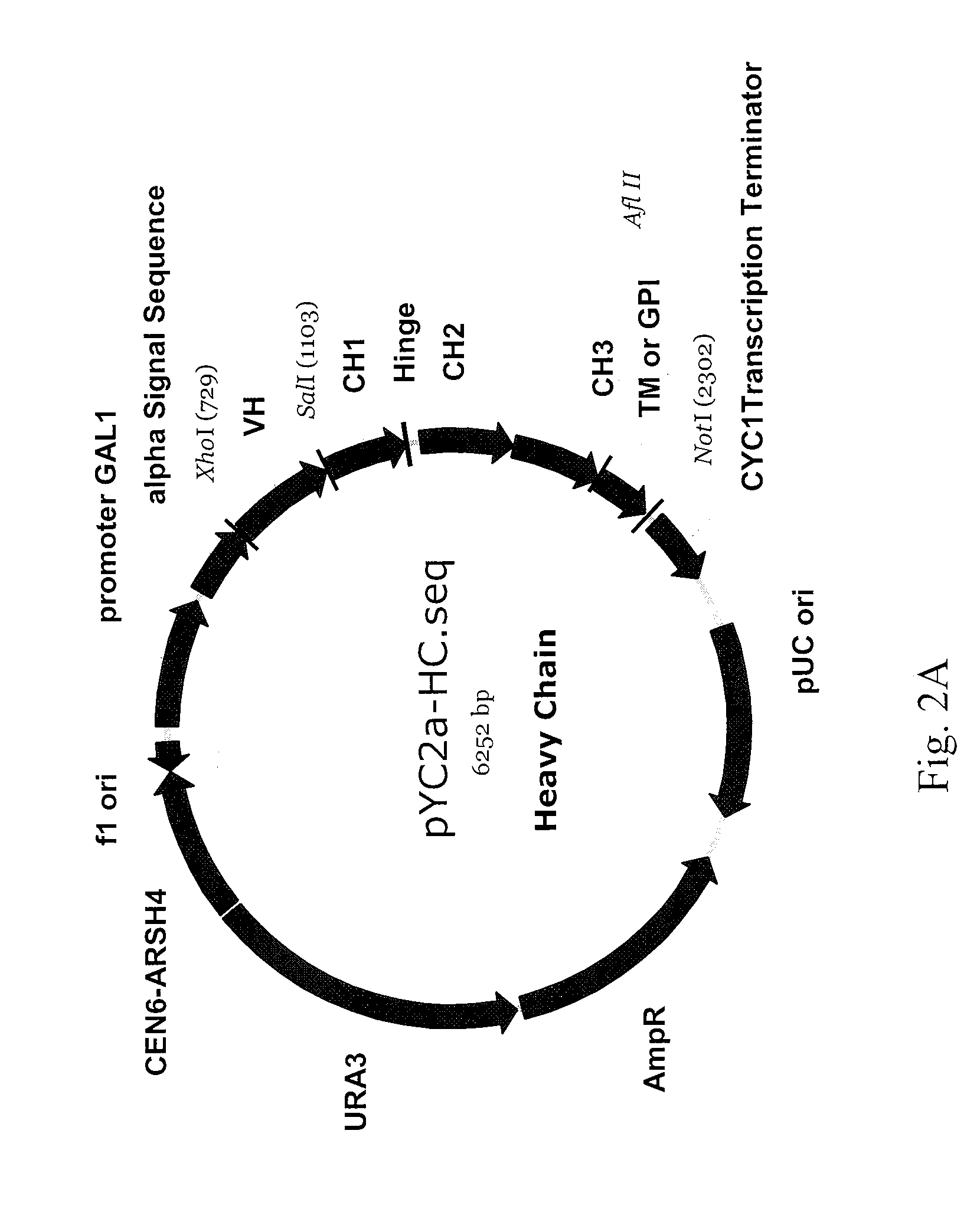
[0302]A novel NheI restriction site was introduced into pYD1 (Invitrogen) at the 3′ end of the nucleotide sequences encoding the Aga2 signal peptide using the QuickChange kit (Stratagene) and the YD1 (SEQ ID NO:26) and YD2 (SEQ ID NO:27) primers. A polynucleotide encoding the full length light chain of the 12G3H11 anti-EphA2 antibody (SEQ ID NO:60) was PCR amplified using the YD3 (SEQ ID NO:28) and YD4 (SEQ ID NO:29) primers, digested with Nhe I restriction endonuclease, and ligated into the NheI PmeI digested modified pYD1 vector described above. The resulting light chain display vector is designated as pYD-LC; a schematic representation of pYD-LC is depicted in FIG. 2B. Primers used herein...
example 2
6.2 Example 2
Antigen Binding Properties of Yeast Plasma Membrane Displayed Antibodies
[0310]The following sections describe experiments demonstrating that antibodies displayed on the extracellular surface of the yeast plasma membrane can efficiently bind their target antigen
6.2.1 A Yeast Plasma Membrane Displayed 10C12 Antibody Efficiently Binds its Target Antigen
[0311]S. cerevisiae cells comprising the pYD-LC-10C12 / pYCa-HC-ThrmTM-10C12 antibody display vector pair are subjected to galactose induction as described in 5.1.3. Following induction, cells are harvested and treated with 100 U / ml lyticase in a 0.5 M sorbitol containing buffer pH 9.6 for 30 minutes at 30° C. Spheroplasts are incubated with biotinylated EphA4-Fc fusion protein (50n / ml) in FACS buffer (PBS (pH 7.2), 0.5% BSA, 1 mM EDTA) for 30 minutes at room temperature. Spheroplasts are subsequently washed with FACS buffer, followed by incubation with 1:200 fold diluted PE conjugated streptavidin (Pierce) on ice for 30 minut...
example 3
6.3 Example 3
. Method for Screening an Artificial Yeast Plasma Membrane Displayed Full Length Antibody Library
[0330]The following section describes a non-limiting example of method for screening and isolating cells displaying a full antibody on their plasma membrane from a population of yeast cells comprising an artificial library. The population of yeast cells comprising an artificial library is generated by mixing yeast cells comprising either the pYD-LC-10C12 / pYCa-HC-ThrmTM-10C12 (full length antibody) or the pYD-LC-3F2 / p3F2Fd-ThrmTM (antibody fragment) antibody display vector pair at a ratio of 1 to 104.
[0331]First round of selection: Yeast cells comprising the pYD-LC-10C12 / pYCa-HC-ThrmTM-10C12 or the pYD-LC-3F2 / p3F2Fd-ThrmTM antibody display vector pair are grown in CAA / 2% glucose medium overnight at about 30° C. on an orbital shaker (about 300 rpm). Cells are harvested, resuspended in CAA-RGD (2% raffinose, 2% galactose, 0.1% glucose) medium and incubated overnight at about 20...
PUM
| Property | Measurement | Unit |
|---|---|---|
| pH | aaaaa | aaaaa |
| pH | aaaaa | aaaaa |
| pH | aaaaa | aaaaa |
Abstract
Description
Claims
Application Information