If not properly and promptly treated, caries could lead to
permanent tooth damage and even to loss of teeth.
There are also hazards associated with conventional detection techniques, including the risk of damaging weakened teeth and spreading infection with tactile methods as well as
exposure to x-
ray radiation.
By the time a caries condition is evident under visual and tactile examination, the
disease is generally in an
advanced stage, requiring a filling and, if not timely treated, possibly leading to
tooth loss.
A recognized drawback with existing techniques that employ fluorescence imaging relates to
image contrast.
The image provided by fluorescence generation techniques such as QLF can be difficult to assess due to relatively poor contrast between healthy and infected areas.
As noted in the '2356 Stookey et al. disclosure, spectral and intensity changes for incipient caries can be very slight, making it difficult to differentiate non-diseased
tooth surface irregularities from incipient caries.
In such cases, detection accuracy using fluorescence techniques may not show marked improvement over conventional methods.
Because of this shortcoming, the use of fluorescence effects appears to have some practical limits that prevent accurate diagnosis of incipient caries.
As a result, a caries condition may continue undetected until it is more serious, requiring a filling, for example.
As noted previously, conventional techniques generally fail to detect caries at a stage at which the condition can be reversed.
More advanced caries, however, grows increasingly more difficult to treat, most often requiring some type of filling or other type of intervention.
In many cases, as is acknowledged in the '2356 Stookey et al. disclosure, this level of detection has been found to be difficult to achieve using existing fluorescence imaging techniques, such as QLF.
As a result, early caries can continue undetected, so that by the time positive detection is obtained, the opportunity for reversal using low-cost preventive measures can be lost.
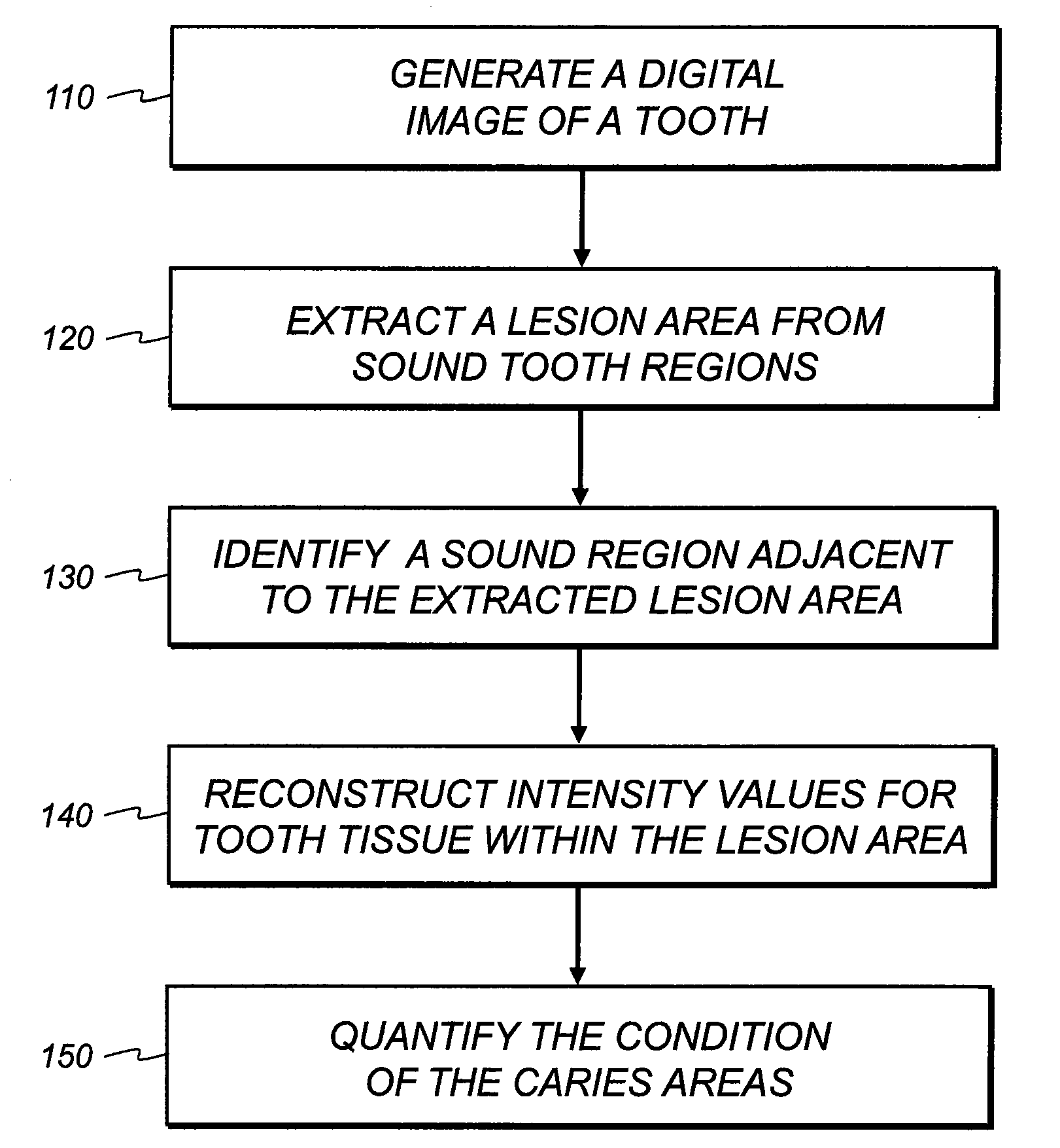
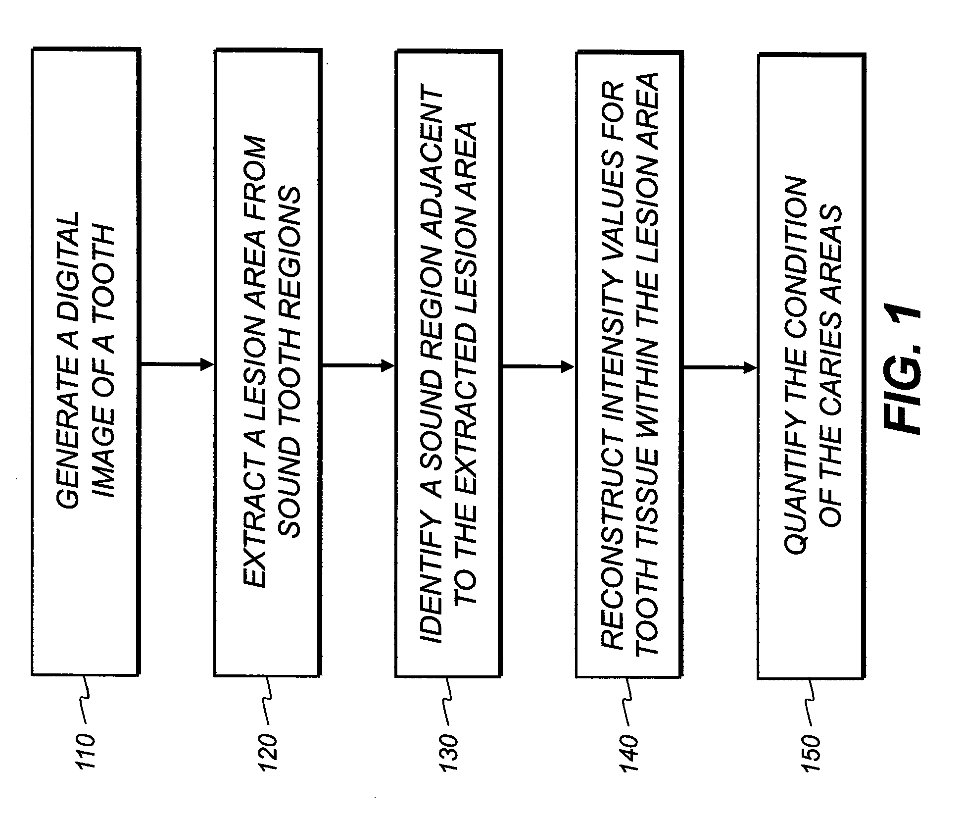
The extraction process is slow, requiring the user to make many mouse clicks or to draw lines on the images to indicate the boundary of a
lesion region.
Secondly,
manual extraction requires considerable caries diagnostic experience on the part of the user and is generally subjective.
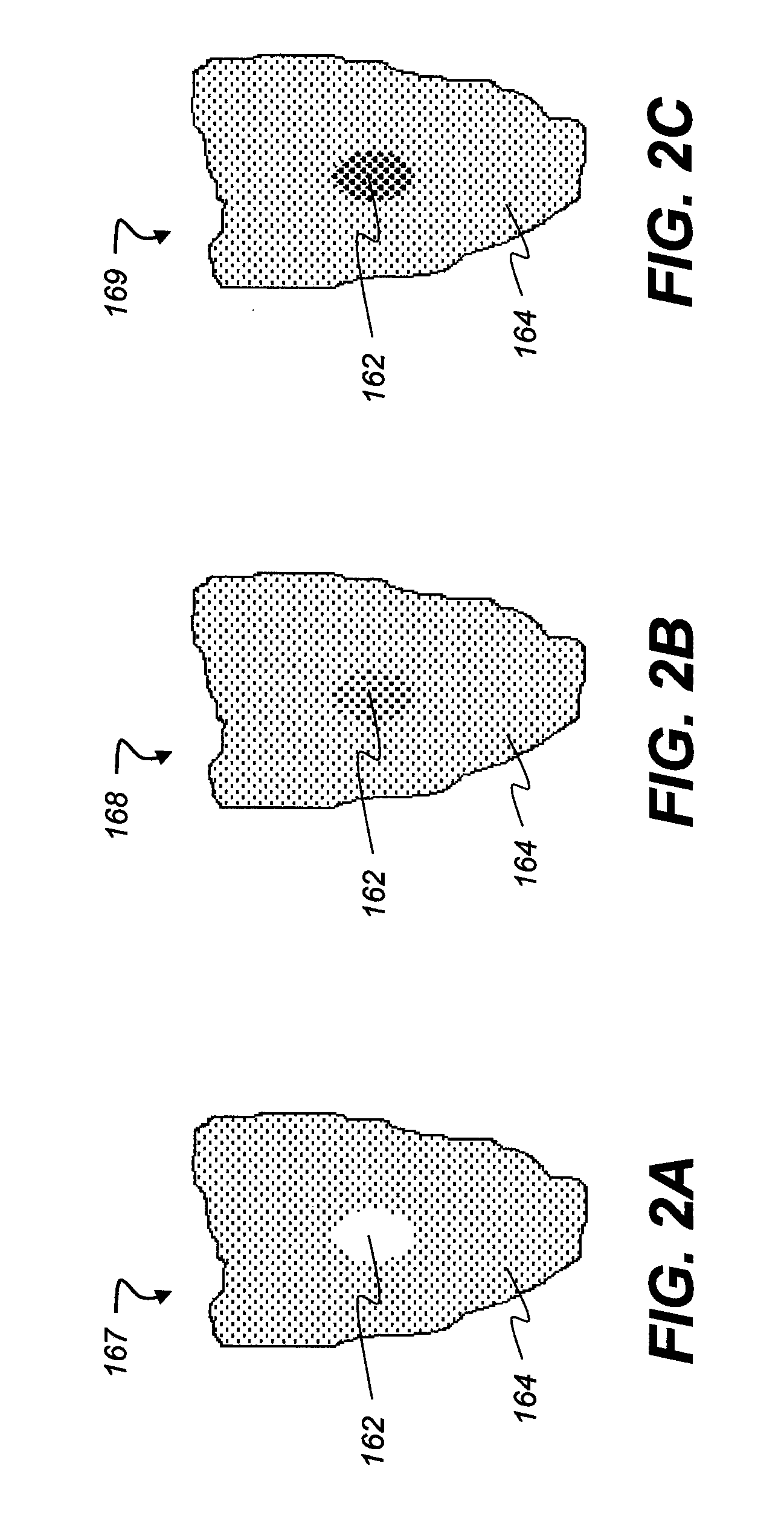
In addition, fluorescence-only images display incipient caries at relatively
low contrast, further adding difficulty to the manual
lesion extraction process.
Therefore, in the disclosed methods, only compromised caries quantification results are achieved, at best.
Additionally, it is difficult to detect or locate caries that are at the interproximal tooth regions.
However, these known methods are not applicable to a fluorescence image, reflectance image, or FIRE image because of significant differences in
image content and characteristics.

