Positron emission tomography and optical tissue imaging

a tomography and optical tissue technology, applied in tomography, material analysis using wave/particle radiation, instruments, etc., can solve the problems of increasing morbidity, increasing the overall cost of the procedure, and adding substantial idle time to the surgery, so as to reduce the total time needed for the procedure and the number of iterations. , the effect of fast feedback

Inactive Publication Date: 2011-04-21
JEFFERSON SCI ASSOCS LLC
View PDF5 Cites 7 Cited by
  • Summary
  • Abstract
  • Description
  • Claims
  • Application Information

AI Technical Summary

Benefits of technology

It is therefore an object of the present invention to provide a method and apparatus for the rapid determination of the boundaries of cancerous tissue and the determination of the presence of adequate margins thereabout after resection during surgery.
According to the present invention there is provided a mobile compact imaging system that combines both PET and optical imaging into a single system w

Problems solved by technology

If the margins are not adequate, this process may be repeated several times before the surgery is concluded, adding substantial idle time to the surgery.
For the patient, the time lost during this proc

Method used


Image

  • Positron emission tomography and optical tissue imaging
  • Positron emission tomography and optical tissue imaging
  • Positron emission tomography and optical tissue imaging

Examples


Embodiment Construction

Positron Emission Tomography with F-18 fluoro-deoxyglucose (FDG) has been shown to be a valuable tool for imaging various cancers. Many cancers demonstrate increased glucose metabolism compared to normal tissues. When FDG is injected intravenously, it concentrates in tumors allowing them to be imaged. In the protocol used in connection with the imaging system of the present invention, one hour before surgery, the patient is injected with a small amount of a positron emitting radiopharmaceutical such as FDG. When the surgeon has removed the cancerous lesion, he or she will mark the resected tissue and the surgical bed with dyes to map orientation for analysis of margins.

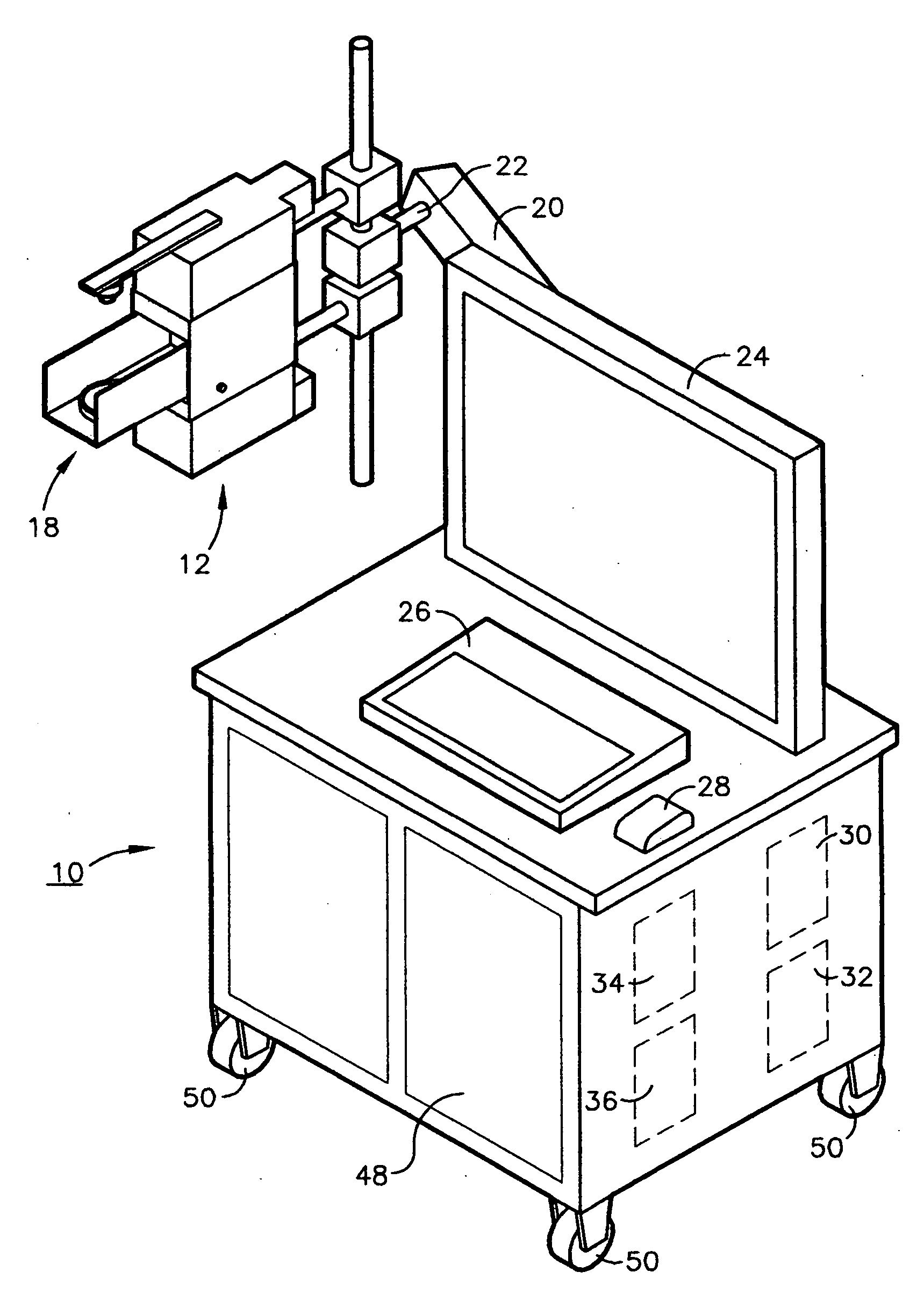
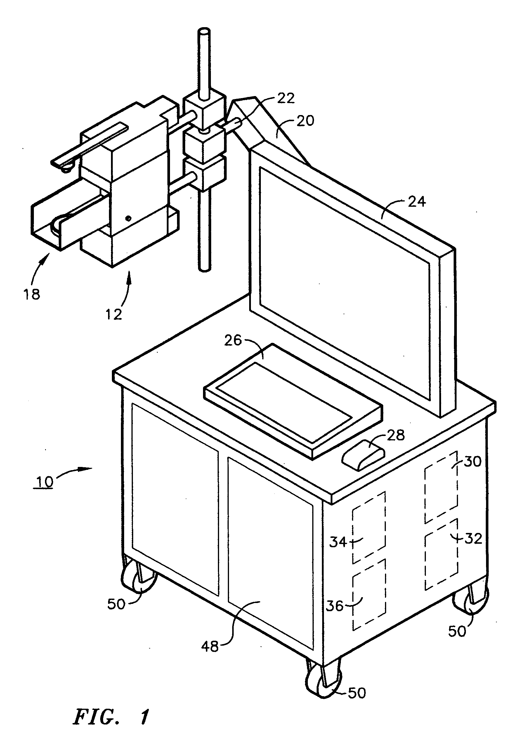
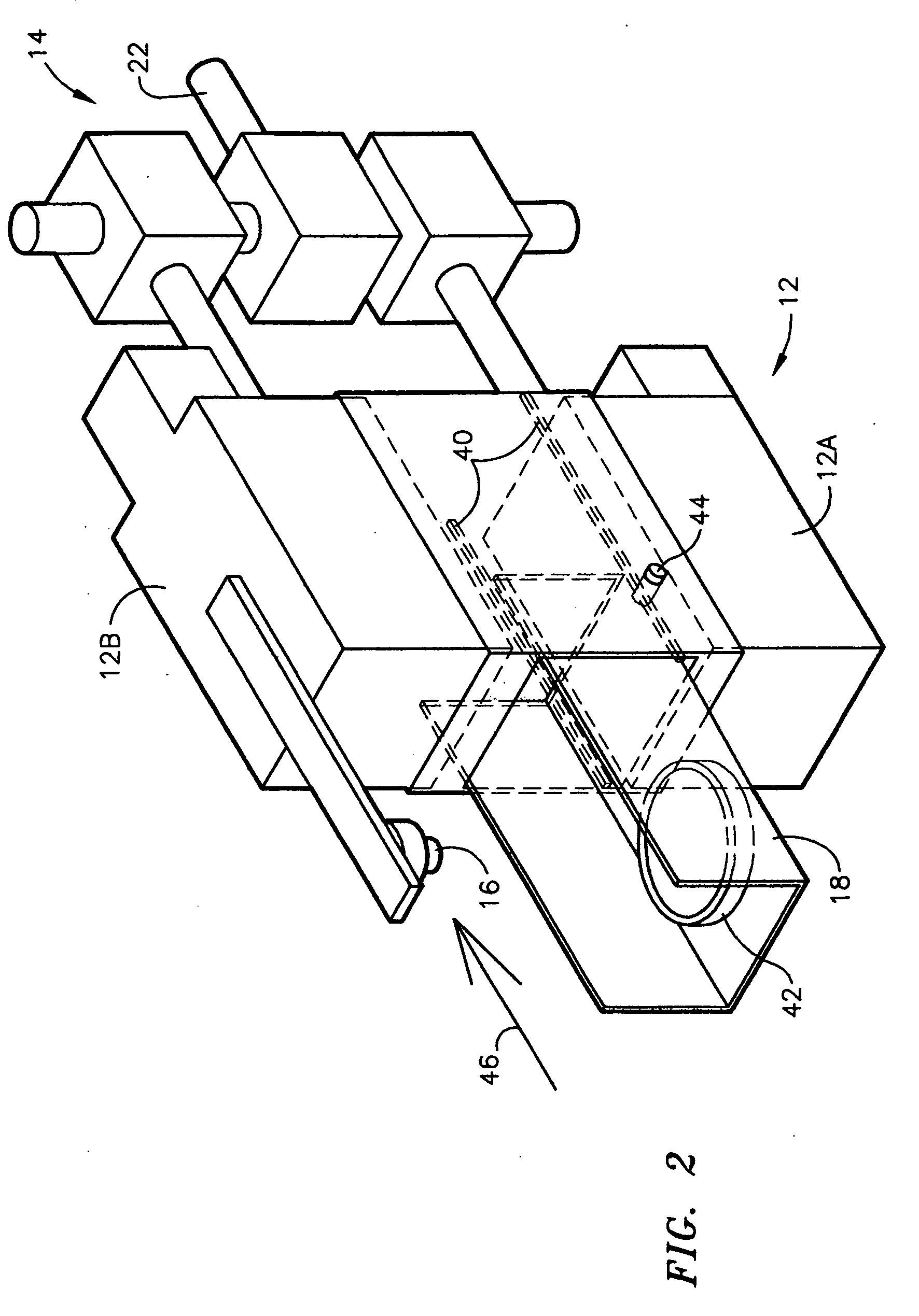
The Positron Emission Tomography (PET) Tissue Sample Imager of the present invention uses a pair of small PET cameras to image radiotracer uptake in the resected tissue sample. The cancerous tissue will typically be identified as a focal area or areas of increased radiotracer accumulation within the sample. The PET Ti...


PUM


Abstract

A mobile compact imaging system that combines both of the imaging system of and optical imaging into a single system which can be located in the operating room (OR) and provides faster feedback to determine if a tumor has been fully resected and if there are adequate surgical margins. While final confirmation is obtained from the pathology lab, such a device can reduce the total time necessary for the procedure and the number of iterations required to achieve satisfactory resection of a tumor with good margins.

Description

FIELD OF THE INVENTION The present invention relates to positron emission tomography (PET) imaging and more particularly to a combined PET and optical imaging system for the rapid determination of adequate cancerous tissue resection and surgical margins during surgical procedures for the removal of cancerous tissue.BACKGROUND OF THE INVENTION During cancer surgeries, one of the important concerns is to determine if there are satisfactory surgical margins around the cancerous tissue. Ideally, the surgeon would like to remove all of the tumor, with adequate margins around the cancerous tissue, but without taking out too much of the healthy surrounding tissue. Typically, after the lesion is surgically removed, the tissue is sent to a pathology lab to determine if the margins around the cancer are sufficient. This process can take 20-30 minutes or more to get a confirmation that the tumor has been excised and that there are adequate surgical margins. During that time, the surgeon(s) mus...

Claims


Application Information

Patent Timeline
IPC IPC(8): G01T1/166
CPCG01N23/046G01T1/1603G01N2223/419A61B6/037G01T1/2985
Inventor FALEN, STEVEN W.HOEFER, RICHARD A.MAJEWSKI, STANISLAWMCKISSON, JOHNKROSS, BRIANPROFFITT, JAMESSTOLIN, ALEXANDERWEISENBERGER, ANDREW G.
Owner JEFFERSON SCI ASSOCS LLC
Why Eureka
Try Eureka