
Devices and Methods for Minimally Invasive Access to Sinuses and Treatment of Sinusitis

a technology of sinuses and minimally invasive access, applied in the field of sinuses and sinusitis treatment, can solve the problems of difficult to execute atraumatically, cumbersome x-ray fluoroscopy and transillumination, and difficulty in advancing to the infundibulum, so as to facilitate broad application, facilitate broad application, and facilitate the effect of learning and execution

Inactive Publication Date: 2011-04-21
COVELLO LEONARD V
View PDF23 Cites 0 Cited by
  • Summary
  • Abstract
  • Description
  • Claims
  • Application Information

AI Technical Summary

Benefits of technology

"The present invention provides devices and methods for refining existing minimally invasive methods of accessing the sinuses for surgery and other treatments. These devices and methods offer a way to perform these procedures in an office setting with minimal anesthesia and little or no postoperative limitations of activity or adverse symptoms. The invention also includes a middle turbinate retractor for retracting the middle turbinate, a hole punch for making a perforation in the uncinate process, a guide-free dilator for dilating the natural ostium of the maxillary sinus, and a probe capable of making a perforation in the wall of the ethmoid bulla. These devices and methods offer improved access to the sinuses for surgery and other treatments."

Problems solved by technology

A problem results, however, if a surgeon creates a new ostium posterior to the natural ostium, which often does cause recirculation, or if a surgeon enlarges an accessory ostium thinking it to be the natural one, which will not achieve the objective and might, as discussed above, cause recirculation.
Both X-ray fluoroscopy and transillumination are cumbersome to a degree, fluoroscopy especially so as it requires large machines that are not normally present at sinus surgery and get in the way of easy, interruption-free operating.
In the transnasal approach, the advance to the infundibulum is awkward.
Those two maneuvers necessitate a 180 degree turn of the guide, guide wire and balloon catheter that is difficult to execute atraumatically in the tight spaces involved.
Another substantial problem with the transnasal approach is that the method is performed blindly because the surgeon's view of the natural ostium and any intervening accessory ostia is blocked by the more medial uncinate.
However, that does not prevent inadvertent introduction into and cannulation of an accessory ostium, rather than the natural ostium, caused by the surgeon's inability to see the position of the instrument.
As discussed above, that is not helpful and often harmful.
Protecting against that eventuality is difficult.
Using a balloon to dilate the now visible natural ostium is of questionable benefit compared to the standard surgical approach of trimming the posterior margin of the ostium—likely a mere victory of style over substance.
Surgeons that would prefer a minimally invasive method will often revert to the unsatisfactory “hybrid” procedure because of frustration with the awkward approach to the natural ostium or because of concern that they will inadvertently and unknowingly dilate the wrong (accessory) ostium.
The anatomic evidence, however, is overwhelmingly against that possibility.
Nonetheless, the problems with the canine fossa approach are substantial.
Thus, it is not easily and naturally combined with intranasal procedures.
Moreover, it is more difficult or impossible to access the ethmoid, frontal, and sphenoid sinuses from the canine fossa access site.
Access to the anterior ethmoid is possible, but cumbersome.
Access to the frontal sinus is quite difficult, and access to the posterior ethmoid and sphenoid is virtually impossible.
Surgeons must utilize another “hybrid” invasive / non-invasive procedure for those sinuses that require a second incision site and a cumbersome combination of nasal and oral procedures.
The primary incision required by the canine fossa approach might be undesirable to patients seeking less rather than more surgery.
The endoscope is understandably fragile with a short (25 procedures) life and therefore carries a rather high cost per use.
Consequently, serious injuries to the eye and brain during sinus surgery can occur in approaches to the ethmoid.
Minimally invasive methods for ethmoid surgery have not progressed as far as those methods for maxillary surgery.
This deficiency is likely due to the significant anatomic variation of the anterior ethmoid anatomy and confusion within the field as to the location of its mucus outlets.
The benefits from these topical agents, however, have been limited.
There is reason to suspect that the limitation is due to an inability to direct the agents to the diseased areas of intact sinuses in sufficient concentrations to be effective, rather than to some more fundamental shortcoming.
There are certainly some drawbacks to this method, however.
Taken together, the cost of the device and the nature of its placement do not lend themselves to repeated use in an office setting; rather, they are more suited to a single treatment or very occasional treatments in a surgical setting.
As many patients have some degree of ongoing medical sinus disease, this is a significant drawback.
This gap in current treatment options is largely because of the presence of the uncinate, which prevents direct atraumatic access to the maxillary sinus, frustrating the guide-based system currently used.

Method used


Image

  • Devices and Methods for Minimally Invasive Access to Sinuses and Treatment of Sinusitis
  • Devices and Methods for Minimally Invasive Access to Sinuses and Treatment of Sinusitis
  • Devices and Methods for Minimally Invasive Access to Sinuses and Treatment of Sinusitis

Examples


first embodiment

[0208]FIG. 42A shows the drug delivery device of the present invention, consisting of a drug containing matrix (120) with an embedded degradable framework (121). The framework (121) is anchored by a spine (122) that follows the axis of the ellipsoid matrix. A series of ribs (123) protrude radially from the spine. Coplanar ribs (123) are depicted here as three in number, but could be more; ideally, no less than three would occupy a single transverse plane of the ellipsoid. Alternatively, the ribs (123) could stack in a spiral. The tip (124) of each rib would ideally protrude just past the surface of the matrix (120) in the nondegraded state. A cross-sectional view of the framework (121) is shown in FIG. 42B. As engineered, the framework (121) should degrade more slowly than the matrix (120) revealing more and more of the ribs (123). Thus, as the matrix resorbs and grows smaller, the retention device remains intact. The tips (124) of the ribs remain in contact with the sinus mucosa, b...

second embodiment

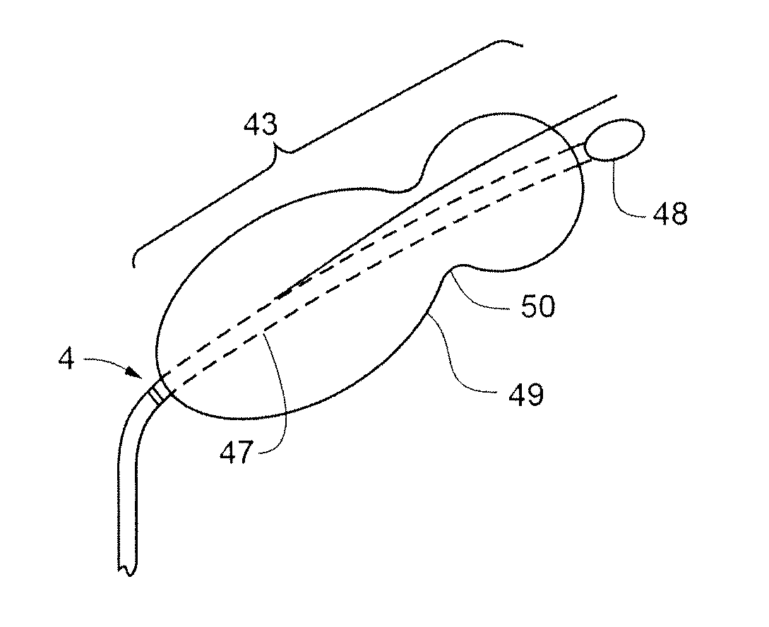
[0209]FIG. 43A shows the drug delivery device of the present invention. FIG. 43B shows a cross-sectional view. The drug delivery device comprises a matrix (130) and an embedded degradable framework (131) design. In addition, this framework (131) is also anchored by a coaxial spine (132). Distinct from the embodiment depicted in FIG. 42A, however, is an expansile “umbrella” of ribs (133) also attached radially to the spine (132); here the attachment is outside the matrix (130) at one apex. The umbrella (134) is collapsed around the surface of the matrix (130) within the receptacle (see FIGS. 38 and 39) of the insertion device. As shown in FIG. 43C (and cross-sectional view FIG. 43D), the umbrella (134) then expands upon extrusion into the sinus. The retention and degradation properties of the drug delivery device are similar to those outlined in the discussion of the embodiment illustrated in FIG. 42A.

third embodiment

[0210]FIG. 44A shows the drug delivery device of the present invention. FIG. 44B shows a cross-sectional view. The drug delivery device shares with the earlier embodiments the matrix (140) and embedded degradable framework (141) design. It, too, can be anchored by a coaxial spine (142), though this is not strictly necessary. Here, the matrix is surrounded at its surface by a cage (143) that is embedded in but projects just above the surface of the matrix (140) to enhance its retention properties at the outset. As the matrix (140) degrades, it remains within the confines of the cage (143) until the matrix (140) fragments become small enough to extrude through the openings in the cage (143). Other retention and degradation properties are similar to those outlined above.

Benefits of the Present Invention

[0211]Unique to the present invention, the combination of the minimally invasive anterior keyhole approach, the described guide-free drug placement devices, and the absence of any retain...


PUM


Abstract

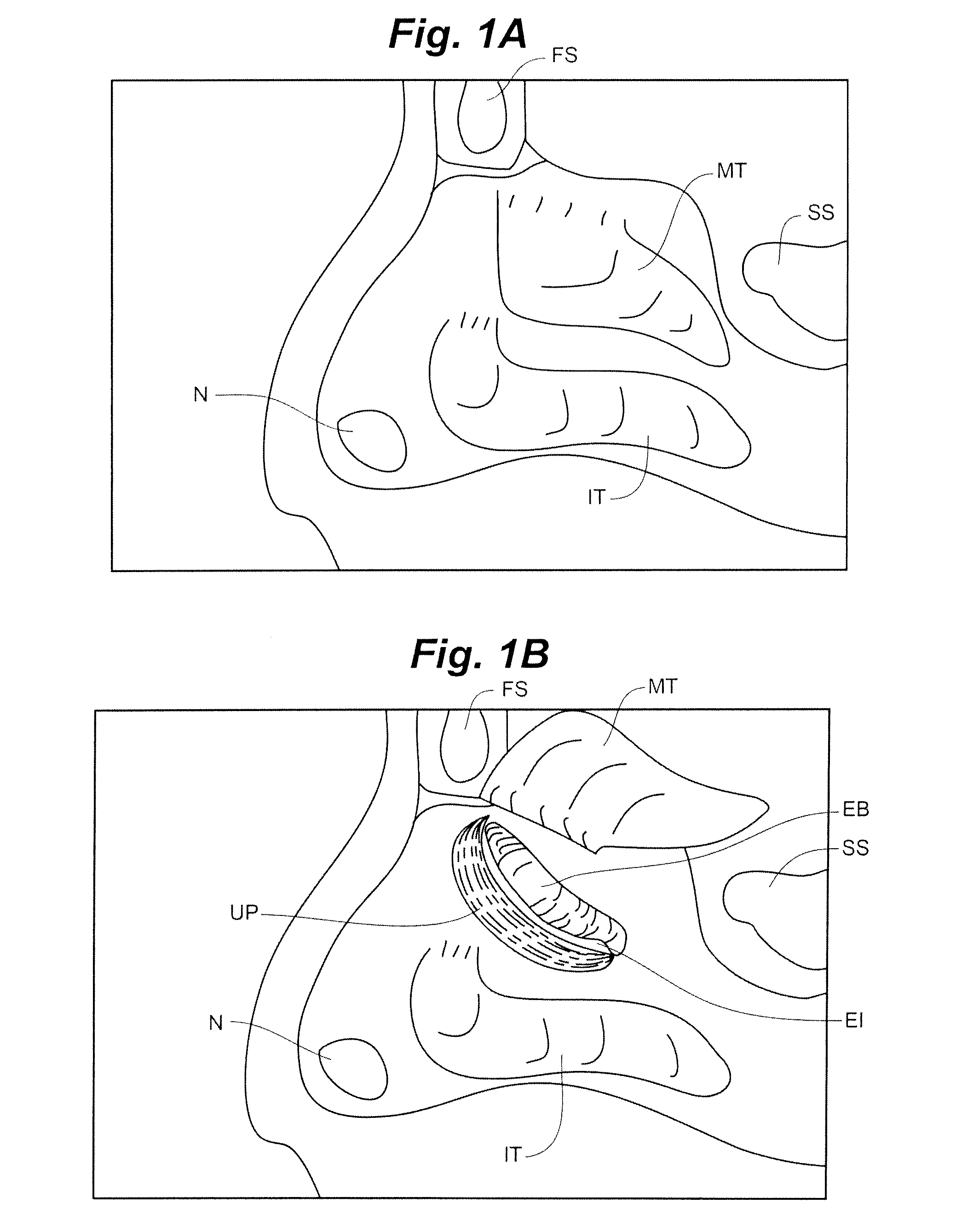
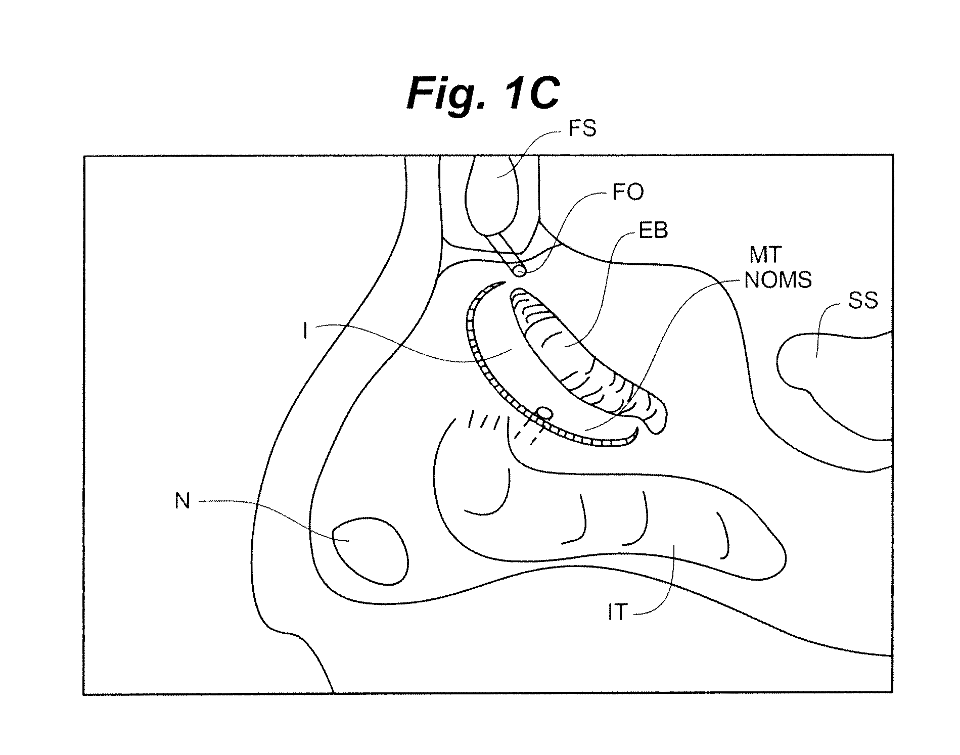
The present invention provides minimally invasive devices and methods for accessing the sinuses and their surrounding structures for surgery and other treatments. The anterior ethmoid and maxillary sinuses are accessed and treated under minimal anesthesia with little or no postoperative limitation of activity or adverse symptoms. Direct visual verification of the sinuses and their natural ostia is utilized. Other paranasal sinuses may be treated by this method as well. The sinuses, in particular the maxillary and anterior ethmoid, are accessed via a direct anterior to posterior axis and the natural ostia of those sinuses is directly visualized for placement of a guide-free dilator, in the desired location within the natural ostia. That access to the maxillary ostium is accomplished by the anterior transuncinate “keyhole” approach in which a hole is punched in the uncinate process with the described devices according to the described methods. The properly placed dilator is expanded to allow drainage of the inflamed sinus and then withdrawn. An analogous ethmoid bulla “keyhole” approach and subsequent dilation are used for the anterior ethmoid sinus ostia. Pharmaceutical agents may be placed at desired locations in the sinuses using the same access technique.

Description

CROSS-REFERENCE TO RELATED APPLICATION[0001]This application is a divisional of application Ser. No. 12 / 804,398, filed Jul. 20, 2010, which claims the benefit of Provisional Application No. 61 / 271,500, filed on Jul. 21, 2009, the contents of which are incorporated herein by reference.FIELD OF INVENTION[0002]The present invention relates to minimally invasive devices, systems and methods for accessing the sinuses of a human patient and for treating sinusitis.BACKGROUND OF INVENTION[0003]There are a substantial number of people with sinus inflammatory diseasesinusitis—that could benefit from sinus surgery. Patients with sinusitis can be grouped according to the severity of their sinusitis into those with mild and those with severe anatomic evidence of sinusitis. The latter category includes those patients with significant anatomic anomalies, patients previously operated on who have substantial postoperative defects in the diseased areas, and those with significant paranasal sinus pol...

Claims


Application Information

Patent Timeline
Patent Type & Authority Applications(United States)
IPC IPC(8): A61M29/02A61M25/10A61F2/958
CPCA61B17/24A61B17/295A61B17/32053A61M25/1002A61B17/3478A61B2019/4889A61B17/3468A61B2090/0817A61B90/08A61B2017/320048A61M29/02A61M2205/04A61M2210/0681
Inventor COVELLO, LEONARD V.
Owner COVELLO LEONARD V
Learn More