
Clinical examination disk, disk pack, and clinical examination device

Inactive Publication Date: 2011-05-05
BRAHMS GMBH
View PDF1 Cites 18 Cited by
  • Summary
  • Abstract
  • Description
  • Claims
  • Application Information

AI Technical Summary

Benefits of technology

[0014]An object of the present invention is to provide a clinical examination disk capable of causing a specimen, such as blood plasma separated by centrifugation from a blood, to react, in a reaction tank, with a reagent held therein with a desired precision.
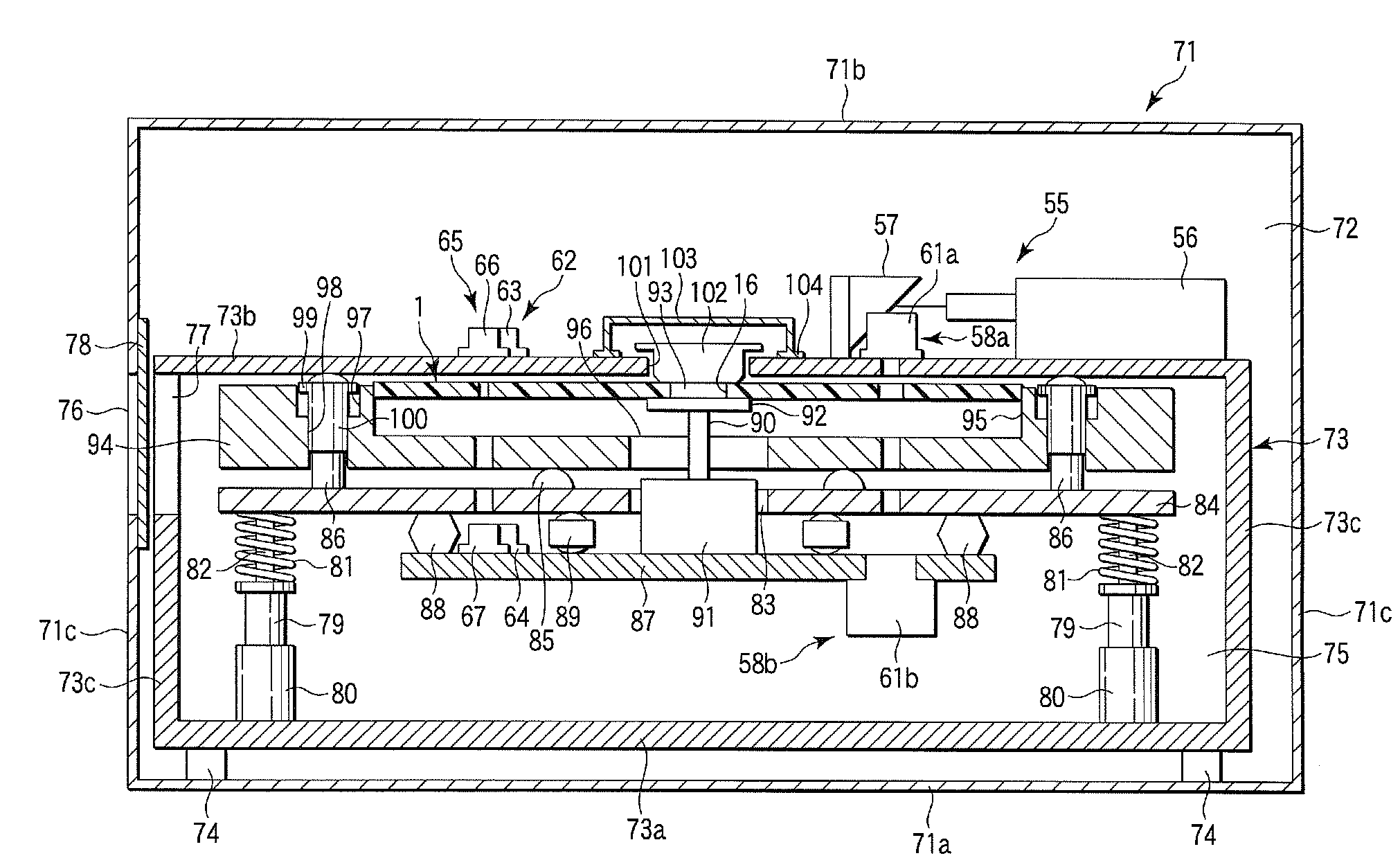
[0016]Still another object of the invention is to provide a clinical examination device capable of maintaining the air-tightness of the inside of a space (chamber) wherein the above-mentioned clinical examination disk is arranged, and further attaining the control of the temperature of the device with a high precision.
[0030]a rotating unit fixed to the linearly-acting unit to cause the disk to be rotated,
[0034]According to the invention, it is possible to provide a clinical examination disk capable of analyzing a specimen, for example, a blood component, such as blood glucose, with a high precision.
[0035]According to the invention, it is possible to provide a disk pack capable of holding stably a gel-form reagent held beforehand in a reaction tank of a clinical examination disk.
[0036]According to the invention, it is possible to provide a clinical examination device that enables precise control over the temperature of reaction between a specimen (for example, blood plasma separated by centrifugation from a blood) and a reagent in a cell of the above-mentioned clinical examination disk which is rotated at a high speed, as well as even liquification of a gelatinized reagent in a reservoir formed in the cell of the disk, before the reaction, thus making it possible to attain a blood examination high in reproducibility.

Problems solved by technology

However, in a clinical examination of a specimen, such as blood, with the rotary cuvette type rotor, for a photometer, described in Jpn. Pat. Appln. KOKOKU Publication No. 55-36937, the reagent-distributing chamber is connected to the sample-analyzing cuvettes through the channels extended linearly in the radial direction; it is therefore difficult for the reagent supplied into the distributing chamber by centrifugal force when the rotor is rotated to be distributed evenly into the individual cuvettes with a desired precision.
However, when the water or buffer liquid is distributed from the reagent-distributing chamber into the individual cuvettes, it is difficult, in the same manner as when the reagent is distributed, for the water or buffer liquid to be evenly distributed into the individual cuvettes with a desired precision.

Method used


Image

  • Clinical examination disk, disk pack, and clinical examination device
  • Clinical examination disk, disk pack, and clinical examination device
  • Clinical examination disk, disk pack, and clinical examination device

Examples


Embodiment Construction

[0060]With reference to some of the drawings, a clinical examination disk and a disk pack according to an embodiment of the invention will be described in detail hereinafter.

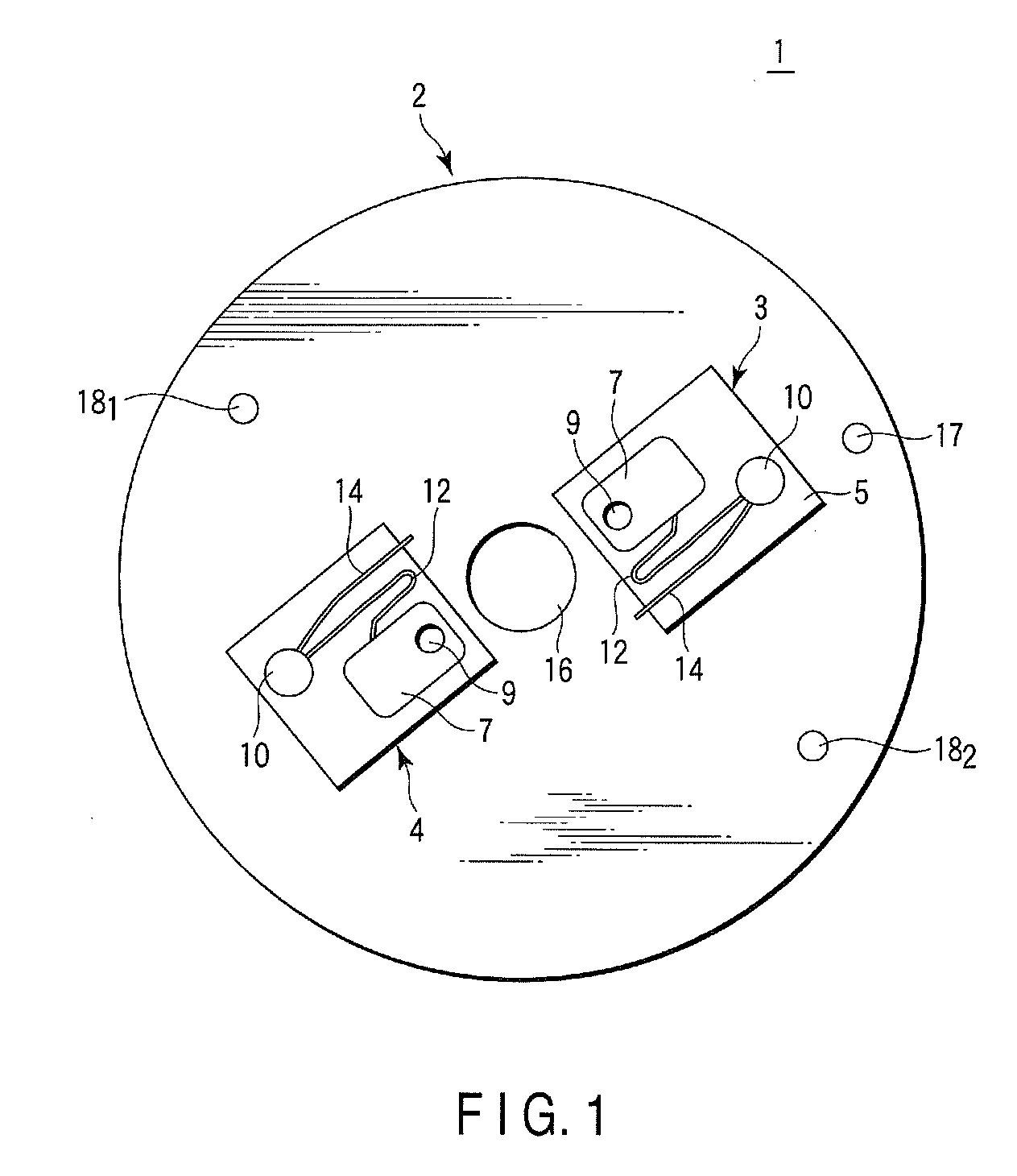
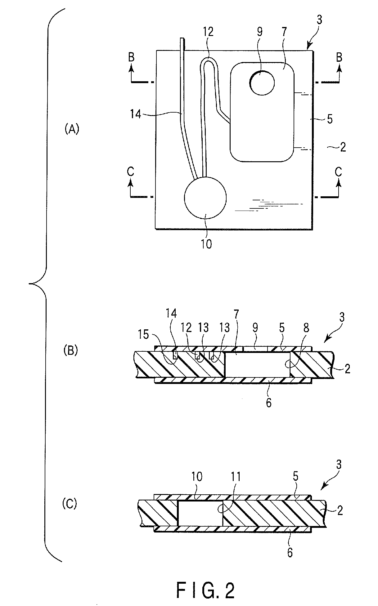
[0061]FIG. 1 is a plan view illustrating the clinical examination disk according to the embodiment. FIG. 2A is an enlarged plan view illustrating one of the cells in FIG. 1, FIG. 2B is a sectional view thereof taken along line B-B in FIG. 2A, and FIG. 2C is a sectional view thereof taken along line C-C in FIG. 2A.

[0062]The clinical examination disk 1 includes a disk body 2 in a disk form. In the disk body 2, for example, two cells (a specimen examining cell 3 and a calibrating cell 4) are formed centrosymmetrically with respect to the center thereof. As illustrated in FIG. 2B and 2C, the specimen examining cell 3 is composed of the disk body 2, and rectangular, front and rear side window plates 5 and 6 which have the same size and are adhered to the front and rear surfaces of the disk body 2, respectively, so as...


PUM


Abstract

A clinical examination disk according to the invention includes a disk body in a disk form, and at least one cell formed in the disk body, wherein the cell comprises a specimen reservoir formed in the disk body and having a specimen injecting port, a reaction tank formed in the disk body so as to position nearer to the outer circumference of the body than a position of the specimen reservoir, and holding a reagent which is reactive with the specimen and is beforehand gelatinized, a first narrow tube formed in the disk body, and communicating to the specimen reservoir and the reaction tank, and a second narrow tube formed in the disk body, and having one end connected to the reaction tank and the other end made open.

Description

CROSS REFERENCE TO RELATED APPLICATIONS[0001]This is a Continuation Application of PCT Application No. PCT / JP2009 / 062382, filed Jul. 7, 2009, which was published under PCT Article 21 (2) in Japanese.[0002]This application is based upon and claims the benefit of priority from prior Japanese Patent Applications No. 2008-180668, filed Jul. 10, 2008; and No. 2008-180669, filed Jul. 10, 2008, the entire contents of both of which are incorporated herein by reference.BACKGROUND OF THE INVENTION[0003]1. Field of the Invention[0004]The present invention relates to a clinical examination disk, a disk pack, and a clinical examination device.[0005]2. Description of the Related Art[0006]In an actual clinical examination, collected blood is centrifuged with a centrifugal separator, and then blood plasma, which is a supernatant, is taken out. The taken-out blood plasma is mixed with a reagent in a reaction vessel to be caused to react therewith. The fluorescence therefrom or the absorbance thereof...

Claims


Application Information

Patent Timeline
IPC IPC(8): G01N33/00
CPCG01N33/491G01N35/00069B01L3/5027B01L2300/0803B01L2400/0409B01L2400/0677B01L2300/0816B01L2300/0887B01L2300/1827B01L2400/0406B01L2200/16
Inventor KAWAMURA, TOSHIMIISHIOKA, SEIICHIROUTONARI, MASAKIKAWAMURA, TAKASHINAKAJIMA, AKIRASEKINO, TETSUOIWAHASHI, KYOICHIUCHIYAMA, KENICHITAKASE, TOMOHIRONAWATA, ISAO
Owner BRAHMS GMBH
Learn More