Novel protein transduction domains derived from secretory leukocyte protease inhibitor

a leukocyte protease inhibitor and protein transduction domain technology, applied in the direction of depsipeptides, peptide/protein ingredients, vector-based foreign material introduction, etc., can solve the problems of ineffective and/or toxic direct microinjection, insufficient cellular localization of slpi, and inefficient intracellular delivery methods such as electroporation, liposome fusion,

Inactive Publication Date: 2011-05-05
RES FOUND THE CITY UNIV OF NEW YORK
View PDF1 Cites 1 Cited by
  • Summary
  • Abstract
  • Description
  • Claims
  • Application Information

AI Technical Summary

Benefits of technology

[0013]The present invention describes for the first time new PTDs derived from secretory leukoprotease inhibitor (SLPI). The present invention provides, in part, SLPI-derived amino acid sequences that function as protein transduction domains (PTDs). These SLPI-derived PTDs may be used to transport proteins, nucleic acids, lipids, carbohydrates, small molecules and other chemical compounds (collectivel

Problems solved by technology

However, existing methods of intracellular delivery, such as electroporation, liposome fusion, gene gun particle bombardment, DEAE-dextran transfection, recombinant viral infe

Method used


Image

  • Novel protein transduction domains derived from secretory leukocyte protease inhibitor
  • Novel protein transduction domains derived from secretory leukocyte protease inhibitor
  • Novel protein transduction domains derived from secretory leukocyte protease inhibitor

Examples


example 1

Recombinant SLPI Localizes to the Cytoplasm and Nucleus of Treated Cerebellar Neurons

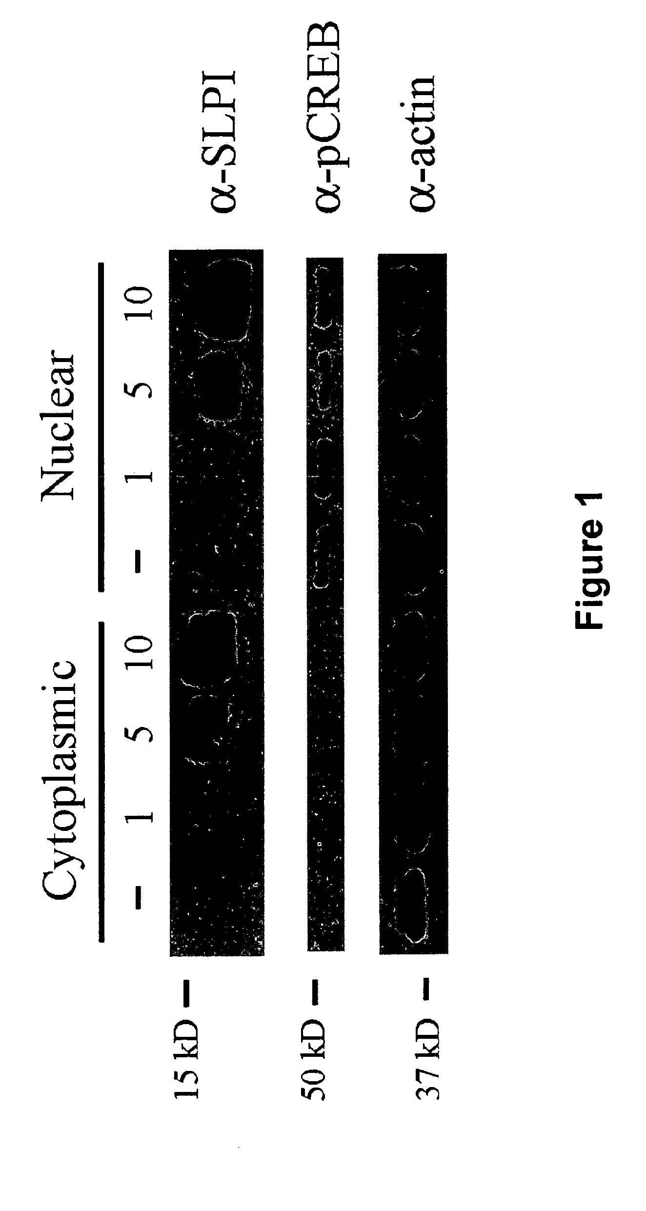
[0122]To characterize the transduction abilities of the SLPI protein, cerebellar neurons were isolated from postnatal day 6 (P6) rats (Mukhopadhyay et al., Neuron 13:757-767 (1994); DeBellard et al., Mol. Cell. Neurosci. 7:89-101 (1996); Cai et al., Neuron 22:89-101 (1999)) and treated with 0, 1, 5, and 10 μg / ml recombinant human SLPI (R&D Systems) in SATO culture medium (Doherty et al., Neuron 5:209-219 (1990); Nature 343:464-466 (1990)). Cells were incubated at 37° C. for 1 hour, and cytoplasmic and nuclear fractions were prepared using NE-PER cytoplasmic and nuclear extraction reagents (Pierce). Protein samples were subjected to SDS-PAGE using 15% SDS polyacrylamide gels and transferred to nitrocellulose. SLPI was detected using a polyclonal antibody to recombinant human SLPI (R&D Systems). SLPI was present in increasing amounts in both the cytoplasmic and nuclear fractions (FIG. 1). The nuclear ...

example 2

SLPI Localizes to the Nuclei of Isolated DRG Neurons

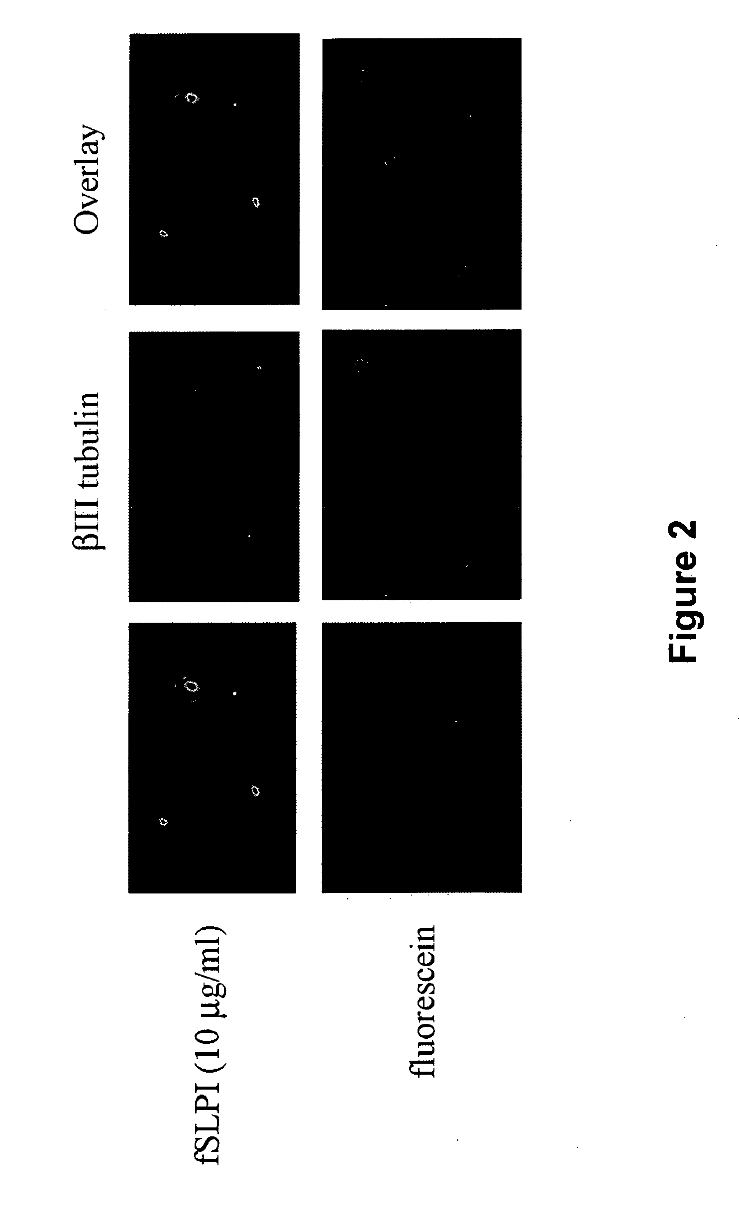
[0123]To visualize internalization of SLPI in living cells, SLPI was labeled with fluorescein using a Fluorescein-EX protein labeling kit (Molecular Probes). The labeling reaction was performed following the protocol provided by the manufacturer. P5 DRG neurons were isolated (Mukhopadhyay et al., 1994; DeBellard et al., 1996; Cai et al., 1999; supra) and treated with either 10 μg / ml of fluorescein-labeled SLPI (fSLPI) or unconjugated fluorescein in SATO culture medium. Cells were then plated in 8 well chamber slides coated with 100 μg / ml poly-D-lysine (PDL) and incubated for 1 hour at 37° C. Cells were then fixed with 4% paraformaldehyde and immunostained using a monoclonal antibody to the neuronal marker βIII tubulin (1:1000 dilution, Covance). After an overnight incubation at 4° C., the cells were rinse and incubated successively with biotinylated anti-mouse antibody (1:500 dilution, Amersham) and Texas Red-conjugated streptavidi...

example 3

Dose-Dependent Internalization of SLPI into Neuronal Nuclei

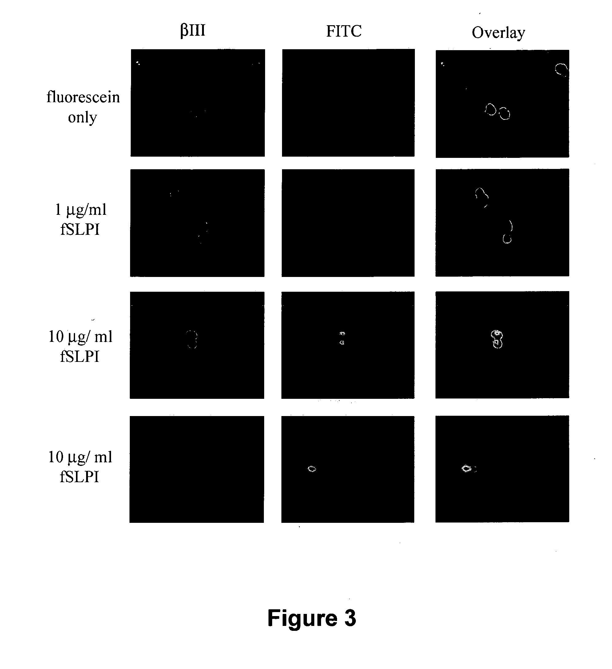
[0124]To further characterize SLPI internalization into living cells, P5 DRG neurons in SATO culture medium were treated with unconjugated fluorescein, 1 μg / ml fSLPI, or 10 μg / ml fSLPI and plated onto PDL-coated slides for 5 minutes at 37° C. Cells were then fixed and immunostained for βIII tubulin as described. In contrast to fluorescein alone, which was not internalized, fSLPI was internalized into nuclei within 5 minutes in a dose-dependent manner (FIG. 3).


PUM

PropertyMeasurementUnit
Compositionaaaaaaaaaa
Therapeuticaaaaaaaaaa

Abstract

This invention relates to novel protein transduction domains (PTDs) derived from secretory leukocyte protease inhibitor (SLPI). SLPI-derived PTDs are able to deliver cargo moieties in vivo and in vitro into the cytoplasm and nucleus of a host cell. The invention also relates to a transduction complex comprising one or more SLPI-derived PTDs linked or fused to one or more cargo moieties, which may comprise, for example, proteins, nucleic acids, lipids, carbohydrates, small molecules and other chemical compounds. The invention also relates to the manufacture of SLPI-derived PTDs, complexes comprising them; compositions comprising SLPI-derived PTDs or complexes; and utilization of SLPI-derived PTDs or complexes comprising them for therapeutic, diagnostic and research methods involving delivery of heterologous molecules across cellular membranes, and especially, across nuclear membranes.

Description

TECHNICAL FIELD OF THE INVENTION[0001]This invention relates to the use of secretory leukocyte protease inhibitor (“SLPI”) and sequences derived from SLPI as protein transduction domains, also known as cell penetrating peptides, useful for transporting heterologous molecules across membranes, such as cellular membranes and especially across nuclear membranes of the cell.BACKGROUND OF THE INVENTION[0002]Cells are generally impermeable to macromolecules such as proteins, nucleic acids, lipids, carbohydrates and other chemical compounds. Methods for efficient translocation of these agents across biological membranes are thus useful for both therapeutic and research applications. However, existing methods of intracellular delivery, such as electroporation, liposome fusion, gene gun particle bombardment, DEAE-dextran transfection, recombinant viral infection, and direct microinjection are often inefficient and / or toxic to cells. Accordingly, there is a continuing need for improved intrac...

Claims


Application Information

Patent Timeline
IPC IPC(8): A01K67/00C07K14/435C07K7/08C07H21/04C12N15/63C12N5/10A01H5/00A61K38/00A61K48/00A61P35/00A61P9/00A61P3/10A61P31/18A61P11/06A61P19/02A61P7/00A61P25/00A61P25/28A61P25/16A61P37/00A61P27/02A61P27/06
CPCA61K47/48246A61K38/00A61K47/64A61P11/06A61P19/02A61P25/00A61P25/16A61P25/28A61P27/02A61P27/06A61P31/18A61P35/00A61P37/00A61P7/00A61P9/00A61P3/10Y02A50/30
Inventor FILBIN, MARIE T.HANNILA, SARI
Owner RES FOUND THE CITY UNIV OF NEW YORK
Why Eureka
Try Eureka