3-d quantitative-imaging ultrasonic method for bone inspections and device for its implementation

a quantitative imaging and ultrasound technology, applied in sonic diagnostics, infrasonic diagnostics, medical science, etc., can solve the problems of unsatisfactory empirical methods, unoptimized ultrasonic imaging systems used for soft tissue study, and unsuitable ultrasonic imaging methods for human body assessmen

Inactive Publication Date: 2011-05-12
GOUREVITCH ALLA
View PDF20 Cites 44 Cited by
  • Summary
  • Abstract
  • Description
  • Claims
  • Application Information

AI Technical Summary

Benefits of technology

[0030]g) Dividing virtually the volume of space containing the layered system into elementary volumes situated in columns and rows; thereby enabling use of a differential approach to processing the obtained data.

Problems solved by technology

Because of the high attenuation coefficient of a bone compared with a soft tissue, ultrasonic imaging methods known in the art are unsuitable for assessing the skeleton of a human body.
Therefore, the conventional ultrasonic imaging systems that are used for studying soft tissue are also not optimized for assessing the skeleton.
Whilst it now forms the basis of clinical QUS bone assessment, this empirical method is not entirely satisfactory.
However, a number of authors argued that the assumption of linear relationship does not have any physical basis.
However the method does not allow estimates of the value of porosity, images of the distribution of porosity values in a considered volume, or 3D imaging of bones in a human body.
But this approach does not provide values for the porosity distribution in a volume being inspected.
Significant mistakes in the values of the parameters are therefore obtained because the method is not sensitive to changes in structure of the object and it ignores a heterogeneous property of the object and its anisotropy.
However, this technique does not apply for investigations of anisotropic and heterogeneous materials and is not used in medicine.
Contrary to X-ray CT, Ultrasonic tomography of elastic wave velocities has not found general practical usage in medical imaging.
The main problems are deviation of the ultrasound beam due to reflection and refraction caused by gas and bone inclusions in the heterogeneous media.
The method cannot account for anisotropy and the heterogeneous nature of the media of which the bone is comprised and thus produces an integral estimation that does not account for the influence of different parts of an inspected heterogeneous media and therefore leads to significant errors.
The method does not provide 3D imaging of the volume of the inspected bone.
The method suffers an additional inaccuracy and inconvenience caused by the necessity of measuring thickness and longitudinal wave velocity in soft tissues by applying an additional Pulse-Echo method.
The method does not estimate the mineral part (matrix) of an inspected bone, porosity values, and porosity distributions in a bone's volume.
The method does not provide the locations and estimations of the risk of fracture.

Method used


Image

  • 3-d quantitative-imaging ultrasonic method for bone inspections and device for its implementation
  • 3-d quantitative-imaging ultrasonic method for bone inspections and device for its implementation
  • 3-d quantitative-imaging ultrasonic method for bone inspections and device for its implementation

Examples


Embodiment Construction

[0109]Before describing at least one embodiment of the invention in detail, it is to be understood that the invention is not limited in its application to the details of construction and the arrangement of the components set forth in the following description or illustrated in the drawings. The invention is capable of other embodiments or of being practiced or carried out in various ways. Also, it is to be understood that the phraseology and terminology employed herein is for the purpose of description and should not be regarded as limiting.

[0110]The drawings are generally not to scale. For clarity, non-essential elements were omitted from some of the drawings.

[0111]As used herein, an element or step recited in the singular and proceeded with the word “a” or “an” should be understood as not excluding plural elements or steps, unless such exclusion is explicitly recited.

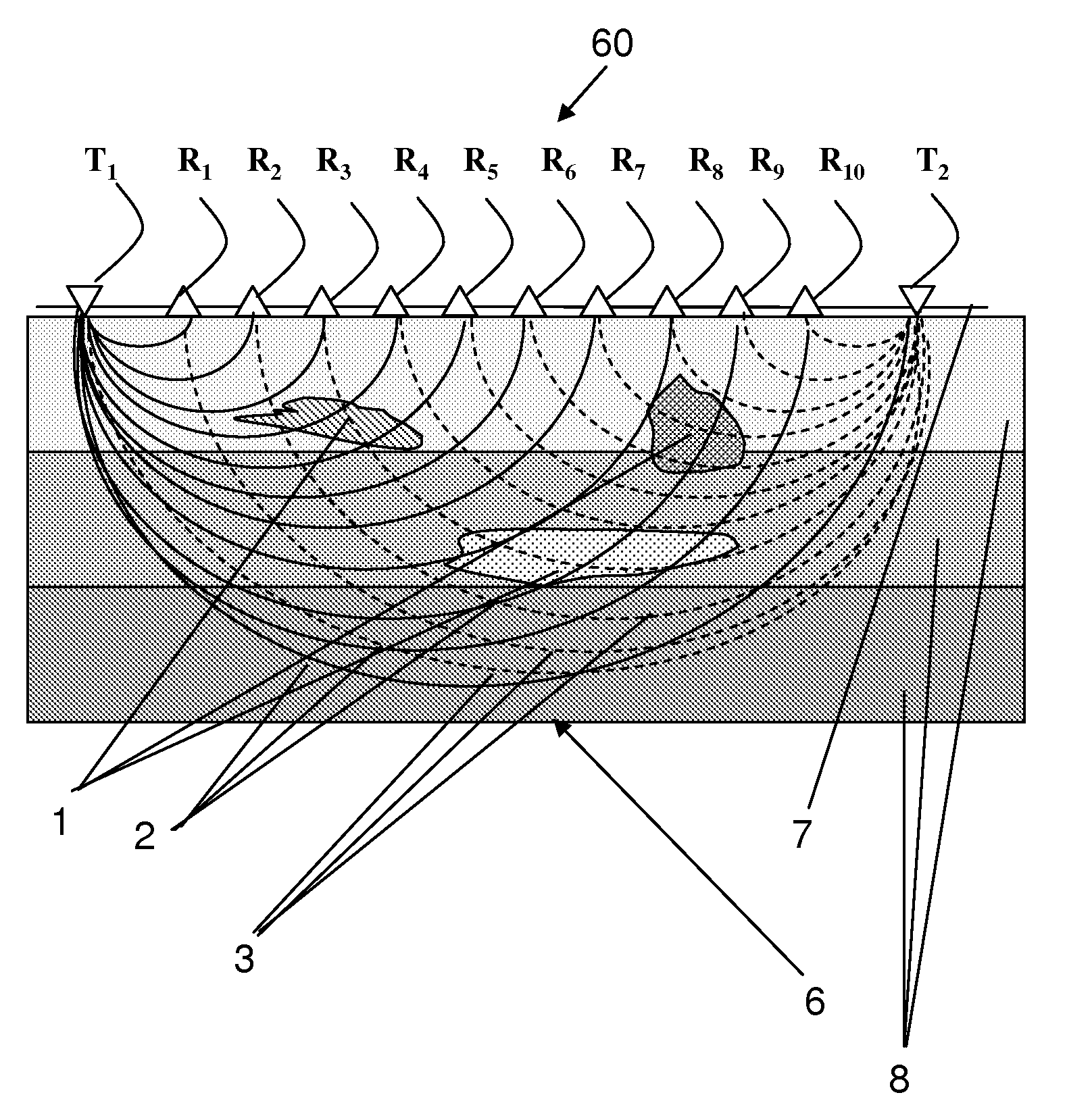
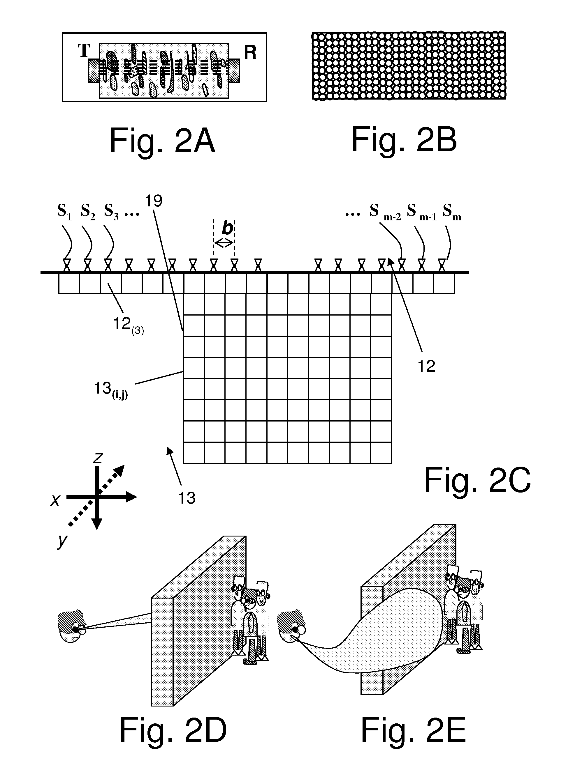
[0112]The invention proposes a novel approach to data measurements and treatments providing the possibility for ins...


PUM


Abstract

The invention is a differential 3D Quantitative-Imaging Ultrasonic Tomography method for inspecting a layered system that is comprised of a heterogeneous object embedded in a volume of space that comprises layers having an acoustic impedance gradient between the layers providing non-linear beams travels. A transducer grid is attached to the surface of the layered system in a unilateral manner. By sequentially changing the distance between transmitter and receiver the fastest travel times of the ultrasonic oscillations are measured. A differential approach provides from the data the longitudinal wave velocities values for each elementary volume that make up an inspected volume. These values are used to construct two and three-dimensional maps of the longitudinal wave velocity values distribution in the inspected volume and the porosity values distribution in the heterogeneous object. By applying statistical treatment to the obtained data the longitudinal wave velocity in the inspected material matrix part is estimated.

Description

[0001]This is a continuation-in-part of U.S. Ser. No. 12 / 444,599 which is a continuation-in-part of PCT / IL2007 / 001283 (WO 2008 / 050333) filed Oct. 24, 2007, which claims priority to 60 / 853,739, filed Oct. 24, 2006, the contents of which are incorporated herewith.FIELD OF THE INVENTION[0002]The present invention relates to the field of quantitative-imaging ultrasound technology and equipment. In particular, the invention relates to the field of as ultrasonic imaging in medical diagnostics and non-medical applications.BACKGROUND OF THE INVENTION[0003]A significant number of ultrasonic methods, which provide for imaging of soft tissues of a human body, exist. They are based for example on the Compound Imaging approach, Stereoscopic imaging, Harmonic imaging, the Spatial Compounding Approach, and other methods. These methods are disclosed for example in: U.S. Pat. No. 6,517,487 and in U.S. Patent Applications 2003 / 0055334, 2003 / 0055337, 2004 / 0193047, 2005 / 0033140, 2005 / 0049479, 2005 / 0124...

Claims


Application Information

Patent Timeline
Patent Type & Authority Applications(United States)
IPC IPC(8): A61B8/14
CPCA61B5/417A61B5/4869A61B8/0875A61B8/485A61B8/4483A61B8/483A61B8/14
Inventor GOUREVITCH, ALLA
Owner GOUREVITCH ALLA
Why Eureka
Try Eureka