Rapid confocal microscopy to support surgical procedures

a confocal microscopy and surgical technology, applied in the field of confocal microscopy with fluorescence to support surgical procedures, can solve the problems of increasing the exposure of patients to infection and undesirable consequences, limiting the availability of surgeons for other procedures, and tedious and time-consuming procedures for preparing histology sections

Active Publication Date: 2011-05-19
SLOAN KETTERING INST FOR CANCER RES +1
View PDF11 Cites 59 Cited by
  • Summary
  • Abstract
  • Description
  • Claims
  • Application Information

AI Technical Summary

Problems solved by technology

However, the procedure for preparing the histology sections is tedious and time consuming, often exceeding 30 minutes per section.
Thus, surgical procedures that rely on such histology sections can extend to two hours and more, increasing the exposure of patient to

Method used


Image

  • Rapid confocal microscopy to support surgical procedures
  • Rapid confocal microscopy to support surgical procedures
  • Rapid confocal microscopy to support surgical procedures

Examples


Embodiment Construction

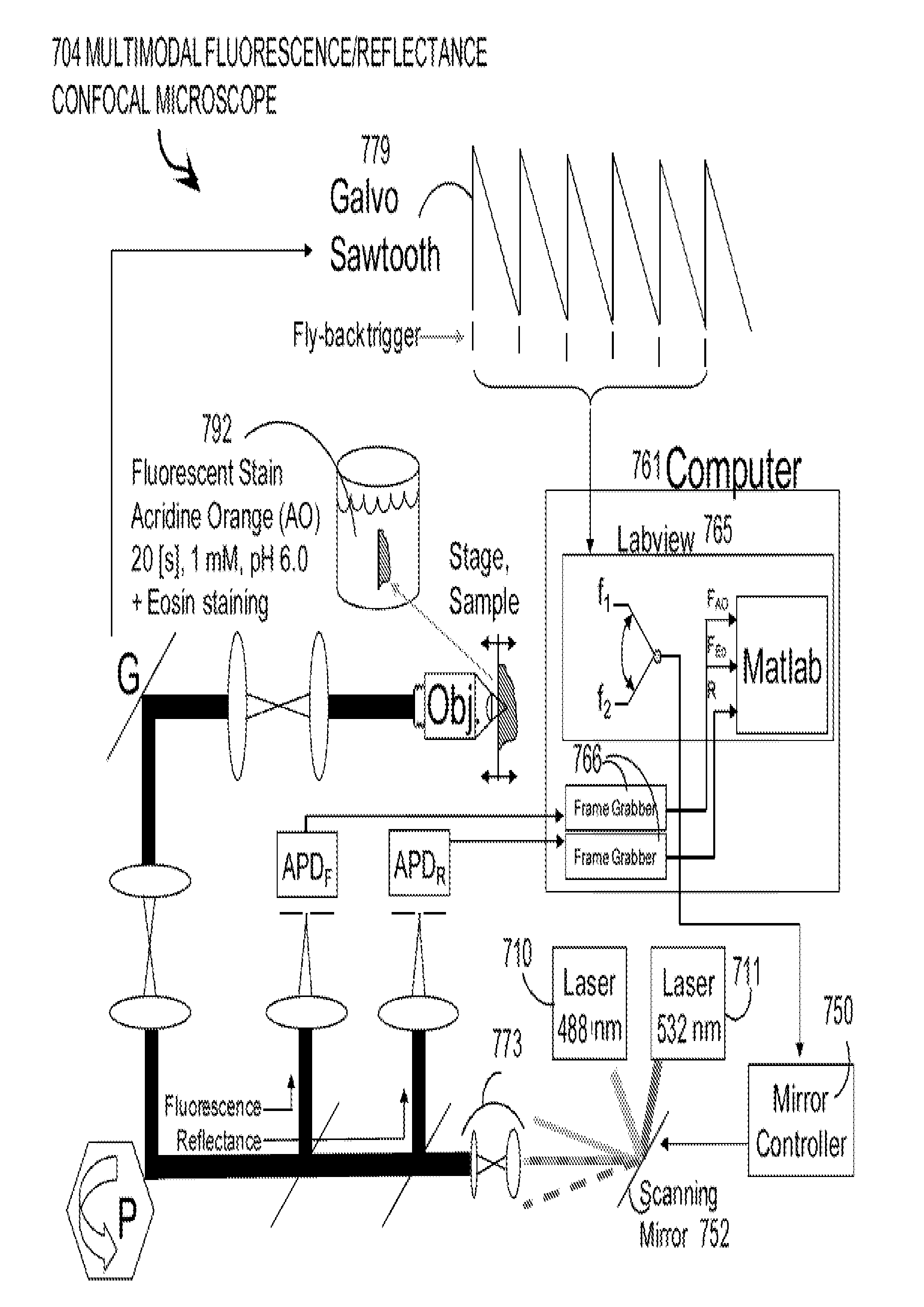
Techniques are provided for confocal microscopy, which offer one or more advantages over prior art approaches.

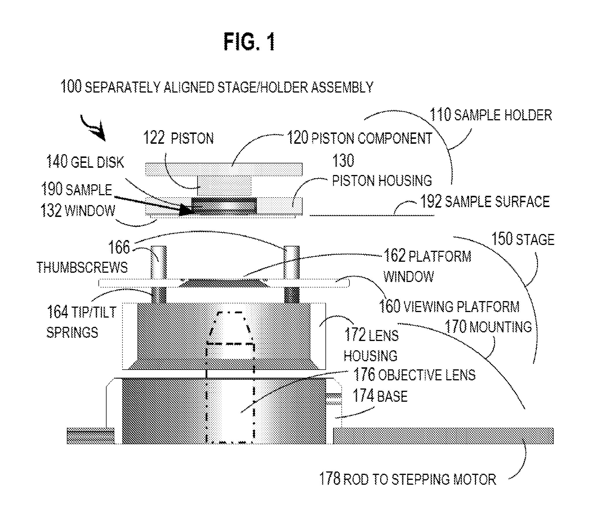
In one set of embodiments, an apparatus for mounting excised tissue for examination by a confocal microscope includes a stage and a sample holder. The stage is configured to be adjusted to align a surface of the stage with a focal plane of a confocal microscope. The sample holder includes a transparent plate configured to compress a sample of excised tissue. The sample holder is removeably mounted to the stage so that the transparent plate is flush with the surface that is aligned with the focal plane of the confocal microscope without further adjustment of the stage.

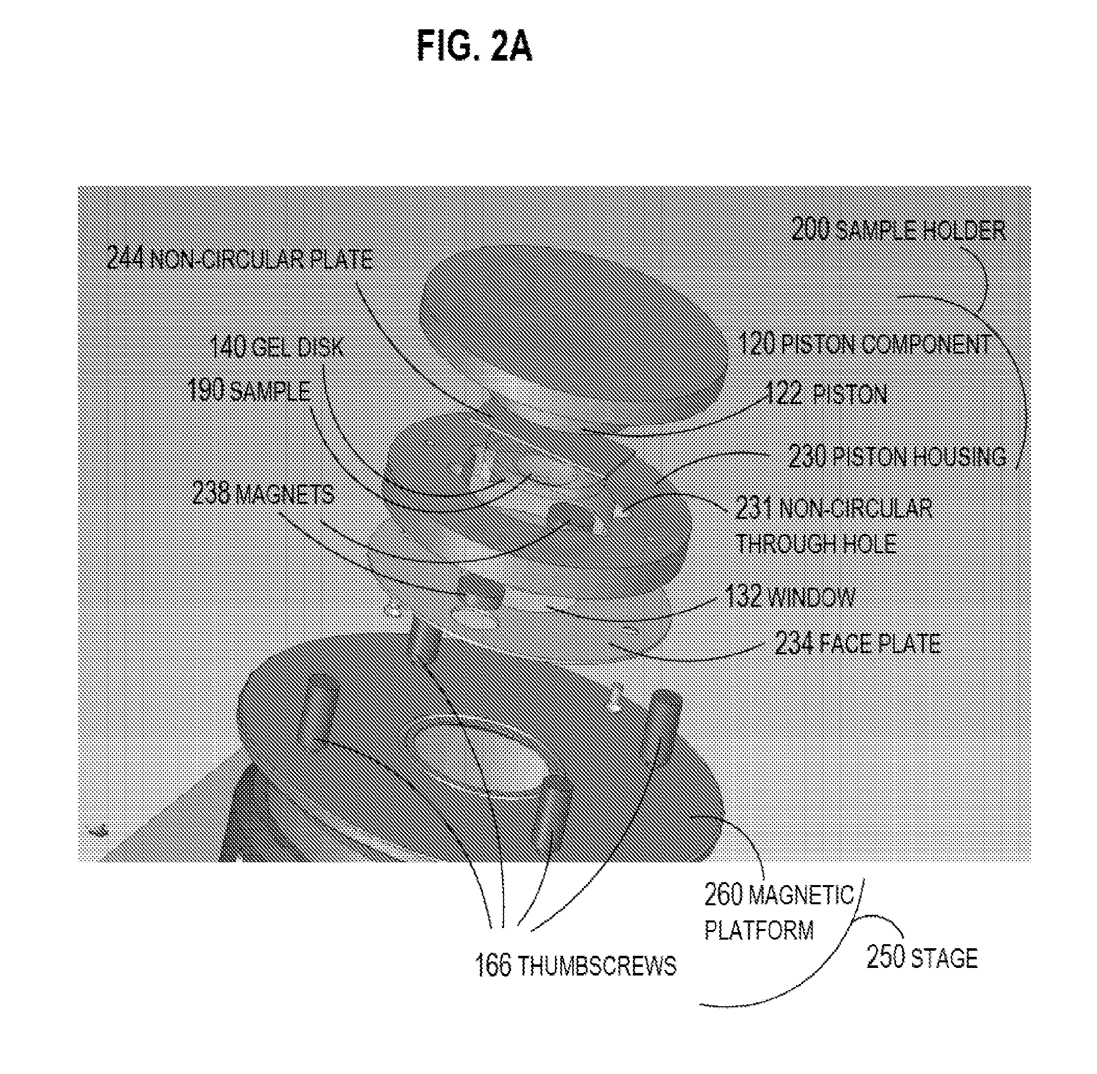
In another set of embodiments, an apparatus includes a support member that includes an axial through hole with a non-circular cross section. A transparent plate is fixed at one end of the axial through hole. A plate has a non-circular cross section that matches the non-circular cross section of the axial through...


PUM

PropertyMeasurementUnit
Lengthaaaaaaaaaa
Fractionaaaaaaaaaa
Nanoscale particle sizeaaaaaaaaaa

Abstract

One embodiment of techniques for confocal microscopy includes illuminating a spot on a surface of a biological sample. A first emission intensity from the spot is detected in a first range of optical properties; and a second emission intensity in a second range. A pixel that corresponds to the spot is colored using a linear combination of the first and second emission intensities. Sometimes, the pixel is colored to approximate a color produced by histology. In some embodiments, a surface of a sample is contacted with a solution of acridine orange. Then, a spot is illuminated with a laser beam of wavelength about 488 nanometers (nm). Fluorescence emission intensity is detected above about 500 nm. Sometimes, a certain illumination correction is applied. In some embodiments, a sample holder that compresses a sample is removable from a stage that is fixed with respect to a focal plane of the microscope.

Description

BACKGROUND OF THE INVENTION1. Field of the InventionEmbodiments of the present invention relate to confocal microscopy with fluorescence to support surgical procedures, such as incremental surgery to remove tumors that are not naturally visible to a surgeon. In some embodiments, multimodal confocal microscopy is used to render images that emulate frozen histology sections.2. Description of the Related ArtSeveral surgical procedures to remove tumors involve cell morphology that is difficult to discern with the unaided eye. In such circumstances, successive incremental excisions are performed, and each sample of excised tissue is subjected to histological examination to determine the remaining extent of the tumor. The position and extent of the tumor evident in the histology sections is used to guide the next excision. However, the procedure for preparing the histology sections is tedious and time consuming, often exceeding 30 minutes per section. Thus, surgical procedures that rely o...

Claims


Application Information

Patent Timeline
IPC IPC(8): G06K9/00G02B21/26G02B21/34G02B21/00G02B26/10C12Q1/68
CPCG01N21/6458G02B21/0064G02B21/367G02B21/008G02B21/0076H04N23/56G01N1/30G01N2001/302G02B21/16H04N9/797
Inventor GAREAU, DANIEL S.
Owner SLOAN KETTERING INST FOR CANCER RES
Why Eureka
Try Eureka