
Balloon catheter with detachable hub, and methods for same

Inactive Publication Date: 2011-05-19
COOK MEDICAL TECH LLC
View PDF6 Cites 24 Cited by
  • Summary
  • Abstract
  • Description
  • Claims
  • Application Information

AI Technical Summary

Problems solved by technology

Early attempts to inspect the biliary and pancreatic ducts endoscopically have been hampered by technical limitations of the scopes.
The mother-baby scope technique can be expensive with regard to personnel and equipment: two endoscopists plus assistants, two image processors (one for each camera), expensive fiber optics in the baby scope that can often be damaged during standard manipulation with resulting image degradation, etc.
The standard 1.2 mm working channel of fiber optic baby scopes limits diagnostic and therapeutic options.
Such scopes are currently available, but they encounter problems of efficient introduction to a patient's biliary duct in a procedure that provides high quality images (e.g., superior to fiber optics imaging) at a desirable procedure cost.
These problems include the difficulty (or impossibility) of navigating a larger fiber optic baby scope having a greater than 1.2 mm working channel through a mother scope (e.g., duodenoscope), out its side-facing distal accessory channel end past and manipulated by the elevator, and then into a patient's biliary duct.
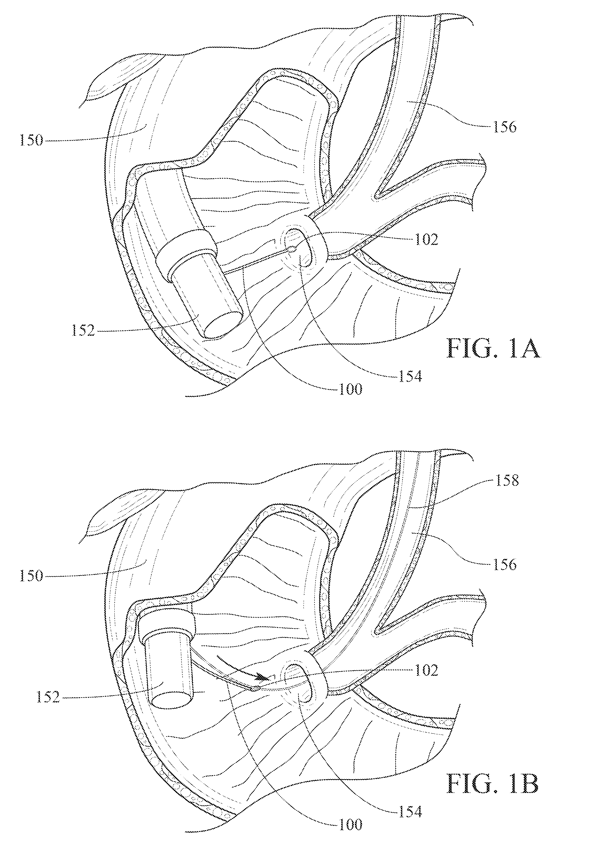
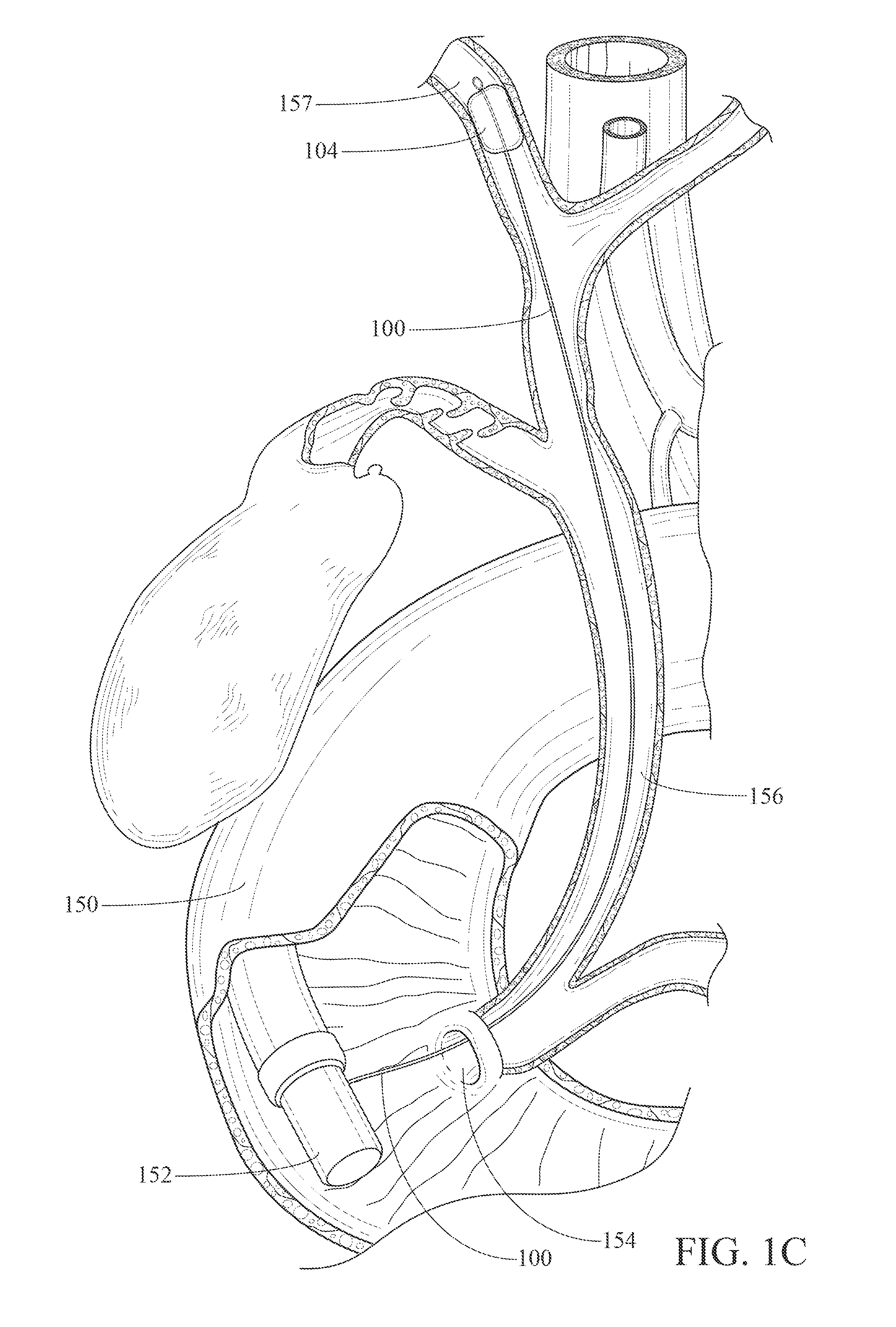
If one is to introduce a small scope (along the size of a “baby scope” or smaller) into the biliary ducts or other patient body structure without a primary (e.g., “mother”) scope, it is necessary to provide some type of “navigating track” because the smaller scopes are not sufficiently rigid / robust to be directed / navigated independently and directly through the esophagus, stomach, and duodenum to, for example, the common biliary duct.

Method used


Image

  • Balloon catheter with detachable hub, and methods for same
  • Balloon catheter with detachable hub, and methods for same
  • Balloon catheter with detachable hub, and methods for same

Examples


Embodiment Construction

Definitions

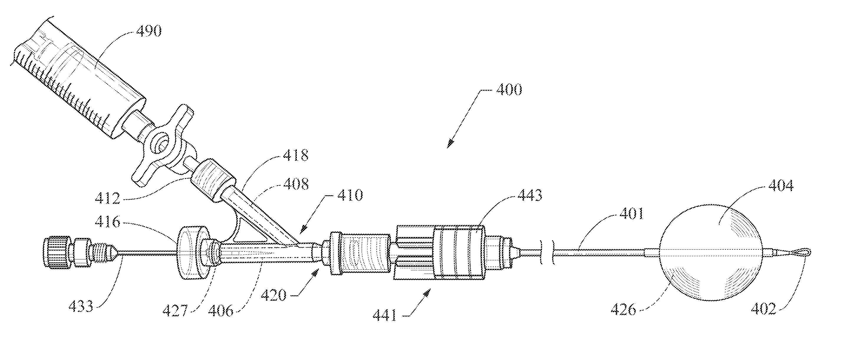
[0025]Ultra-slim endoscopes, as that term is used herein, refer to endoscopes having an outer diameter of about 6.0 mm or less (including less than 5.0 mm). The term “hub” refers to the proximal end structure of a balloon catheter including a connection structure (e.g., Luer-type or other fluid-patent connection) configured for effective connection to provide a path of fluid communication between a source of inflation fluid, a catheter inflation lumen, and a balloon lumen, and includes manifold-style hubs that may have more complex or ancillary structures. The terms “distal” and “proximal” are to be understood with their standard usages, referring to the direction away from and the direction toward the handle / user end of a tool or device, respectively (i.e., away from and toward the patient, respectively).

[0026]A cholangioscopy procedure using a scope-exchange facilitated by a balloon catheter including a proximal actuatable sealing valve and removable hub is described wi...


PUM


Abstract

A balloon catheter may include a distal anchoring balloon and a proximal hub that is removable from the catheter body. The catheter body may include a valve structure providing for maintaining the balloon in an inflated state during and after removal of the proximal hub. The valve preferably is constructed such that removal of the hub provides a low-profile proximal catheter end that will allow that proximal catheter end to pass through an endoscopic surgical device such as, for example, through an accessory channel of a standard duodenoscope and / or an ultra-slim endoscope / cholangioscope, facilitating a scope-exchange for use during, for example, a cholangioscopy or pancreatoscopy procedure. A method useful for scope exchange and / or introducing another elongate surgical device may utilize a balloon catheter with a distal anchoring balloon and a proximal hub that is removable from the catheter body.

Description

CROSS-REFERENCE TO RELATED APPLICATIONS[0001]This application claims priority to U.S. Provisional Application Ser. Nos. 61 / 256,773 and 61 / 256,755, both filed Oct. 30, 2009, and 61 / 329,243, filed Apr. 29, 2010, each of which is incorporated herein by reference in its entirety.TECHNICAL FIELD[0002]Embodiments of the claimed invention relate to a medical balloon catheter device configured for passage through an ultra-slim endoscope. More particularly, embodiments of the claimed invention relate to a balloon catheter including a proximal actuatable catheter lumen seal and a detachable hub, and methods of use.BACKGROUND[0003]Intraductal endoscopes have an increasingly important role in the diagnosis and nonsurgical treatment of biliary and pancreatic diseases. Early attempts to inspect the biliary and pancreatic ducts endoscopically have been hampered by technical limitations of the scopes. More recently, the development of fine-caliber flexible scopes known as fiber optic miniscopes has...

Claims


Application Information

Patent Timeline
IPC IPC(8): A61B1/00A61B1/018A61F2/958
CPCA61M25/0075A61M25/0097A61M25/10186A61M25/10A61M2025/0076A61M25/04
Inventor DILLON, TRAVIS E.AGUIRRE, ANDRES F.CHMURA, KEVIN
Owner COOK MEDICAL TECH LLC
Learn More