Transnasal method and catheter for lacrimal system

a transnasal method and lacrimal system technology, applied in the field of transnasal method and lacrimal system treatment, can solve the problems of no medical treatment for acquired nasolacrimal duct obstruction, significant morbidity, swelling of the lacrimal sac, etc., to avoid the trauma associated with insertion through the delicate canaliculi, avoid trauma, and high surgical success rate

Inactive Publication Date: 2007-01-30
BECKER BRUCE
View PDF22 Cites 241 Cited by
  • Summary
  • Abstract
  • Description
  • Claims
  • Application Information

AI Technical Summary

Benefits of technology

[0010]A balloon catheter of the invention can be introduced transnasally into the area of a planned DCR ostium. The catheter has a larger deflated profile and, thus, a larger inflated diameter, than a balloon catheter introduced through the delicate canaliculi. The balloon catheter of the invention does not need a guide wire; and, therefore, there is no chance that a guide wire will damage the delicate canaliculi. A larger diameter balloon DCR ostium is less likely to stenose after surgery and results in a better surgical success rate.
[0011]The balloon catheter of the invention comprises a hypotube formed of stainless steel of sufficient stiffness and column strength to enable the deflated balloon catheter to be pushed from the nasal cavity through a prepared small, tight opening in the lateral nasal wall and lacrimal fossa into the lacrimal sac. A distal end segment of the hypotube has a rounded bend, placing the distal segment at an angle of 70° to 115°, preferably 90°, to a long proximal segment or shaft. This bend allows the surgeon to rotate or shift the position of the long proximal catheter shaft, thus placing the distal balloon catheter segment in position to enter from the nasal cavity into the lacrimal sac at various angles appropriate to each individual patient. Due to the stiffness and strength of the hypotube, neither a guide wire nor a curve retention element are necessary.
[0013]The balloon is formed of inflatable material with a first neck bonded with adhesive to the very distal portion of the distal segment of the hypotube and a second neck bonded with adhesive to the distal end of the proximal shaft, the bend, and the proximal end of the distal segment. This permits a longer working segment of balloon to be used, because the area of adhesion of the balloon includes the bend and the adjacent portion of the proximal long segment of the hypotube. The proximal end of the catheter tube has a luer lock with wings or an expansion to allow the catheter to be attached to tubing from the inflation device. The wings or expansion allow the surgeon to more easily hold, manipulate, and push the balloon catheter.
[0016]Since the method of the insertion requires insertion of the balloon catheter transnasally, the trauma associated with insertion through the delicate canaliculi is avoided. The catheter and method of the invention provide a satisfactory endoscopic DCR and thus avoid the trauma associated with an external (incisional) DCR. The catheter and method of the invention achieve a high surgical success rate.

Problems solved by technology

Dacryocystitis causes the lacrimal sac to become swollen, red and painful.
At present, there is no medical therapy for acquired nasolacrimal duct obstruction other than antibiotics to temporarily suppress infection.
This procedure has significant morbidity, a prolonged recovery, and the threat of scarring and hemorrhage.
A laser endoscopic DCR requires expensive and time-consuming lasers, and has a low success rate.
An endoscopic DCR with traditional instruments places the eye and surrounding structures at risk because tissue is removed from the lacrimal sac and lateral nasal wall, with the instruments in the nasal cavity going toward the eye and orbit.
Bleeding and edema may make it difficult to identify the relevant structures.
However, a 5 mm diameter ostium is much smaller than the 17 mm plus diameter ostium of an external DCR and leads to a higher stenosis rate of the balloon DCR ostium after surgery.
Although this technique is useful, it involves a number of time-consuming steps, including the insertion of a guide wire through the lacrimal system, and then separately advancing the balloon catheter over the guide wire.
However, there are problems with this method.
First, there may be difficulty locating and grasping the guide wire in the nose, especially if even mild bleeding is present.
There is often resistance to pulling the balloon from the nasal cavity into the lacrimal sac and considerable force is required to pull the balloon and guide wire into the lacrimal sac.
This pull on the guide wire can cause it to slice through the delicate canaliculi, which may lead to secondary fibrosis and obstruction of the canaliculi after surgery.
This catheter is not suitable for transnasal insertion.
The tube does not have sufficient stiffness and column strength to enable the deflated balloon catheter to be pushed from the nasal cavity through a small, tight opening in the lateral nasal wall and lacrimal fossa into the lacrimal sac.

Method used


Image

  • Transnasal method and catheter for lacrimal system
  • Transnasal method and catheter for lacrimal system
  • Transnasal method and catheter for lacrimal system

Examples


Embodiment Construction

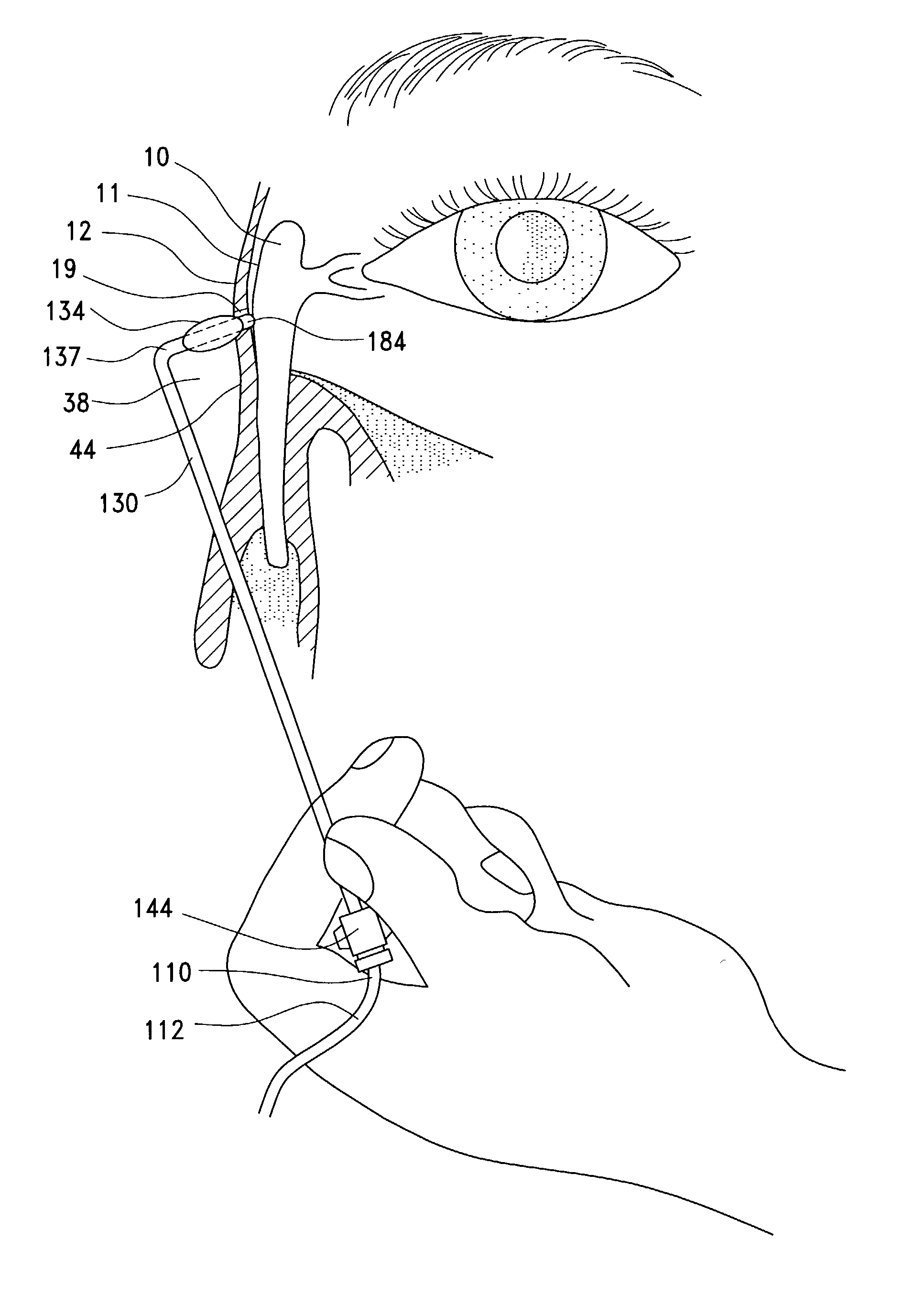
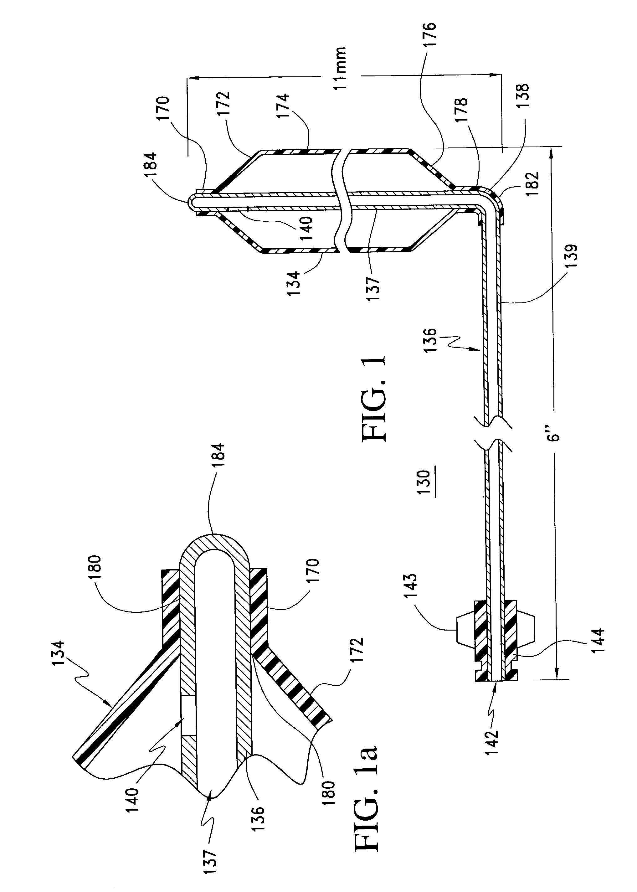
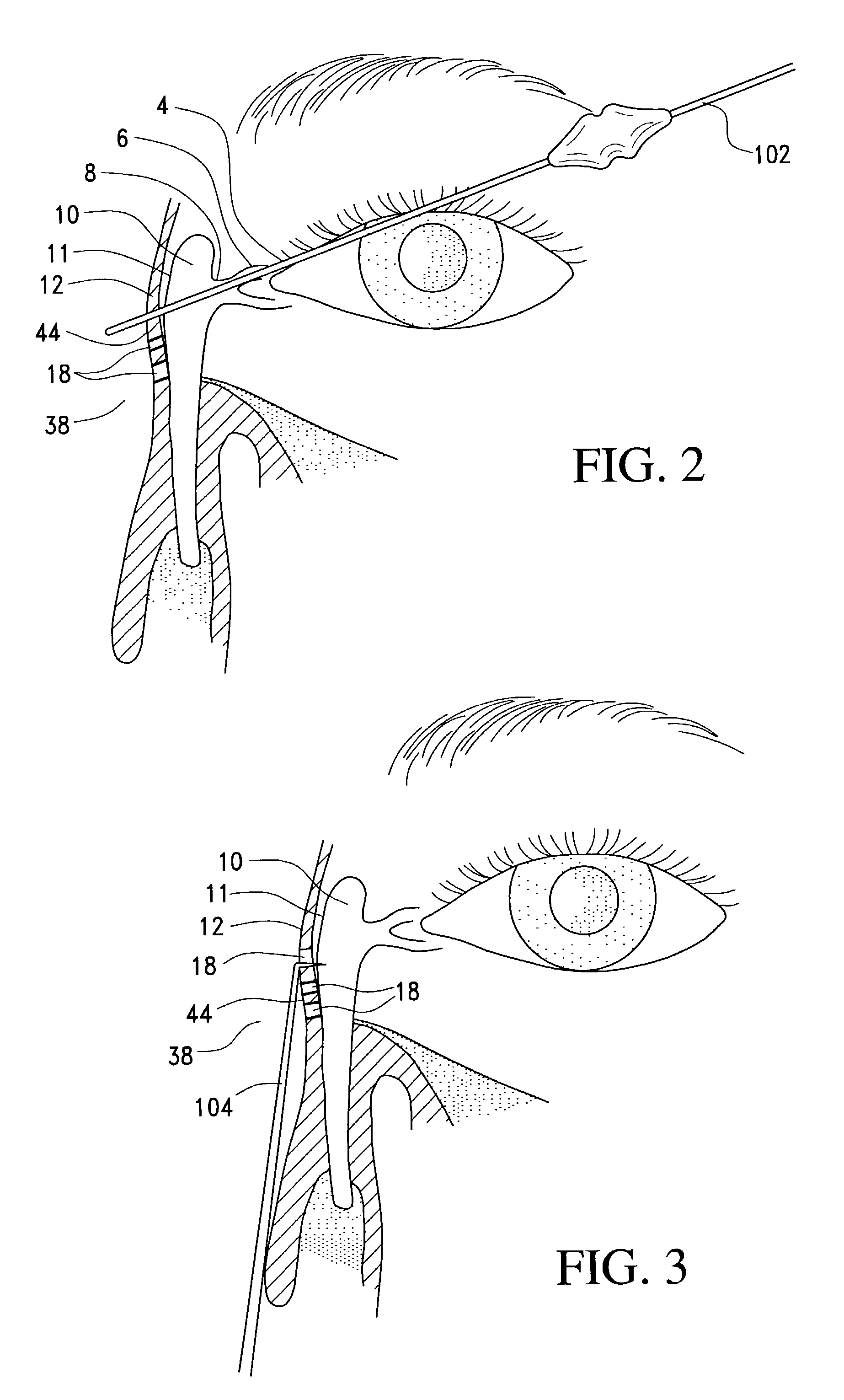
[0025]As shown in FIGS. 1 and 1a, a balloon catheter 130 of the invention is assembled from a tube 136, preferably a stainless steel hard tempered hypotube which has a circular bend 138 of 0.13″ radius such that distal segment 137 is oriented 70° to 115°, preferably 90°, to proximal segment 139. The distance from the distal tip 184 of distal segment 137 to the outer wall of proximal segment 139 of hypotube 136 is 4 mm to 30 mm, preferably 14 mm, as shown in FIG. 1. The distal tip 184 of the hypotube 136 is closed, whereas the proximal end 142 is open. However, the lumen of tube 136 may be closed in the distal segment 137, up to 10 mm from the distal tip 184 allowing the distal tip 184 to remain open for engagement with a probe, as shown in FIGS. 6 and 7. The proximal end 142 of hypotube 136 is inserted into a mold for forming luer 144. Heated plastic is injected into the mold to form luer 144 attached to proximal end 142. The inner diameter of the luer 144 matches the external diame...


PUM


Abstract

A balloon catheter for treatment of a patient's lacrimal system is applied transnasally without the use of a guide wire or a curve retention member. The catheter uses a stainless steel hypotube of sufficient stiffness and column strength to be pushed from the patent's nasal cavity through an opening-formed through the lateral nasal wall and lacrimal fossa, into the lacrimal sac. The catheter has an inflatable member mounted about a rigid bent distal segment. The opening is first formed by pushing small holes through the medial sac, lacrimal fossa, and lateral nasal wall with an instrument and coalescing the holes. The catheter is then introduced into the nasal cavity and pushed laterally through the opening by manipulating its proximal end. Pressurized fluid is then applied to the catheter to inflate the inflatable member and dilate the opening.

Description

FIELD OF THE INVENTION[0001]This invention relates to a method and catheter for treating the lacrimal system and, more particularly, to a transnasal method of treating the lacrimal system and a balloon catheter used in this method.BACKGROUND AND DESCRIPTION OF THE PRIOR ART[0002]To fully understand the invention, it is necessary to consider the anatomy and physiology of the lacrimal system. The orbital portion of the lacrimal gland is located in the superotemporal orbit and produces the aqueous layer of the tear film. Ductules from the orbital portion of the lacrimal gland pass through the adjacent palpebral lacrimal gland to empty in the superior conjunctival cul-de-sac. Smaller accessory lacrimal glands in the upper and lower lids also contribute to tear production The tears bathe the surface of the eye and then drain into the puncta and canaliculi in the medial upper and lower lids. The tears flow from the canaliculi into the lacrimal sac down the nasolacrimal duct into the nose....

Claims


Application Information

Patent Timeline
Patent Type & Authority Patents(United States)
IPC IPC(8): A61M29/00A61F9/007A61F2/958
CPCA61F9/00772A61M29/02A61B17/24A61M2210/0618A61M2025/1093
Inventor BECKER, BRUCE
Owner BECKER BRUCE
Why Eureka
Try Eureka