Methods And Systems For Spatially Modulated Ultrasound Radiation Force Imaging

a radiation force imaging and spatial modulation technology, applied in the field of ultrasonic imaging, can solve the problems of introducing a time delay between each spatial peak of the push beam, the viscoelastic nature of the tissue, and the dispersion effect complicating the application of smurf in strongly viscoelastic media, so as to achieve the effect of convenient detection

Active Publication Date: 2011-07-28
UNIVERSITY OF ROCHESTER
View PDF3 Cites 55 Cited by
  • Summary
  • Abstract
  • Description
  • Claims
  • Application Information

AI Technical Summary

Benefits of technology

[0015]A challenge for the SMURF method is the viscoelastic nature of tissues. Our previous development of the SMURF method assumed a linear elastic model that neglected shear wave dispersion. We have found that dispersion effects complicate the application of SMURF in strongly viscoelastic media, e.g. liver tissue. We have dev

Problems solved by technology

A challenge for the SMURF method is the viscoelastic nature of tissues.
We have found that dispersion effects complicate the application of SMU

Method used


Image

  • Methods And Systems For Spatially Modulated Ultrasound Radiation Force Imaging
  • Methods And Systems For Spatially Modulated Ultrasound Radiation Force Imaging
  • Methods And Systems For Spatially Modulated Ultrasound Radiation Force Imaging

Examples


Embodiment Construction

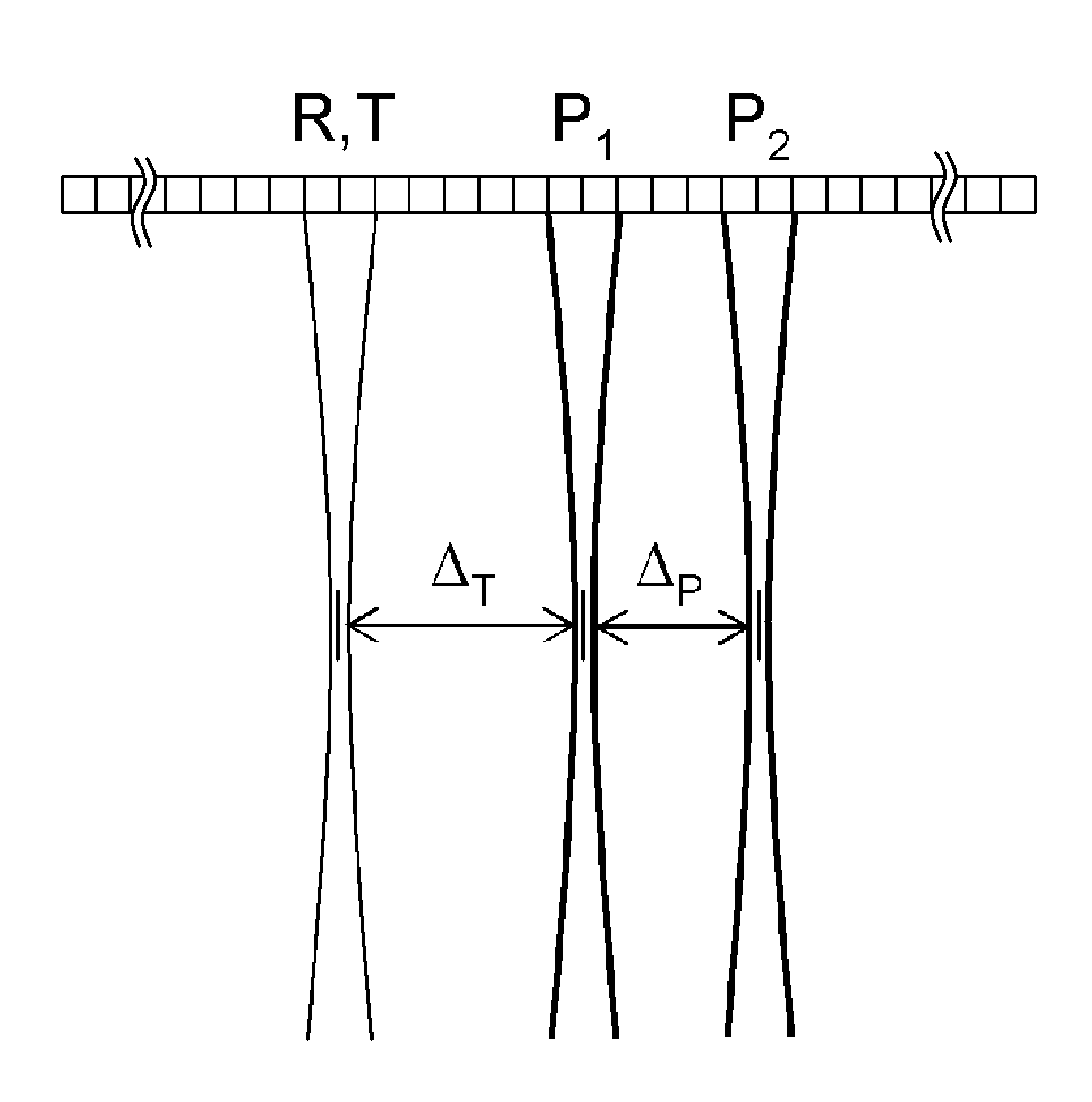
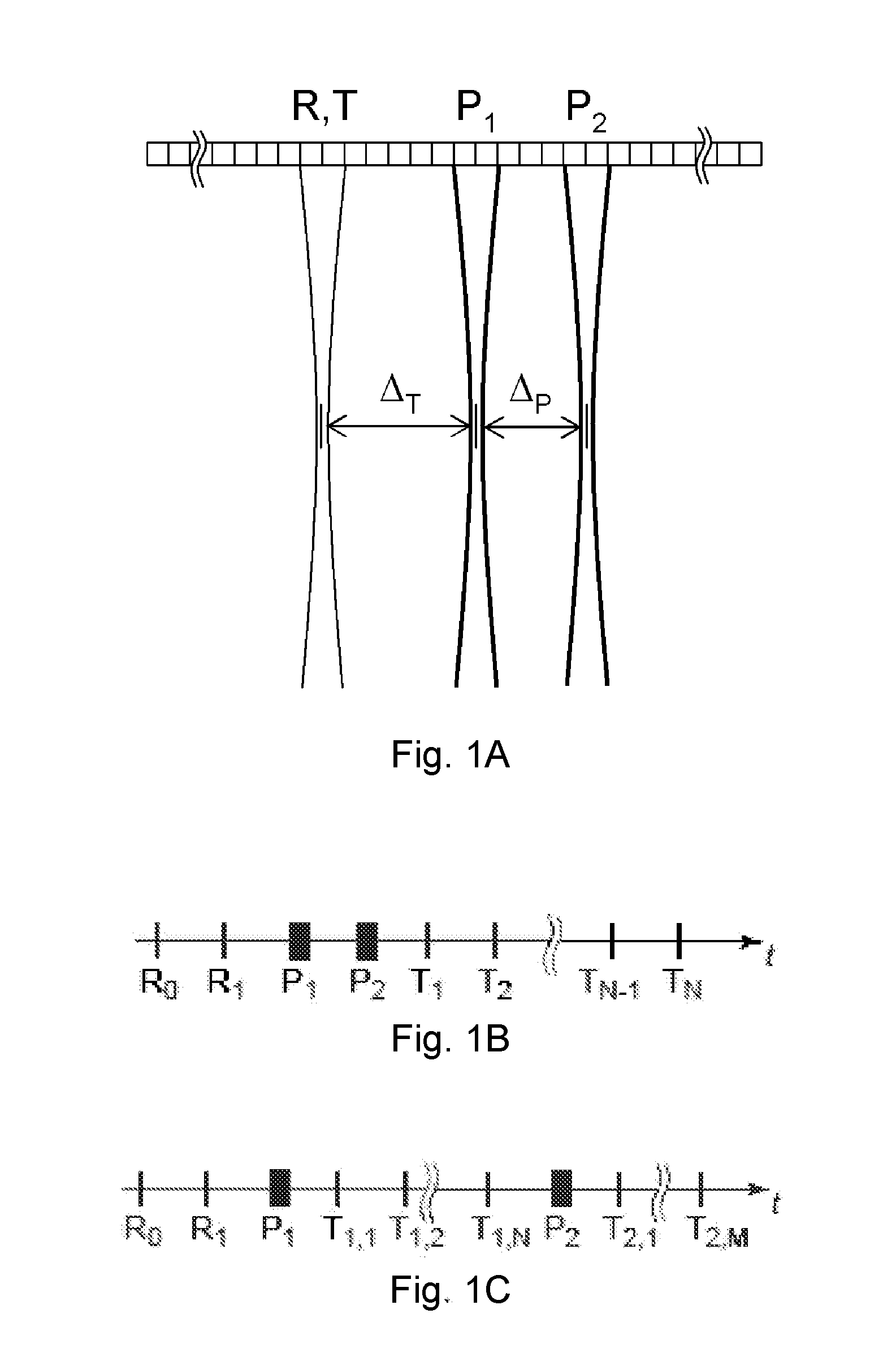
diagram representation of the signal flow for the data acquisition procedure described in the text.

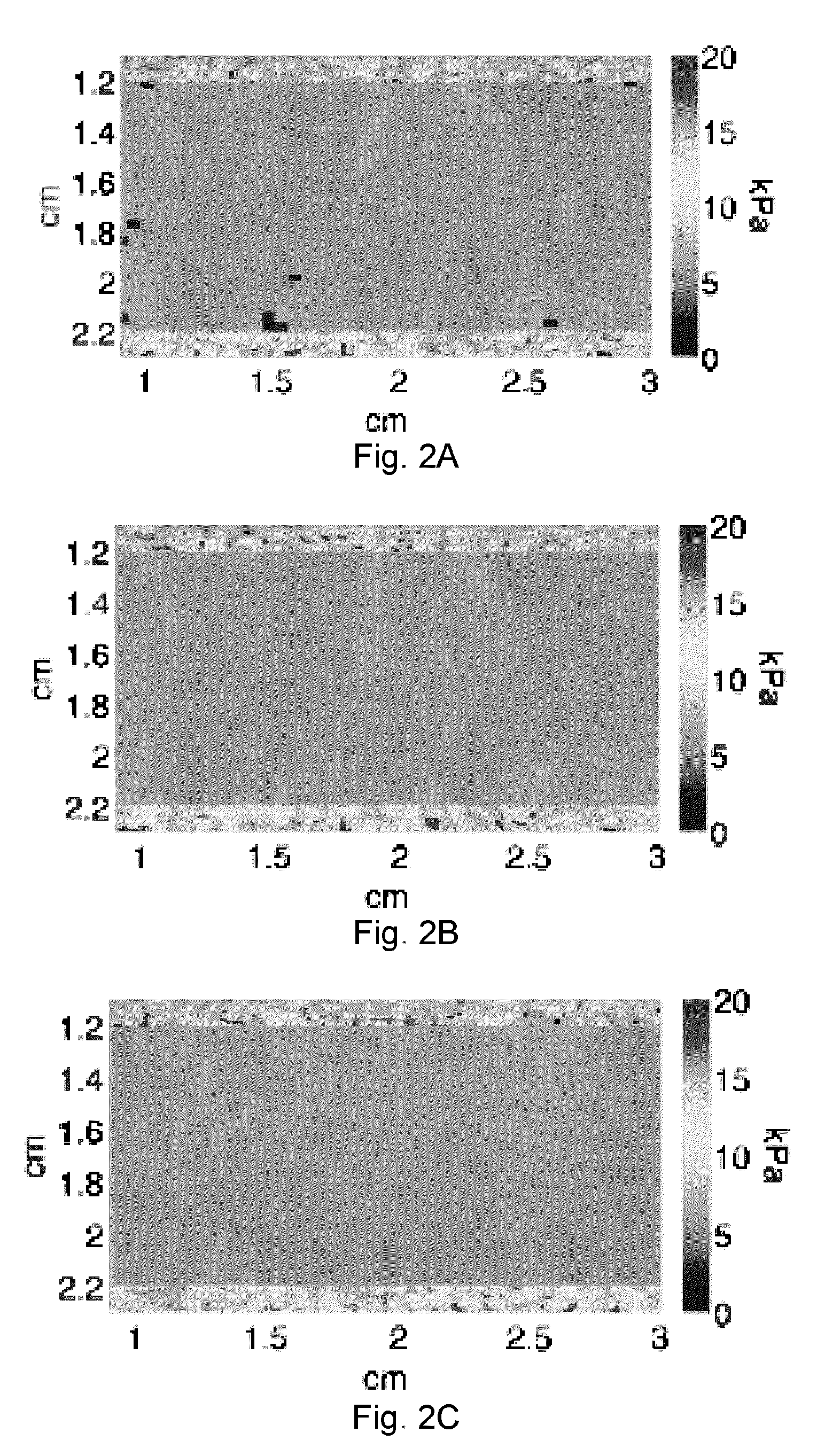
[0037]FIGS. 15A-15F: Left column 20 dB grayscale. Right column 40 dB grayscale. Top row: Boxcar (rect.) window applied. Center row: Hamming window applied. Bottom row: raised hamming window.

[0038]FIG. 16A is a simulated image of a 1 cm diameter lesion where the shear modulus of the lesion and the background are both 5 kPa. Image is on a 40 dB grayscale.

[0039]FIG. 16B is a simulated image of a 1 cm diameter lesion where the shear modulus of the lesion is 7.5 kPa and the shear modulus of the background is 5 kPa. Image is on a 40 dB grayscale.

[0040]FIGS. 17A-17B are images of nylon monofilament wires in a gelatin block as imaged by the shear wave method. The images are displayed on a 20 dB (FIG. 17A) or 40 dB (FIG. 17B) dynamic range grayscale.

[0041]FIG. 18 is an image of the hyperechoic cylinder in the gelatin block phantom reconstructed from experimental shear wave modulated data. The d...


PUM


Abstract

Methods for determining the shear modulus of a tissue of an individual are disclosed. In one embodiment, an acoustic pulse is transmitted and a reference echo signal is received. A first push pulse is transmitted at a second location of the individual. A second push pulse is transmitted at a third location of the individual. A series of tracking pulses are transmitted at the first location and a tracking echo signal is received from each tracking pulse. The effect of the first and second push pulses on the tissue is determined through analysis of the tracking echo signals. The shear modulus of the tissue is calculated based on the effect of the push pulses on the tissue. A system for imaging a region of tissue is presented.

Description

CROSS-REFERENCE TO RELATED APPLICATION[0001]This application claims the benefit of priority to U.S. provisional patent application Ser. No. 61 / 285,339 filed Dec. 10, 2009, now pending, the disclosure of which is incorporated herein by reference.[0002]This invention was made with government support under 1R21 EB008724 and 1R01 EB008368 awarded by the National Institutes of Health. The government has certain rights in the invention.FIELD OF THE INVENTION[0003]The invention relates to the field of ultrasound, and more particularly to ultrasonic imaging.BACKGROUND OF THE INVENTION[0004]Methods for quantitative imaging of tissue shear modulus using acoustic radiation force induced shear waves have been proposed. Tissue shear modulus is thought to have significant diagnostic utility, and provides an image contrast mechanism distinct from that of conventional imaging modalities, e.g. B-mode ultrasound, X-ray CT. The ability to reliably quantify tissue modulus may reduce the need for tissue...

Claims


Application Information

Patent Timeline
IPC IPC(8): A61B8/00
CPCA61B8/4483A61B8/485G01S7/52042G01S7/52022
Inventor MCALEAVEY, STEPHEN A.
Owner UNIVERSITY OF ROCHESTER
Why Eureka
Try Eureka