Medical image diagnosis assisting apparatus and method, and computer readable recording medium on which is recorded program for the same

a technology of medical image diagnosis and recording medium, which is applied in the field of medical image diagnosis assisting apparatus and method, can solve the problems of difficult to interpret the evaluation value representing perfusion or regional lung volume in each section of the lung, and achieve the effect of high-quality pulmonary image diagnosis

Active Publication Date: 2011-08-04
FUJIFILM CORP
View PDF5 Cites 17 Cited by
  • Summary
  • Abstract
  • Description
  • Claims
  • Application Information

AI Technical Summary

Benefits of technology

[0035]According to the present invention, a lung field area and a branch structure area are extracted from a three-dimensional medical image, then a branch structure local area representing a portion of the branch structure area is divided into a plurality of branch structure local sub-areas and a lung field local sub-area in the lung field area functionally associated with each divided branch structure local sub-area is estimated based on the branch structure area, a pulmonary evaluation value of pulmonary function and / or morphology in each estimated lung field local sub-area is obtained, and, in a morphological image representing morphology of at least a portion of the branch structure local area generated from the three-dimensional medical image, a pulmonary evaluation value of each lung field local sub-area functionally associated with each branch structure local sub-area in the morphological image is displayed superimposed such that correspondence relationship between the pulmonary evaluation value and the branch structure local sub-area in the morphological image is visually recognizable. This allows local observation / evaluation of a lung to be made based on the functional relationship between a local area of the branch structure and an adjacent lung field area. That is, highly accurate pulmonary image diagnosis may be made by considering the association with a branch structure.

Problems solved by technology

But, a bronchus or a blood vessel is complexly distributed over the entire lung field by repeating branching and thinning, and it is extremely difficult to interpret an evaluation value representing a perfusion or a regional lung volume in each section of a lung by considering the association with a bronchus or a blood vessel in an image representing the entirety of a lung as in the method described in each patent document above.

Method used


Image

  • Medical image diagnosis assisting apparatus and method, and computer readable recording medium on which is recorded program for the same
  • Medical image diagnosis assisting apparatus and method, and computer readable recording medium on which is recorded program for the same
  • Medical image diagnosis assisting apparatus and method, and computer readable recording medium on which is recorded program for the same

Examples


first embodiment

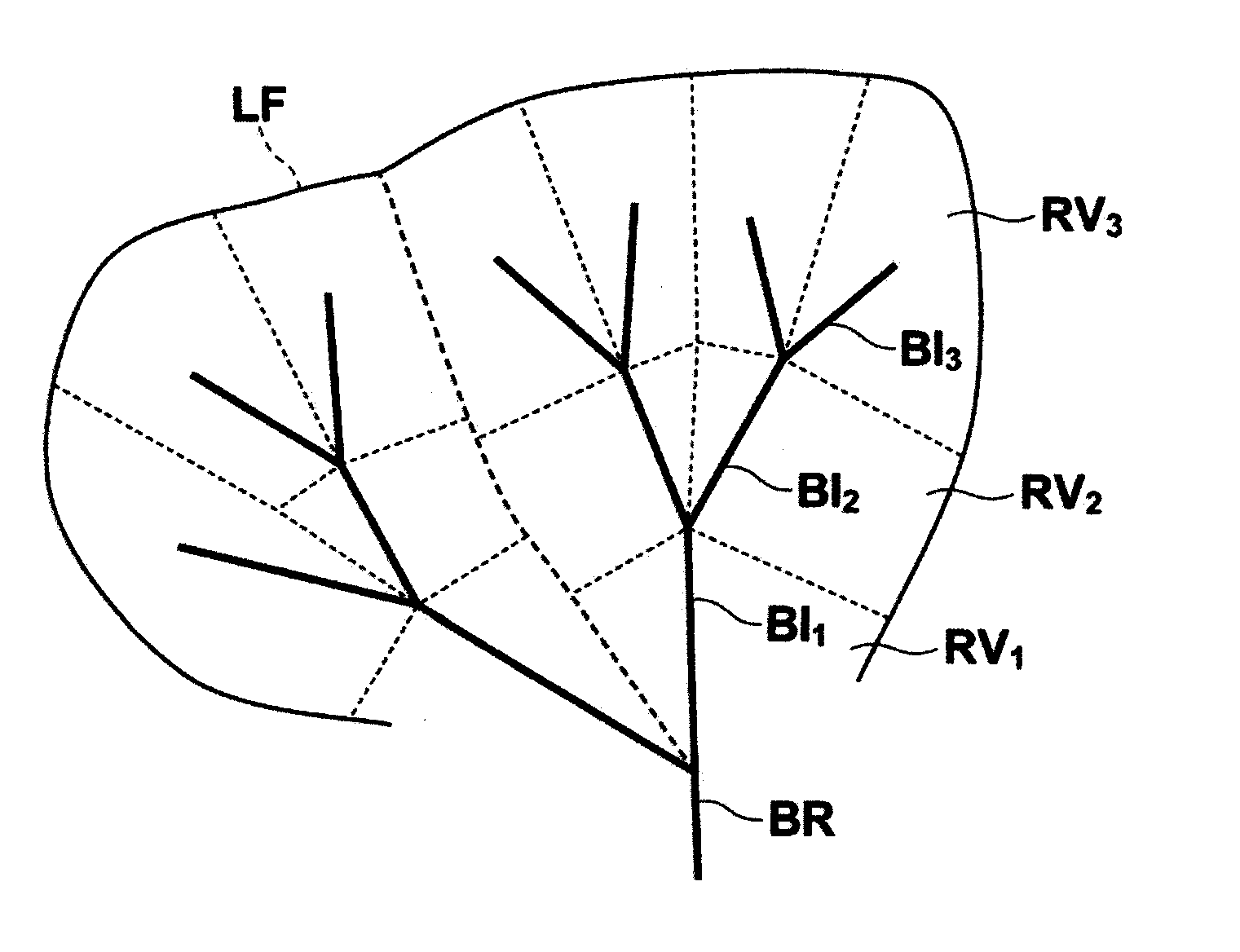
[0070]In the present invention, by examining the superimposed image IM generated by the series of processing described above, the lung may be locally evaluated based on the functional relationship between the attention bronchus structure BI, which is a local region of the bronchus, and the adjacent lung field area. That is, highly accurate pulmonary image diagnosis may be made by considering the association with a bronchus.

[0071]Further, as shown in FIG. 5, display control unit 36 causes a color map of pulmonary evaluation values ELn to be displayed on a position where the color map does not overlap with the attention bronchus structure BI in the CPR image IS, so that, while checking pulmonary evaluation values ELn, the state of each attention bronchus sub-area BIn corresponding to each pulmonary evaluation value ELn may be observed in the CPR image IS, thereby contributing to the improvement of diagnostic efficiency and accuracy.

[0072]Still further, the CPR image IS may extensively...

second embodiment

[0077]As described above, in the present invention, by examining the superimposed image IM generated by the series of processing described above, the relationship between the evaluation of emphysema degree based on the pulmonary evaluation value ELn and the inner diameter of the bronchus based on the bronchus evaluation value EBn may be understood easily while examining the state of attention bronchus sub-areas BIn in the CPR image IS, whereby highly accurate diagnosis may be performed more efficiently.

[0078]The embodiments described above are illustration purposes only and should not be construed as limiting the scope of the technical scope of the present invention.

[0079]It should be appreciated that various modifications and changes made to the system configurations, processing flows, module structures, specific processing contents, and the like in the embodiments described above without departing from the spirit of the present invention are included in the scope of the present in...


PUM


Abstract

Extracting a lung field area and a branch structure area from a three-dimensional medical image, dividing a branch structure local area representing a portion of the branch structure area into a plurality of branch structure local sub-areas and estimating a lung field local sub-area in the lung field area functionally associated with each divided branch structure local sub-area based on the branch structure area, obtaining a pulmonary evaluation value in each estimated lung field local sub-area, and displaying, in a morphological image representing morphology of at least a portion of the branch structure local area, the pulmonary evaluation value in each lung field local sub-area functionally associated with each branch structure local sub-area in the morphological image superimposed such that correspondence relationship between the pulmonary evaluation value and the branch structure local sub-area in the morphological image is visually recognizable.

Description

BACKGROUND OF THE INVENTION[0001]1. Field of the Invention[0002]The present invention relates to an apparatus and method for assisting image diagnosis using a three-dimensional medical image representing a chest region of a subject. The invention also relates to a computer readable recording medium on which is recorded a program for the same.[0003]2. Description of the Related Art[0004]A technology for assisting pulmonary image diagnosis using a three-dimensional medical image representing a chest region is known.[0005]For example, a method in which segmentation of pulmonary parechymal tissue is performed on a CT image representing both lungs, then a perfusion map of the segmented image is generated, and an abnormally underperfused area is rendered and displayed comparatively opaque is proposed as described, for example, in U.S. Pat. No. 7,715,608.[0006]Another method in which lung areas are extracted from CT images representing a lung in insipiratory phase and in expiratory phase, ...

Claims


Application Information

Patent Timeline
Patent Type & Authority Applications(United States)
IPC IPC(8): A61B6/03G06T11/00
CPCG06T7/0012G06T7/0083G06T2207/10072A61B6/507G06T2207/30061G06T2207/30101G06T2207/20221G06T7/12
Inventor MIZUNO, OSAMU
Owner FUJIFILM CORP
Why Eureka
Try Eureka