Medical image processing apparatus and method, and computer readable recording medium on which is recorded program for the same

Active Publication Date: 2011-10-06
FUJIFILM CORP
View PDF6 Cites 37 Cited by
  • Summary
  • Abstract
  • Description
  • Claims
  • Application Information

AI Technical Summary

Benefits of technology

[0035]According to the present invention, a branch structure and a surrounding structure are extracted from a three-dimensional medical image representing a subject's internal structure, an attention branch structure is set in the branch structure, then an attention surrounding structure area functionally associated with the attention branch structure is estimated from the surrounding structure area, an evaluation value in the attention surrounding structure area is obtained, and a mapped image, which is an image generated by mapping the evaluation value in the attention surrounding structure area to a morphological image representing morphology of at least one of the attention branch structure and the attention surr

Problems solved by technology

The method described in U.S. Pat. No. 7,583,829, however, only visualizes a lung area affected by embolization of a pulmonary blood vessel and does not indicate how the pulmonary function has been changed in the area.
In the mean time, it is

Method used


Image

  • Medical image processing apparatus and method, and computer readable recording medium on which is recorded program for the same
  • Medical image processing apparatus and method, and computer readable recording medium on which is recorded program for the same
  • Medical image processing apparatus and method, and computer readable recording medium on which is recorded program for the same

Examples


Embodiment Construction

[0050]Hereinafter, a medical image diagnosis system that employs a medical image processing apparatus according to an embodiment of the present invention will be described by taking, as example, the case in which influence of embolization of a pulmonary blood vessel on a surrounding lung parenchymal area is observed or evaluated based on a CT image of a human chest.

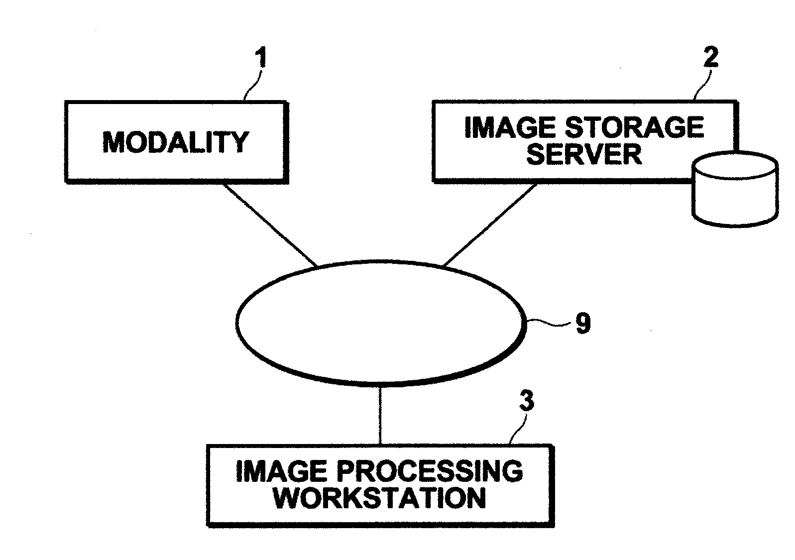
[0051]FIG. 1 is a hardware configuration diagram of the medical image diagnosis system, illustrating an overview thereof. As shown in FIG. 1, the system includes modality 1, image storage server 2, and image processing workstation 3 which are communicatably connected to each other via network 9.

[0052]Modality 1 includes an apparatus for imaging an inspection target region of a subject to generate image data representing a three-dimensional medical image of the region and outputting the image data by attaching auxiliary information defined in DICOM (Digital Imaging and Communication in Medicine) standard as image informati...


PUM


Abstract

Extracting a branch structure and a surrounding structure area in which a surrounding structure of the branch structure exists from a three-dimensional medical image, setting an attention branch structure in the branch structure, estimating an attention surrounding structure area functionally associated with the attention branch structure from the surrounding structure area based on the branch structure, obtaining an evaluation value in the attention surrounding structure area, and generating a mapped image which is an image generated by mapping the evaluation value in the attention surrounding structure area to a morphological image representing morphology of at least one of the attention branch structure and the attention surrounding structure area.

Description

BACKGROUND OF THE INVENTION[0001]1. Field of the Invention[0002]The present invention relates to an apparatus and method for performing image processing on a three-dimensional medical image representing a subject's internal structure having a branch structure and a surrounding structure, such as a lung. The invention also relates to a computer readable recording medium on which is recorded a program for the same.[0003]2. Description of the Related Art[0004]An image processing method for diagnosing embolization of a pulmonary blood vessel based on a three-dimensional medical image representing a chest region is known.[0005]For example, U.S. Pat. No. 7,583,829 proposes a method in which a pulmonary blood vessel system is divided into segments, then an embolization area in the pulmonary blood vessel system after the segmentation is identified manually or automatically, a blood vessel sub-tree affected by the identified embolization is analyzed to identify a lung area affected by the em...

Claims


Application Information

Patent Timeline
IPC IPC(8): G06K9/00
CPCG06T7/0012G06T2200/04G06T2207/10072A61B6/507G06T2207/30061G06T2207/30101G06T2207/20101
Inventor MIZUNO, OSAMU
Owner FUJIFILM CORP
Why Eureka
Try Eureka