Ultrasound monitoring systems, methods and components

a monitoring system and ultrasound technology, applied in the field of ultrasound monitoring systems and components, can solve problems such as difficult identification of desired target sites using tcd probes, and achieve the effect of convenient and stable positioning of ultrasound emitting faces

Inactive Publication Date: 2011-10-13
PHYSIOSONICS
View PDF7 Cites 75 Cited by
  • Summary
  • Abstract
  • Description
  • Claims
  • Application Information

AI Technical Summary

Benefits of technology

[0016]An ultrasound probe mount may be provided as part of the framework component or may be provided as a separate component mountable to the framework component and is configured to receive an ultrasound probe. The ultrasound probe mount is generally adjustable with respect to the framework component and a subject's anatomical surface in at least two dimensions to provide convenient and stable positioning of an ultrasound emitting face of an ultrasound probe at desired anatomical locations on a subject. In some embodiments, the ultrasound probe mount may be adjustable along at least three adjustment paths. In some embodiments, the probe mount is adjustable along at least two linear paths and at least one rotational path. In some embodiments, the probe mount has at least one curved, at least partial

Problems solved by technology

One drawback of measuring physiological parameters using a standard TCD probe is that identifying a desired target site using a TCD probe is challenging and generally requires a trained, experienced sonographer to find and (acoustically) illuminate a desired target site, such as the middle cerebral artery (MCA).
When longer term

Method used


Image

  • Ultrasound monitoring systems, methods and components
  • Ultrasound monitoring systems, methods and components
  • Ultrasound monitoring systems, methods and components

Examples


Embodiment Construction

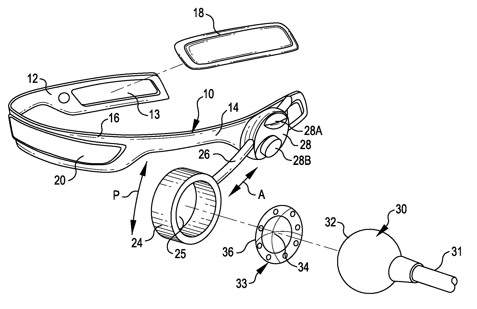
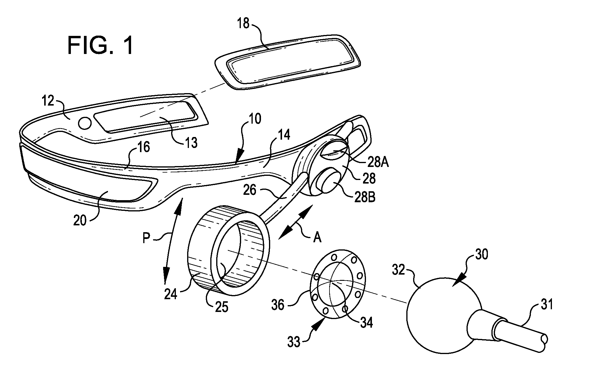
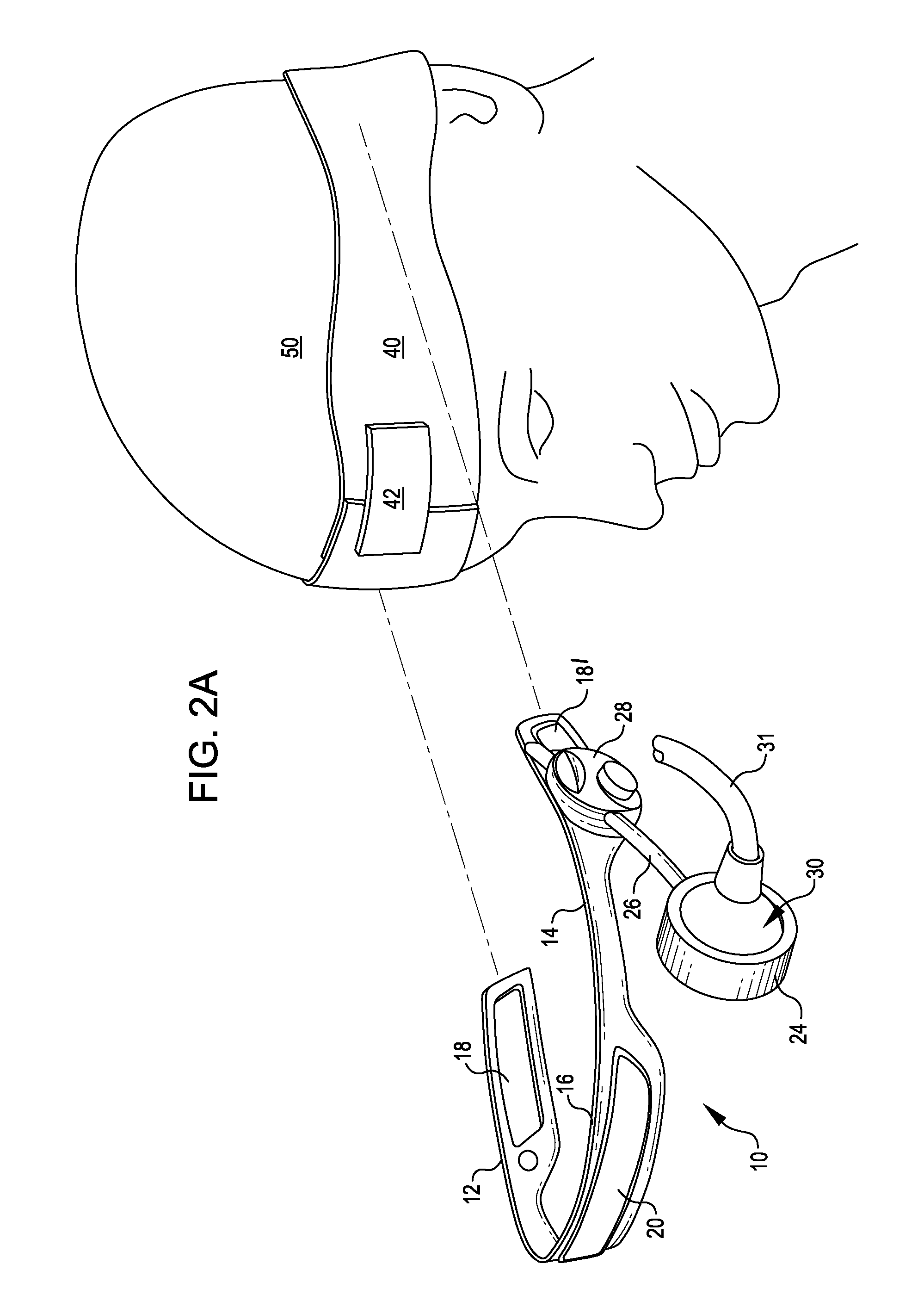
[0033]In one embodiment, illustrated schematically in FIG. 1, a framework structure for use with ultrasound monitoring systems requiring interface of an ultrasound probe with a subject's anatomy, such as an anatomical surface at a cranial window, (e.g., at a temporal window), comprises a generally U-shaped frame member 10 sized and configured for placement on a subject's skull. Frame member 10 comprises two framework legs 12, 14 positioned opposite one another for placement on opposite sides of a patient's skull and a connecting member 16 positioned to provide a bridge between the framework legs. In some embodiments, connecting member 16 may be configured to contact and generally conform to the shape of a subject's forehead. In some embodiments, the frame member 10 may be configured for positioning connecting member 16 adjacent to or contacting a subject's forehead; in alternative embodiments, frame member 10 may be configured for positioning connecting member 16 adjacent to or cont...


PUM


Abstract

Ultrasound monitoring systems and components used in ultrasound monitoring systems, such as Transcranial Dopper (TCD) systems, are disclosed. Components include framework systems for mounting, locating and maintaining one or more ultrasound probes in contact with an anatomical surface, adjustable probe mounting systems, and probe interface components providing an acoustically transmissive interface between a probe mounting system and the emissive face of the ultrasound probe.

Description

TECHNICAL FIELD OF THE INVENTION[0001]The present invention relates to ultrasound monitoring systems and components used in ultrasound protocols and monitoring systems, such as transcranial Doppler (TCD) systems, including framework systems for mounting, locating and maintaining one or more ultrasound transducer(s), or probe(s), in contact with an anatomical surface (e.g., skin, skull) of a subject, adjustable probe mounting systems, and probe interface components providing an interface between an ultrasound probe mounting system and the probe and, optionally, providing an acoustically transmissive coupling for contacting a subject's skin or another anatomical surface. Methods for using the probe mounting systems, interface components and / or framework structure, and for adjusting the acoustic illumination area of ultrasound probes with respect to a target site are also disclosed.BACKGROUND OF THE INVENTION[0002]In the field of medical imaging, ultrasound systems may be used in vario...

Claims


Application Information

Patent Timeline
IPC IPC(8): A61B8/00
CPCA61B8/0808A61B8/4472A61B8/4227
Inventor ZHANG, JIMINSERROELS, RANDYLIN, INGRIDHUBLER, ROBERT BRUCESULLIVAN, JOSEPH PATRICKLEONARD, PAUL C.ARAGON, JOELFRYER, LUKEBROWN, HAROLD A.LONG, CLAREDALE, NATHAN J.
Owner PHYSIOSONICS
Why Eureka
Try Eureka