Medical examination device for CT imaging and for nuclear medical imaging

a medical imaging and examination device technology, applied in the field of medical examination devices for ct imaging and nuclear medical imaging, can solve the problems of insufficient fluoroscopy imaging c-arm x-ray devices, insufficient functional medical imaging, and insufficient x-ray power to achieve a sufficiently good image quality with more complex interventions, and achieve good anatomical and functional imaging

Inactive Publication Date: 2011-11-17
SIEMENS HEALTHCARE GMBH
View PDF4 Cites 45 Cited by
  • Summary
  • Abstract
  • Description
  • Claims
  • Application Information

AI Technical Summary

Benefits of technology

[0009]The underlying object of the invention is therefore to specify a medical examination device in which good access to a pati

Problems solved by technology

The previous mobile C-arm X-ray devices with fluoroscopy imaging are not sufficient for this, since these only deliver a two-dimensional image representation, no adequate soft tissue resolution and no functional medical imaging.
It can also occur that the X-ray power is too weak to achieve a sufficiently good image quality with more complex interventions

Method used


Image

  • Medical examination device for CT imaging and for nuclear medical imaging
  • Medical examination device for CT imaging and for nuclear medical imaging
  • Medical examination device for CT imaging and for nuclear medical imaging

Examples


Embodiment Construction

[0039]It should be pointed out in advance that although the exemplary embodiments depicted here all show a PET recording arrangement as a nuclear medical recording arrangement, all the exemplary embodiments can naturally also be provided with an SPECT recording arrangement.

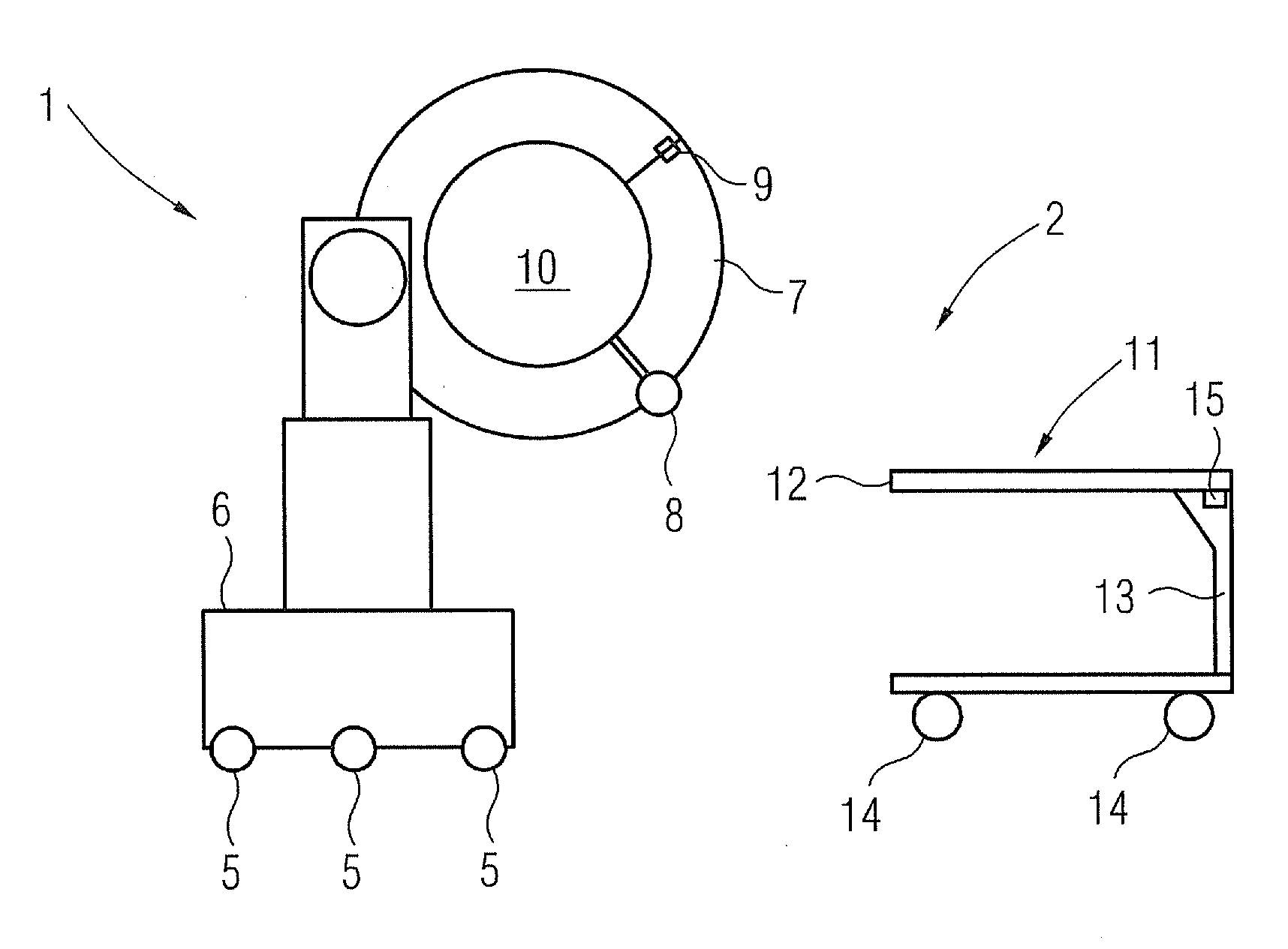
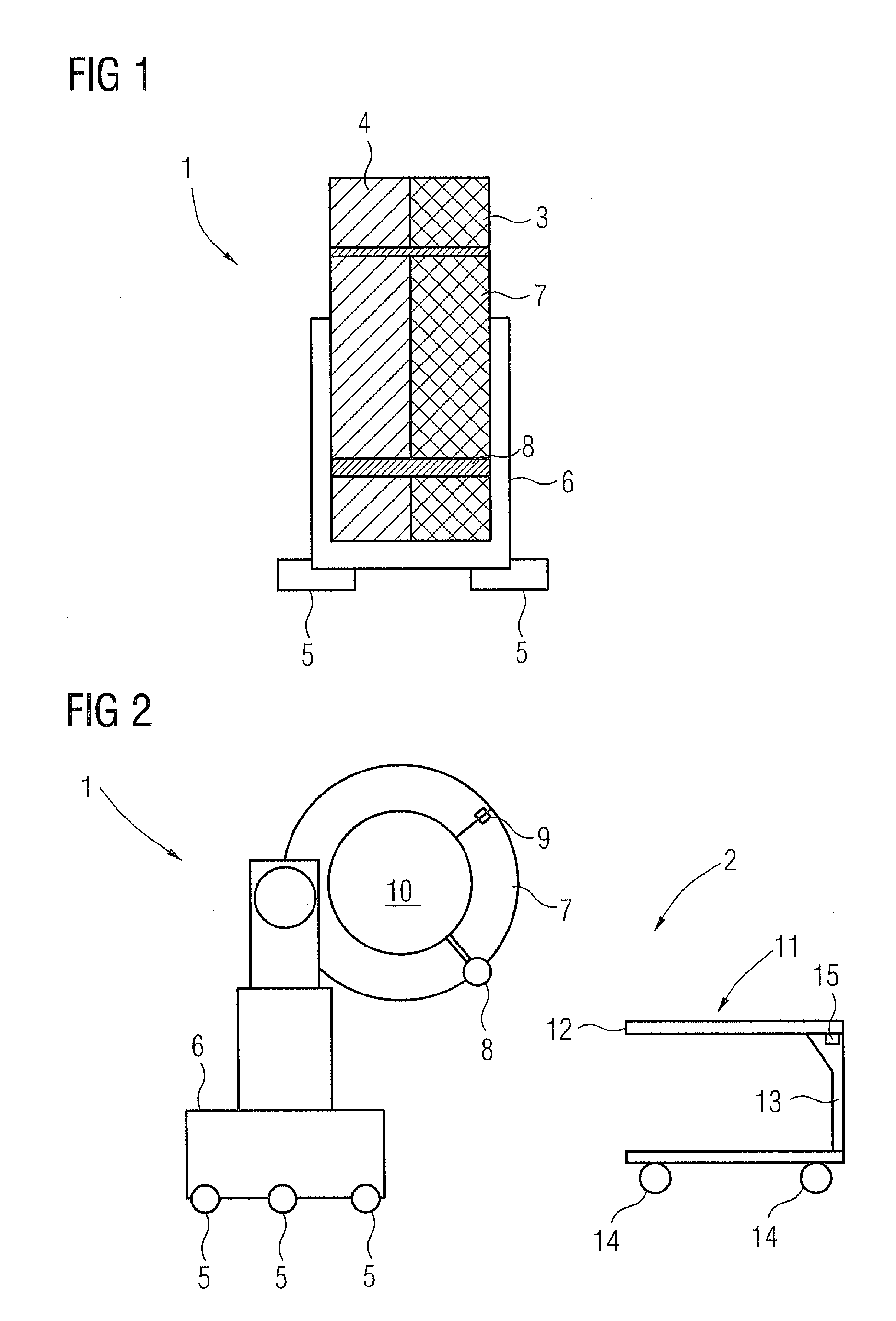
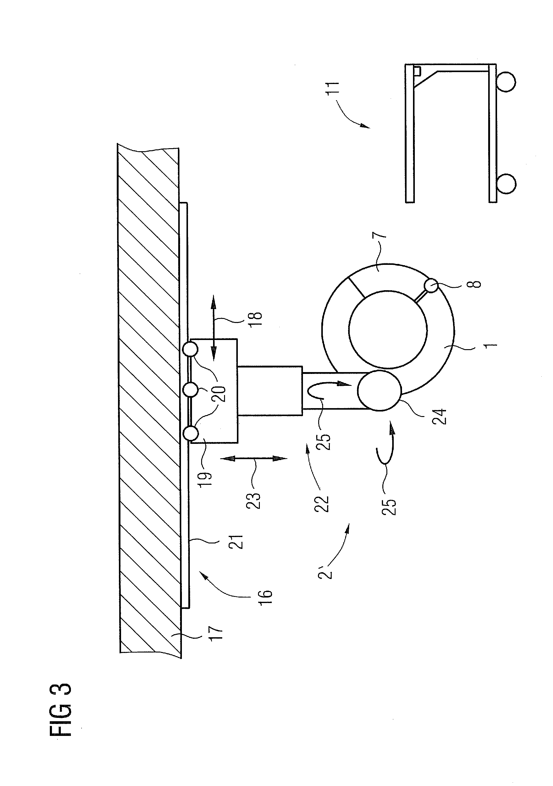
[0040]FIG. 1 shows the gantry 1 of a first embodiment of an inventive examination device 2 (FIG. 2). The gantry 1 comprises a CT recording arrangement 3 and a PET recording arrangement 4, as are basically known and do not need to be explained in greater detail here. It is basically embodied in the form of a ring, cf. FIG. 2 and is supported on a mount 6 able to be moved in this case by means of rollers 5. The internal diameter of the gantry can amount to 60 cm or 70 cm for example.

[0041]The gantry 1 with the two recording arrangements is now characterized by the fact that it includes a fold-out segment 7 which encloses the side facing away from the mount 6 by 90° and is arranged on a hinge 8 on the lower end of th...


PUM


Abstract

A medical examination device for CT imaging and for nuclear medical imaging is provided. The medical examination device has an essentially ring-shaped gantry with a CT imaging arrangement and a nuclear medical imaging arrangement. The gantry has an especially laterally arranged, fold-out or removable segment for creating an access opening to the interior of the gantry.

Description

CROSS REFERENCE TO RELATED APPLICATIONS[0001]This application claims priority of German application No. 10 2010 020 605.9 filed May 14, 2010, which is incorporated by reference herein in its entirety.FIELD OF THE INVENTION[0002]The invention relates to a medical examination device for CT imaging and for nuclear medical imaging, especially for PET imaging or SPECT imaging, comprising a substantially ring-shaped gantry with a CT imaging arrangement and a nuclear medical imaging arrangement.BACKGROUND OF THE INVENTION[0003]In many cases of medical treatment, in the field of cancers for example, minimally-invasive interventions and minimal surgical interventions on a patient are increasingly replacing the classical surgical operations. A decisive factor in such cases is a good anatomical and functional medical imaging, especially of soft tissue. The previous mobile C-arm X-ray devices with fluoroscopy imaging are not sufficient for this, since these only deliver a two-dimensional image ...

Claims


Application Information

Patent Timeline
IPC IPC(8): A61B6/03
CPCA61B6/037A61B6/4405A61B6/4417A61B6/4464A61B6/5229A61B6/5235A61B6/4458A61B6/4429
Inventor MASCHKE, MICHAEL
Owner SIEMENS HEALTHCARE GMBH
Why Eureka
Try Eureka