Method for detecting gynecologic cancer

a gynecologic cancer and detection method technology, applied in the field of gynecologic cancer detection, can solve the problems of difficult to find cancer, low positive rate of ca125 in ovarian cancer patients at an early stage, and extremely low survival rate of such patients, so as to achieve high detection rate and high detection rate. , the effect of significant increase in the detective rate of ovarian cancer

Inactive Publication Date: 2011-12-15
LSIP
View PDF1 Cites 6 Cited by
  • Summary
  • Abstract
  • Description
  • Claims
  • Application Information

AI Technical Summary

Benefits of technology

[0039]Hitherto, as a detective marker of ovarian cancer, CA125 or CA546 which are glycoproteins was widely used. According to the detecting method of the present invention, ovarian cancer patients can be detected at high rates in early stages i.e. stage I or II, in comparison with the above conventional detective marker of ovarian cancer. That is, according to the detecting method of the present invention, β3Gal-T4 and / or β3Gal-T5, or GlcNAc6ST-2, which are secreted from relatively-small cancer tissue, can also be detected, and therefore, ovarian cancer patients of stage I, who have no subjective symptoms, can be detected in a health examination. In addition, according to the detecting method wherein β3Gal-T4 and / or β3Gal-T5 is analyzed, of the present invention, mucinous adenocarcinoma, endometrioid adenocarcinoma, and clear cell adenocarcinoma can be detected in high rate in comparison with the conventional method, in particular, mucinous adenocarcinoma and clear cell adenocarcinoma, which are intractable and resistive to the chemotherapy, can be detected at high rates.
[0040]A detective rate of ovarian cancer can be significantly increased by a combination of a detecting method wherein β3Gal-T4 and / or β3Gal-T5 is analyzed of the present invention, and a detecting method wherein GlcNAc6ST-2 is analyzed of the present invention. In particular, in the patients at an early stage i.e. stage I or II of ovarian cancer, the combination thereof is significantly effective. In addition, in the patients of histological types of endometrioid adenocarcinoma and clear cell adenocarcinoma, the detection rate of ovarian cancer can be increased by the combination thereof.
[0041]As a detective marker of endometrial cancer, CA125, CA602, or CA19-9 was used. According to the detecting method of the present invention, endometrial cancer can be detected at high rates at an early stage i.e. stage I, in comparison with the above conventional detective markers. That is, according to the detecting method of the present invention, β3Gal-T4 and / or β3Gal-T5, or GlcNAc6ST-2, which are secreted from relatively-small amount of cancer tissue, can also be detected. Therefore, endometrial cancer, which may not be detected by cytodiagnosis in conventional gynecologic examination, can be detected.
[0042]Further, the detection rate of endometrial cancer can be significantly increased by the combination of a detecting method wherein β3Gal-T4 and / or β3Gal-T5 is analyzed of the present invention, and a detecting method wherein GlcNAc6ST-2 is analyzed of the present invention.
[0043]Furthermore, the detective method of the present invention can be also used in monitoring for a recurrence of cancer, and monitoring effects of irradiation therapy and chemotherapy.

Problems solved by technology

In these many cancers, it is difficult to find the cancer by symptoms thereof at an early stage.
However, it is difficult to completely prevent the metastatic cancer by irradiation therapy and chemotherapy, and thus, five years survival rate of such patients is extremely low in comparison with those of the patients which are found at stage I or II.
However, positive rate of CA125 in the ovarian cancer patients at an early stage is low.
Although an attempt of early diagnosis by means of CA125 assay in a health examination has been made in Europe and the United states, effectiveness thereof could not be revealed.
However, the detection rates of the endometrial cancer patients by the diagnostic markers are extremely low, and thus, these diagnostic markers have no diagnostic benefit.
However, these reports merely disclose that the glycosyl / sulfotransferases are expressed in cancer cells or cancer tissues, but do not disclose usefulness of the glycosyl / sulfotransferases as a tumor marker in blood.
Glycosyltransferases are not generally secreted to the outside of cells, and thus, it was thought that these glycosyltransferases are not useful as the tumor marker in bloods.

Method used


Image

  • Method for detecting gynecologic cancer
  • Method for detecting gynecologic cancer
  • Method for detecting gynecologic cancer

Examples


example 3

Preparation of GlcNAc6ST-2 Antibody

[0113](A) Preparation of Soluble form of GlcNAc6ST-2 (Preparation of Antigen for Immunization)

(A-1) Construction of GlcNAc6ST-2 Expression Vector

[0114]A cDNA fragment encoding a peptide lacking cytoplasma and transmembrane domains was amplified by PCR. The PCR was carried out using a full-length cDNA prepared previously (Glycobiology, 2002, 12, p. 379-388) and the following Forward primer for GlcNAc6ST2 and Reverse primer for GlcNAc6ST2. The obtained cDNA fragment encodes a peptide consisting of the 28th to 386th amino acids in the amino acid sequence of GlcNAc6ST-2.

Forward primer for GlcNAc6ST2:5′-tttgtcgacAGCCACAACATCAGCT-3′(SEQ ID NO: 17)Reverse primer for GlcNAc6ST2:5′-tttaagcttAGTGGATTTGCTCAG-3′(SEQ ID NO: 18)

[0115]In the above sequences, the sequences in lowercase letters contain appropriate restriction sites to be fused to an expression vector. The obtained cDNAs were inserted into a cloning site of pQE9 vector (QIAGEN GmbH, Hilden, Germany)...

example 4

Construction of Sandwich ELISA of β3Gal-T5 and GlcNAc6ST-2

[0124]Construction of an immunoassay system for β3Gal-T5 and GlcNAc6ST-2 were performed by a sandwich ELISA method using the monoclonal antibody and polyclonal antisera obtained by immunizing the β3Gal-T5, and by using the monoclonal antibody and polyclonal antisera obtained by immunizing the GlcNAc6ST-2.

(A) β3Gal-T5 Detection System

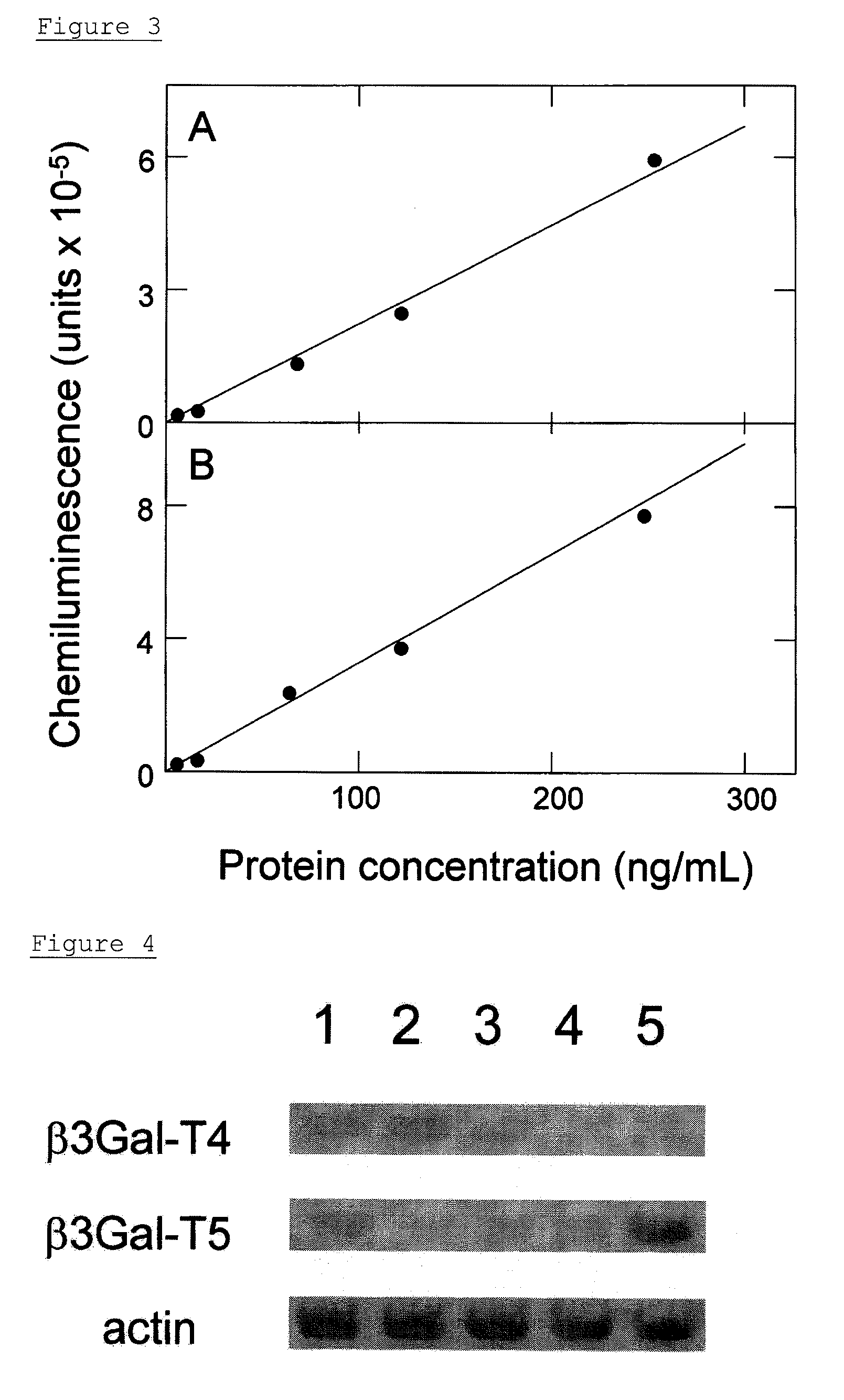
[0125]50 μL of b3Gal-T5 monoclonal antibody, which had been diluted to a concentration of 1 μg / mL with a PBS, was added to 96-well black high-binding plates (Corning Inc., Corning, N.Y.), and was immobilized at 4° C. for 16 hr. The surface of each well was blocked with PBS containing 1% BSA at 25° C. for 1 hr. The non-denatured β3Gal-T5 (E. coli) as reference materials, was diluted to a concentration of 250 ng / mL, 125 ng / mL, 62.5 ng / mL, 16 ng / mL, and 4.0 ng / mL with a diluent wherein 1% BSA had been added to PBS-0.1% Tween-20 (PBS-T). Then 50 μL of the non-denatured β3Gal-T5 (E. coli) was added to ...

example 5

Measurement in Sera of Ovarian Cancer Patients Using β3Gal-T5 Detection System

[0130]Sera of 60 cases of ovarian cancer patients and sera of 8 samples of healthy adult women were measured using the β3Gal-T5 detection system. The sera of the ovarian cancer patients to be used in the measurement were obtained from ovarian cancer patients of stages I to IV which were confirmed by abdominal operation. In the 57 cases, it was possible to determine the histological types of ovarian cancer by using histological examination. Any cancer developments other than ovarian cancer were not confirmed in these ovarian cancer patients.

[0131]Step (A) in Example 4 was repeated, except that the sera of patients and of healthy persons diluted with PBS-T by a factor of 10 were used instead of non-denatured β3Gal-T5 (E. coli). The amount of β3Gal-T5 in the sera was determined by the standard curve (A) in FIG. 3. The value of an average+2SD in healthy adult women was defined as a cut-off point, and it was de...


PUM

PropertyMeasurementUnit
pHaaaaaaaaaa
v/vaaaaaaaaaa
volumeaaaaaaaaaa

Abstract

The object of the present invention is to provide a method for detecting gynecologic cancer and a kit for detecting the same.
The object can be solved by a method for detecting gynecologic cancer characterized in that β1,3-galactosyltransferase-5 and/or β1,3-galactosyltransferase-4, and GlcNAc 6-O-sulfotransferase-2 are analyzed. According to the detecting method, ovarian cancer and endometrial cancer patients can be detected at an early stage i.e. stages I and II, wherein patients have no subjective symptoms.

Description

TECHNICAL FIELD[0001]The present invention relates to a method for detecting gynecologic cancers, particularly ovarian cancer or endometrial cancer, characterized in that β1,3-galactosyltransferase-5 and / or β1,3-galactosyltransferase-4, and GlcNAc 6-O-sulfotransferase are analyzed. The term “analyzing” or “analysis” as used herein includes a quantitative or semiquantitative measurement of an amount of a compound to be analyzed and a detection used to determine the presence or the absence of a compound to be analyzed.BACKGROUND ART[0002]A gynecologic cancer includes, for example, endocervical cancer, endometrial cancer, ovarian cancer, valvar cancer, vaginal cancer, uterine sarcoma, and chorionic cancer (trophoblastic disease). In these many cancers, it is difficult to find the cancer by symptoms thereof at an early stage. However, five years survival rate is significantly increased by carrying out a surgical therapy such as operation at an early stage. Thus, it is desired to develop...

Claims


Application Information

Patent Timeline
Patent Type & Authority Applications(United States)
IPC IPC(8): C12Q1/68C07K16/40G01N33/574
CPCC07K16/40C12Q1/48G01N2333/91097G01N33/57449G01N33/57442
Inventor YAMASHITA, KATSUKOSEKO, AKIRAAOKI, DAISUKESAKAMOTO, MASARU
Owner LSIP
Why Eureka
Try Eureka