Retractor cannula system for accessing and visualizing spine and related methods

a cannula system and rectal cannula technology, applied in the field of rectal cannula system for accessing and visualizing spine, can solve the problems of lack of real-time visualization capabilities, inability to provide tactile sensation to the surgeon, and difficulty in accurately diagnosing back pain, etc., to achieve the effect of increasing the field of view

Active Publication Date: 2012-01-19
EXPANDING INNOVATIONS INC
View PDF3 Cites 12 Cited by
  • Summary
  • Abstract
  • Description
  • Claims
  • Application Information

AI Technical Summary

Benefits of technology

[0010]In one embodiment, a retractor cannula system may comprise at least two interlocking jaws that have a substantially rounded or curved geometry when in the closed configuration. Following positioning of the retractor cannula system about the target site, the jaws of the retractor assembly may be placed in the open configuration and may be used as an atraumatic tool for dissection and / or a displacing tissue to create working space, thereby enhancing visualization of other surrounding structures. In one embodiment, the retractor assembly is a forward-looking structure so that the distal tip of the retractor assembly may push obstructive tissue away from the scope, and the distal tip of the retractor assembly may provide a depth of view between the scope and the targeted sites to be treated.
[0020]In another embodiment, a retractor cannula device for assessing a target site within the body may include a multi-lumen elongate shaft and a retractor assembly attached at a distal end of the shaft, wherein the retractor assembly is comprised of at least two jaws coupled to the shaft via any suitable articulation, including hinge structures, such that the jaws may have a closed configuration and an open configuration. In the closed configuration, the jaws may mate with one another such that a substantially smooth and rounded tip is formed. In the open configuration, the jaws are moved outward, increasing the angle between their inner edges, which may be used to dilate tissue and to increase the field of view.
[0026]In another embodiment, a method for minimally invasively accessing a body site is provided, comprising providing a tubular body with a retractor assembly located at a distal end of the tubular body and protruding from the distal end of the tubular body, where retractor assembly has a closed and an open configuration, and the tubular body comprises a viewing lumen to retain an endoscope and a working space distal to the viewing lumen, inserting a tubular body toward a non-vascular target site in a body, urging the retractor assembly of the tubular body into an open configuration while in the body, and visualizing the non-vascular target site from the tubular body and through the lumen of the retractor assembly. The method may also optionally comprise inserting an endoscopic device into the tubular body. The method may also include advancing the distal end of the tubular body toward a neural structure in contact with a non-neural structure, and displacing the neural structure from the non-neural structure using the retractor assembly. The method may also comprise orienting the working space of the retractor assembly with the non-vascular target site. In some embodiments, the method may additionally or alternatively comprise urging the retractor assembly from a closed to an open configuration while in the body, and visualizing the non-vascular target site from the tubular body and through a working space of the retractor assembly.

Problems solved by technology

However, these devices also provide little in the form of tactile sensation for the surgeon or allow the surgeon to atraumatically manipulate surrounding tissue.
In general, these conventional systems rely on external visualization for the approach to the disc and thus lack any sort of real time, on-board visualization capabilities.
Accurately diagnosing back pain is often more challenging than expected and often involves a combination of a thorough patient history and physical examination, as well as a number of diagnostic tests.
A major problem is the complexity of the various components of the spine, as well as the broad range of physical symptoms experienced by individual patients.
These anatomical elements make it difficult to treat or diagnose conditions within the epidural area because they tend to collapse around any instrument or device inserted therein.
This may reduce visibility in the epidural space, and may cause inadvertent damage to nerve roots during device insertion.
Also, the insertion of a visualization device may result in blocked or reduced viewing capabilities.
As such, many anatomical elements within the epidural space may limit the insertion, movement, and viewing capabilities of any access, visualization, diagnostic, or therapeutic device inserted into the epidural space.

Method used


Image

  • Retractor cannula system for accessing and visualizing spine and related methods
  • Retractor cannula system for accessing and visualizing spine and related methods
  • Retractor cannula system for accessing and visualizing spine and related methods

Examples


Embodiment Construction

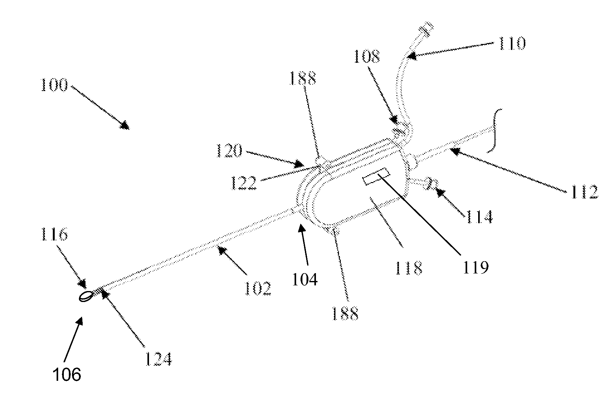
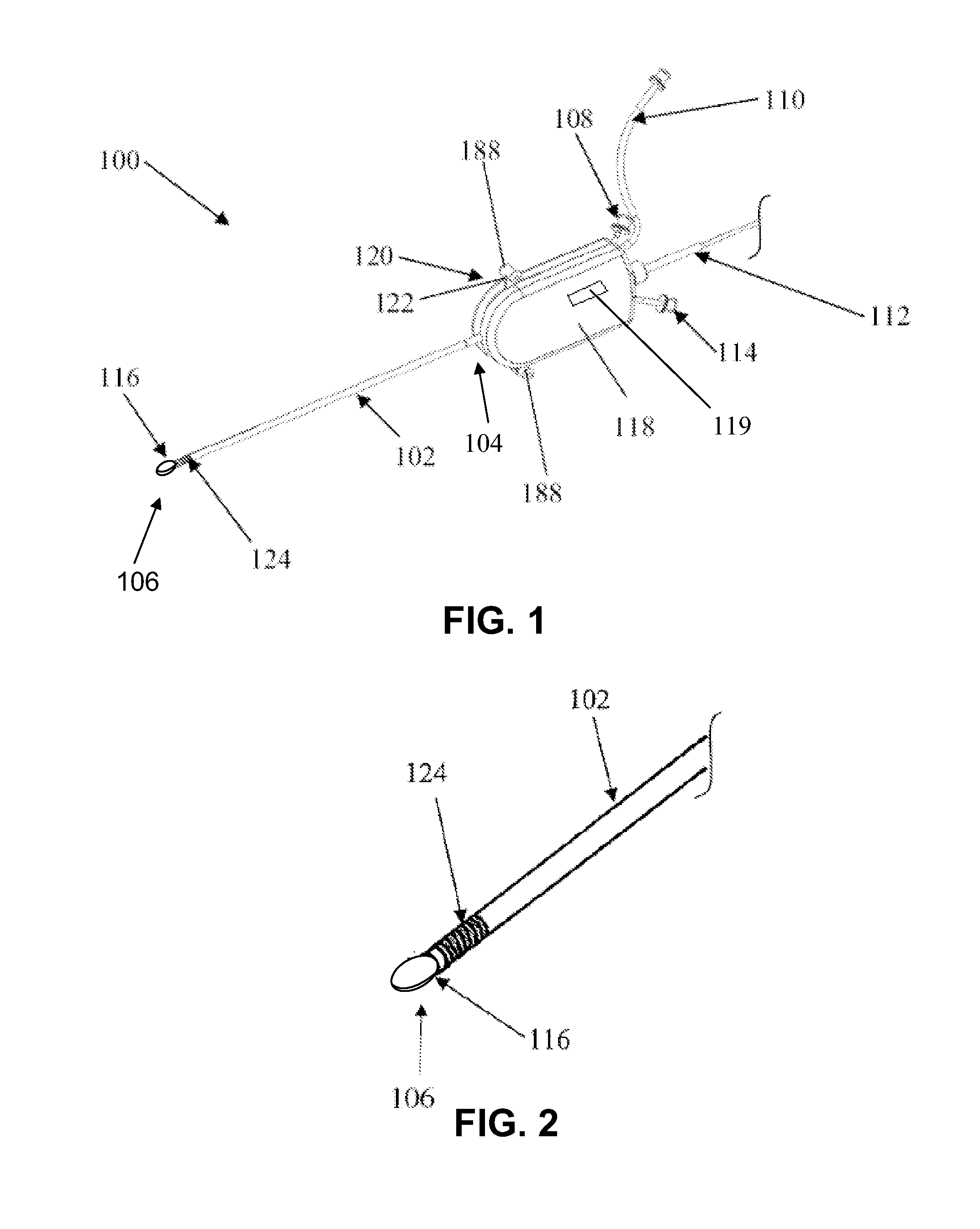
[0054]Conventional systems often rely on external visualization such as fluoroscopy and CT scanning for the approach to the disc, and thus lack any sort of real time, on-board visualization capabilities. Also, existing devices provide little in the form of tactile sensation for the surgeon and do not allow the surgeon to atraumatically manipulate surrounding tissue.

[0055]There is a need, therefore, for minimally invasive techniques and systems that provide the capability to diagnose or repair the spine using direct visualization while minimizing damage to surrounding anatomical structures and tissues. There is also a need for a method and device that allows a physician to effectively enter the epidural space of a patient, clear an area within the space to enhance visualization and use the visualization capability to diagnose and treat the disc injury.

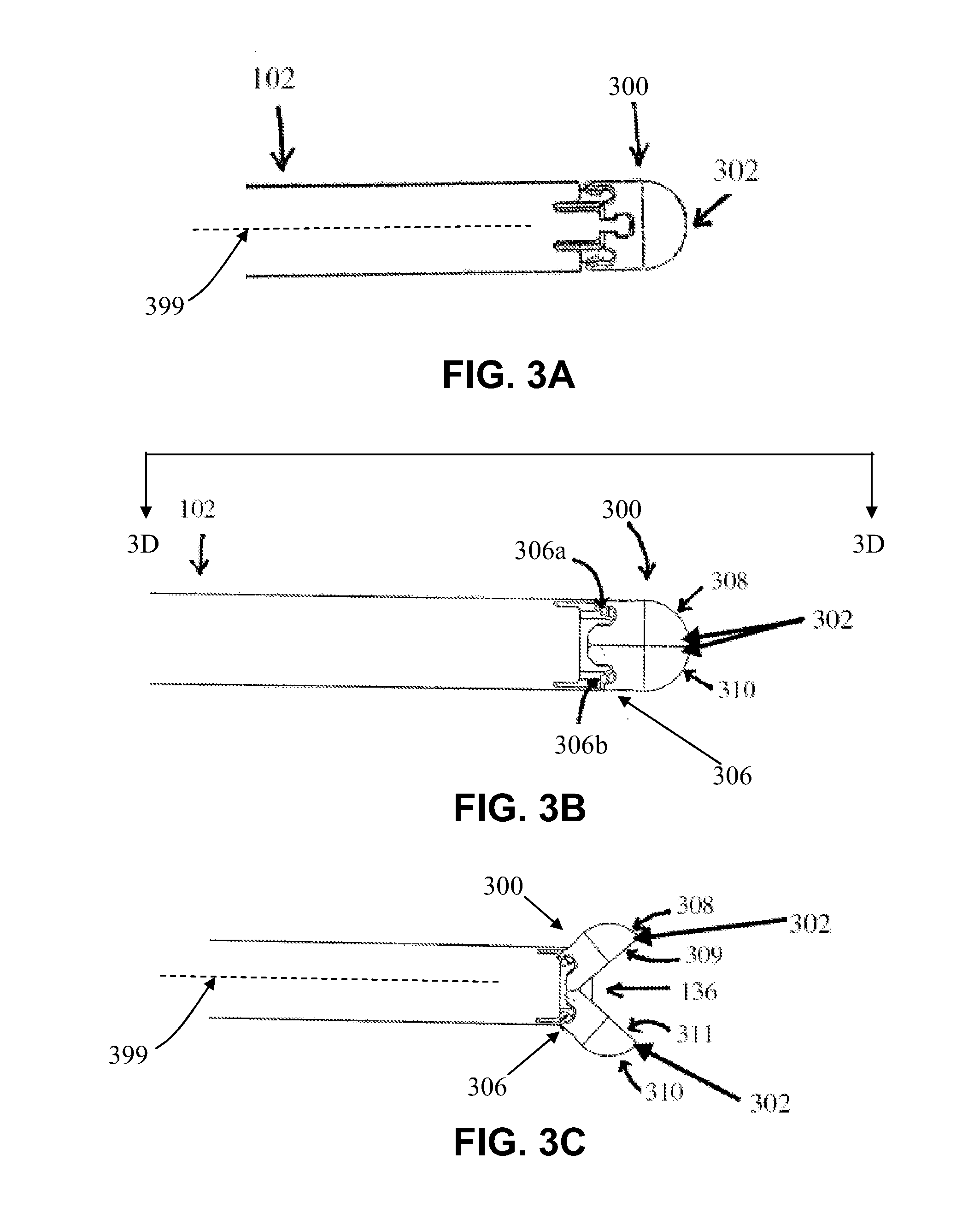
[0056]The embodiments disclosed herein will be more clearly understood and appreciated with respect to the following Detailed Descript...


PUM


Abstract

Retractor cannula systems may be used for accessing and visualizing the spine and related methods of treatment, including a forward-looking retractor cannula system for creating a working space and the retractor cannula system having atraumatic dissection capability to allow visualization in spine. The devices and methods described may be used, for example, to perform annulus repair, herniated disc excision, and denervation of neurological tissue; to dispense pharmacological agents and / or cell or tissue therapy agents; to diagnose disc degeneration and bony degeneration, spinal stenosis, and nucleus decompression, and to perform disc augmentation.

Description

CROSS-REFERENCE TO RELATED APPLICATIONS[0001]This application is a continuation of U.S. patent application Ser. No. 12 / 582,638, filed Oct. 20, 2009, which claims priority under 35 U.S.C. §119(e) to U.S. Provisional Application No. 61 / 106,914, filed Oct. 20, 2008, the disclosures of which are hereby incorporated by reference in their entirety. This application is also related to U.S. application Ser. No. 12 / 199,706, filed Aug. 27, 2008, which is also hereby incorporated by reference in its entirety.BACKGROUND[0002]Injured intervertebral discs are generally treated with bed rest, physical therapy, modified activities, and pain medications for substantial treatment durations. There are also a number of treatments that attempt to repair injured intervertebral discs and to avoid surgical removal of injured discs. For example, disc decompression is a procedure used to remove or shrink the nucleus, thereby decompressing and decreasing the pressure on the annulus and nerves. Less invasive p...

Claims


Application Information

Patent Timeline
Patent Type & Authority Applications(United States)
IPC IPC(8): A61B10/00A61B1/00
CPCA61B1/3135A61B5/0084A61B8/12A61B10/06A61B2017/320064A61B17/3417A61B19/5212A61B2017/00261A61B2017/003A61B17/0218A61B90/361A61B2017/00907A61B2017/3419
Inventor TO, JOHN T.
Owner EXPANDING INNOVATIONS INC
Why Eureka
Try Eureka