Portable pet scanner for imaging of a portion of the body

a scanner and portability technology, applied in tomography, instruments, x/gamma/cosmic radiation measurement, etc., can solve the problems of spatial resolution degradation, higher cost, identification error,

Inactive Publication Date: 2012-03-22
INTRAMEDICAL IMAGING LLC
View PDF2 Cites 48 Cited by
  • Summary
  • Abstract
  • Description
  • Claims
  • Application Information

AI Technical Summary

Problems solved by technology

This effect degrades spatial resolution since an uncertainty in the interaction location results in an error in the identification of the proper line-of-response (LOR) for that event.
This remedy has two major disadvantages:1) An increased number of scintillators and PMTs with associated electronics results in a higher cost, and2) A reduction in sensitivity, which is directly proportional to the system diameter, requires longer scan duration and/or produces increased image noise.
Shortening the depth of each detector element can also reduce the DOI effect; however, this will also reduce the system sensitivity.
In general, despite the improvement in spatial resolution, the sensitivity of scanners has not improved.
There are two reasons for this: 1) Typically the detector ring is substantially larger than the port to reduce the effect of the “Depth of Interaction problem”, 2) The use of ...

Method used


Image

  • Portable pet scanner for imaging of a portion of the body
  • Portable pet scanner for imaging of a portion of the body
  • Portable pet scanner for imaging of a portion of the body

Examples


first embodiment

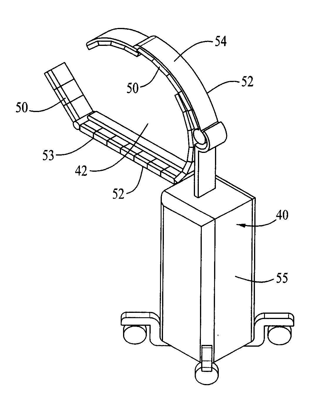
[0057]FIG. 9 shows one embodiment of a mobile PET scanner 40 incorporating features of the invention. The detector blocks 50 in the embodiment shown are 8 cm long, and 1.6 cm thick. The frame 52 that supports the blocks are stainless steel 3 mm thick. The upper portion 54 slides to open and close the patient enclosing space 42 defined by the surrounding detector blocks 50. The cart is designed as a mobile PET unit and is adjustable so that the bottom frame 53 with detector blocks 50 of the PET unit can be fit through a gap or space 66 below a patient 72 placed on an operating table 70, such as shown by the arrow 56 in FIGS. 10 and 11.

Placement of the Detector Inside the Gap in the Operating Room Table

[0058]All clinical PET scanners are based on the use of photomultiplier tubes, which are relatively large (several centimeters). Because Solid-State Photo-Multipliers (SSPM) have dimensions of only a few millimeters, it is now possible to build a PET scanner with detectors that can fit ...

second embodiment

[0066]A “spot-PET scanner” is proposed to provide PET images of a portion of the body in the operating room. These further embodiments of the detector design for a PET scanner for spot imaging, are based on fast SiPM or Digital Photon Counter—DPC, and the Ca-dopped LSO scintillator that enable “time-of-flight assisted limited-angle tomography”. The theory of the “time-of-flight assisted limited-angle tomography” was simulated for a dedicated breast imaging system (ref. Karp et al. 2009).

[0067]We performed simulations to determine if a time-of-flight assisted, limited-angle tomography Spot-PET could obtain images of a torso-sized object that were comparable to those generated by commercial PET scanners. Simulations were performed with GRAY (P. Olcott, S. Buss, C. Levin, G. Pratx, and C. Sramek, “GRAY: High Energy Photon Ray Tracer for PET Applications,” Nuclear Science Symposium Conference Record, 2006. IEEE, vol. 4, 2006, pp. 2011-2015), an in house fast Monte Carlo package for simu...


PUM


Abstract

A mobile PET scanner for use in bed side or a surgical environment comprises a mobile support base, with first and a second arm arms extending therefrom. The first arm is configured for placement under a table supporting an individual while the second arm is substantially parallel to and above said first arm with the individual being located between the first and second arms. Multiple module blocks are positioned along the length of the first and second arm. Each modules block comprises scintillators with solid state silicone multipliers or multi-pixel photon counters attached thereto. Positrons emitted from radiation labeled tissue within the individual's body impinge on the multiple scintillators to generate. The photons from each of the scintillator are received by each of a solid state silicone multipliers or multi-pixel photon counters associated therewith and an electrical signal representative of the received photons is then generated. The electrical signal output from each of the solid state silicone multipliers or multi-pixel photon counters is then transmitted to a computerized data collection and analysis system, which substantially instantaneously generates a visual image on a screen showing the location within the individuals body emitted the photons. This image can be coordinated with a photo image or a CT image showing the same portion of the individual's body.

Description

[0001]This application claims benefit of Provisional Application 61 / 302,452 and is a Continuation-In-Part of U.S. application Ser. No. 12 / 776,777, filed May 10, 2010, which is a Divisional of application Ser. No. 11 / 929,349, filed Oct. 30, 2007, now U.S. Pat. No. 7,750,311.[0002]The devices described herein include novel detector modules for positron emission tomography (PET) that utilizes a novel photodetector referred to as solid state photomultiplier. (SSPM). These SSPM enable development of novel detector configurations for PET scanners that are more compact and therefore portable. Also, the fast response times of solid state photomultipliers enable time-of-flight assisted limited-angle tomography, and therefore imaging of only that part of the body that is of interest.BACKGROUND[0003]Positron emission tomography (PET) is becoming a powerful modality to image cancer and other disease. It is the most accurate non-invasive method for measuring the concentrations of radiolabeled tr...

Claims


Application Information

Patent Timeline
IPC IPC(8): G01T1/164
CPCA61B6/037A61B6/4405A61B6/5235A61B6/5247A61B17/3403A61B2019/5291A61B2017/3413A61B2019/5251A61B2019/5255A61B2019/5259A61B2019/5276A61B19/5244A61B2034/2055A61B34/20A61B2034/2051A61B2034/2059A61B2090/378A61B2090/365
Inventor DAGHIGHIAN, FARHAD
Owner INTRAMEDICAL IMAGING LLC
Why Eureka
Try Eureka