System and method for characterization of oral, systemic and mucosal tissue utilizing raman spectroscopy

Inactive Publication Date: 2012-04-12
PRESIDENT & FELLOWS OF HARVARD COLLEGE +2
View PDF9 Cites 24 Cited by
  • Summary
  • Abstract
  • Description
  • Claims
  • Application Information

AI Technical Summary

Benefits of technology

[0014]In accordance with the invention, the probe can include a second laser radiation source that can be projected from the first end of the probe. The wavelength of the second laser radiation source can produce radiation that is known to cause diseased tissue to fluoresce and be visible with the use of a filter. The second laser radiation can be used to illuminate an area to identify potentially diseased tissue and then using the Raman system according to the invention, capture Raman spectra of the tissue, compare the Raman spectra of the potentially diseased tissue with the Raman spectra of healthy tissue to determine whether the tissue is diseased. This can be accompli

Problems solved by technology

Although oral cancer is the most serious of oral cavity disease, and is often life threatening, it makes up only a small fraction of the total number of oral diseases.
However benign oral diseases can also be severe and debilitating if not treated properly and at an early stage.
Despite its desirability as a means to provide a definitive diagnosis, logistical, psychological, and economic hurdles often negatively impact

Method used


Image

  • System and method for characterization of oral, systemic and mucosal tissue utilizing raman spectroscopy
  • System and method for characterization of oral, systemic and mucosal tissue utilizing raman spectroscopy
  • System and method for characterization of oral, systemic and mucosal tissue utilizing raman spectroscopy

Examples


Embodiment Construction

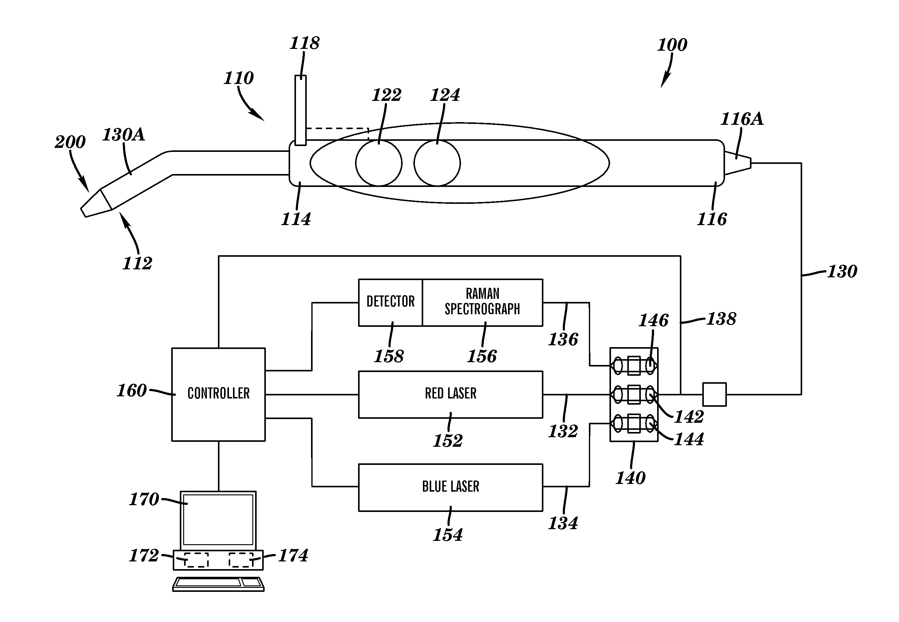
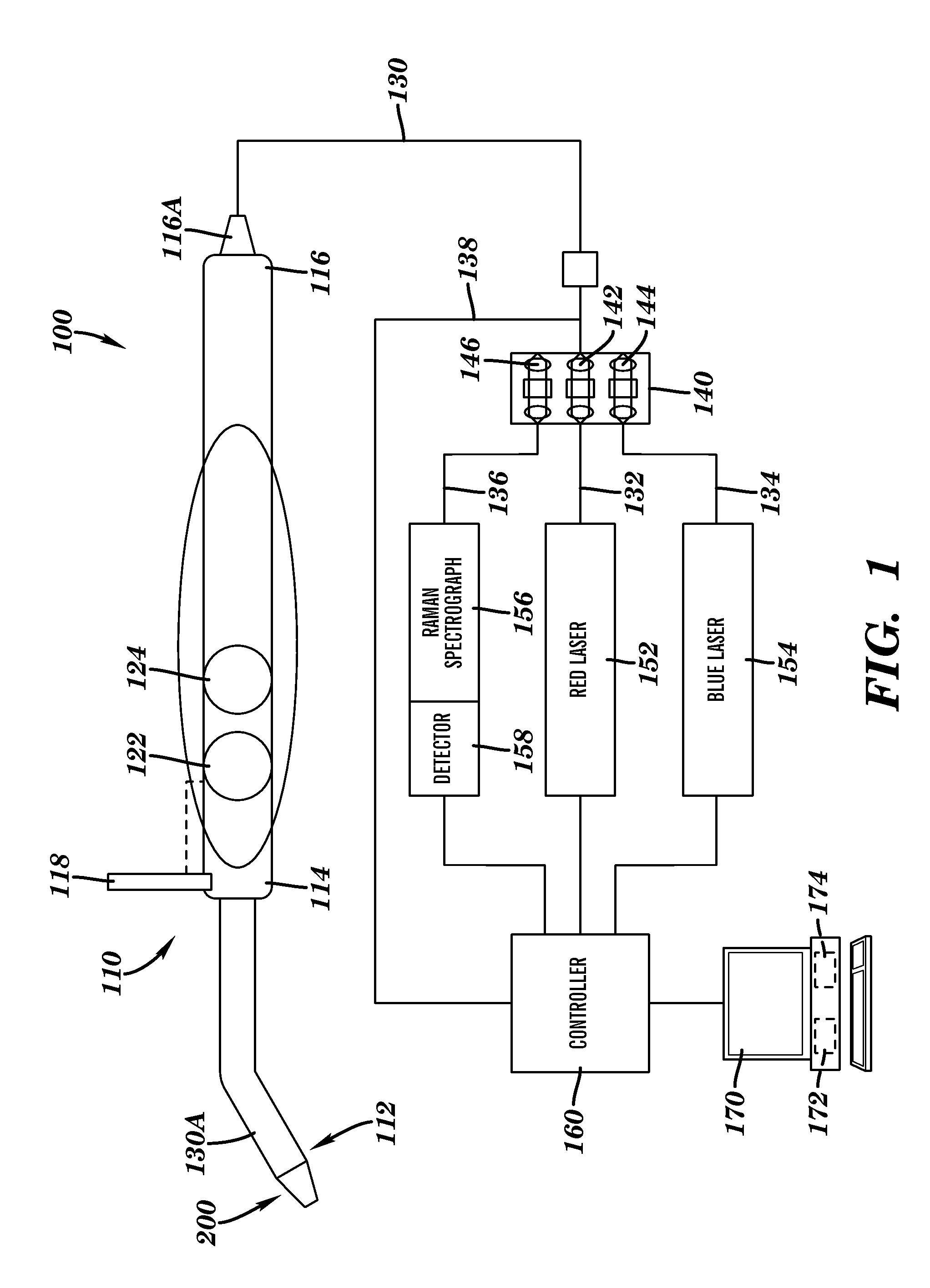
[0028]Example embodiments are described herein in the context of a system and method for characterization of tissue utilizing Raman Spectroscopy. Those of ordinary skill in the art will realize that the following description is illustrative only and is not intended to be in any way limiting. Other embodiments will readily suggest themselves to such skilled persons having the benefit of this disclosure. Reference will now be made in detail to implementations of the example embodiments as illustrated in the accompanying drawings. The same reference indicators will be used throughout the drawings and the following description to refer to the same or like items.

[0029]In the interest of clarity, not all of the routine features of the implementations described herein are shown and described. It will, of course, be appreciated that in the development of any such actual implementation, numerous implementation-specific decisions must be made in order to achieve the developer's specific goals...


PUM


Abstract

A method and system for characterizing tissue includes a probe connected to a red LASER source and a Raman spectroscope. The probe includes at least excitation fiber and one or more emission fibers that connect the probe with the LASER source and the Raman spectroscope. The excitation fiber is connected to the red LASER source and terminates in the first end of the probe adjacent the tip of the probe. The emission fibers are connected to the Raman spectroscope and terminate in the first end of the probe adjacent the tip of the probe. In one embodiment, the excitation fiber extends through the central portion of the probe and one or more emission fibers are arranged around the excitation fiber. The tip of the probe is intended to come in contact with the tissue to be examined. The tip includes a central opening to allow red LASER radiation to project out of the end of the red excitation fiber on to the tissue and to permit Raman spectra to enter the emission fiber(s) and travel to the Raman spectroscope. The tip is constructed to have a predefined focal length to position the first end of the probe a predefined distance from the surface of the tissue being examined. The tip can be removable and tips having different focal lengths can be used to accommodate different types of tissues and examinations. A detector can convert the Raman spectra into signals and data for analysis by a computer system. The Raman spectra for tissue in a predefined location can be profiled such that the system can distinguish between healthy and diseased tissue.

Description

CROSS-REFERENCE TO RELATED APPLICATIONS[0001]This application claims any and all benefits as provided by law of U.S. Provisional Application No. 61 / 145,362 filed Jan. 16, 2009, which is hereby incorporated by reference in its entirety.STATEMENT REGARDING FEDERALLY SPONSORED RESEARCH[0002]Not ApplicableREFERENCE TO MICROFICHE APPENDIX[0003]Not ApplicableBACKGROUND[0004]1. Technical Field of the Invention[0005]The present invention is directed to methods and systems for characterizing and diagnosing tissue and tissue disease using Raman spectroscopy. Specifically, the invention is directed to a Raman spectrometer system including a Raman spectrometer probe adapted for non-invasive diagnosis of tissue.[0006]2. Description of the Prior Art[0007]In 2008, in the US alone, it was estimated that about 34,000 individuals were to diagnosed with oral cancer. 66% of the time these will be found as late stage three and four disease. Low public awareness of the disease is a significant factor, bu...

Claims


Application Information

Patent Timeline
IPC IPC(8): A61B6/00
CPCA61B5/0088G01N2021/656G01N21/65A61B5/682
Inventor GUZE, KEVINSHORT, MICHAELZENG, HAISHANSONIS, STEPHEN
Owner PRESIDENT & FELLOWS OF HARVARD COLLEGE
Why Eureka
Try Eureka