Portable bio-magnetic imager and method

a bio-magnetic imager and portable technology, applied in the field of bio-magnetic imaging, can solve the problems of undiagnosed mild to moderate tbi, the gcs is a manual approach, and the likelihood of soldiers returning from combat to remain significant problems,

Inactive Publication Date: 2012-05-03
LOCKHEED MARTIN CORP
View PDF8 Cites 7 Cited by
  • Summary
  • Abstract
  • Description
  • Claims
  • Application Information

AI Technical Summary

Benefits of technology

[0010]The present invention is directed to imaging methods and apparatuses. According to a first aspect of the present invention, an Imager comprises: a field source capable of generating a magnetic field directed to a subject; a contrast agent applied to the subject, the contrast agent selectively seeking out an area in the subject, wherein the area also receives the magnetic field; and a low magnetic field detector arranged downstream from the field source, the low magnetic field detector being capable of detecting a low magnetic field associated with the area indicated by the contrast agent.
[0011]According to a second aspect of the present invention, an Imager comprises: a field source capable of generating a magnetic field directed to an area in a subject; a low magnetic field detector arranged downstream from the field source, the low magnetic field detector being capable of detecting a low magnetic field signature associated with the area in the subject; and a metamaterials lens arranged downstream from the field source, the metamaterials lens concentrating the magnetic field produced by the field source to the area in the subject, and / or concentrating back the magnetic signature from the area in the subject to the low magnetic field detector.
[0012]According to a third aspect of the present invention, an imaging method comprises: generating a magnetic field directed to a subject; indicating an area in the subject using a contrast agent including nanoparticles which selectively seek out the area in the subject; and detecting a low magnetic field associated with the area indicated by the contrast agent.
[0013]According to a fourth aspect of the present invention, an imaging method comprises: generating a magnetic field directed to an area in the subject; concentrating, using a metamaterials lens, the generated magnetic field to the area in the subject; and detecting a low magnetic field signature associated with the area in the subject.

Problems solved by technology

With the prevalence of Improvised Explosive Devices (IEDs) used by adversaries on the battlefield, undiagnosed mild to moderate TBI is likely to remain a significant problem for soldiers returning from combat.
However, the qualitative exam is unlikely to detect mild to moderate TBI.
However, the GCS is a manual approach and cannot reliably detect mild to moderate TBI.
Currently, there is no technique that is portable and can also give an early diagnosis of mild to moderate TBI.
There are, in fact, no diagnostic devices that can detect brain injury within a few hours of the injury.
With traditional MRI and CT imaging techniques, detection of mild to moderate TBI within 24 hours after exposure to trauma can be difficult to achieve, due to the relatively small injury size at start of injury or soon after injury, as compared to the large sampling size / volume of traditional imaging techniques, and because it takes more than 24 hours after exposure for brain damage to meet resolution of conventional MRI equipment which uses magnetic fields of the order of 3-5 Tesla.
These systems cannot be used for brain imaging, for TBI diagnosis, or for full body imaging, and cannot detect details of superimposed organs.
These systems also suffer from poor imaging quality and an inability to scan over a wide area of interest, as they have a low imaging volume. FIG. 12 illustrates the image acquisition for a conventional clinical MRI system, which uses a magnetic field source 520, a contrast agent 530 and a magnetic field / RF detector 540 (to detect medium to high Tesla magnetic fields).
In this configuration, cooling and infrastructure are required to acquire an image, and they significantly increase the size and weight of the MRI system.
While this portable version does not require the cooling or infrastructure that typical clinical MRIs require, its functionality is extremely limited.
Traditional clinical MRI systems are limited by cryogenic cooling and magnetic shielding requirements, while state-of-the-art portable versions suffer from a lack of sensitivity and poor image quality.
The MRI signal is usually proportional to the magnetic field applied to the subject, so lower field MRIs have poorer signal to noise ratio.
This conventional MRI system can only image dedicated extremities (only the knee, or only the arm), which reduces its shielding requirements.
Because the portable system is only for dedicated extremities, no contrast agent is used, since the portable system cannot be used to image organs, which are usually obscured by other organs.
The poor image quality of this conventional MRI device is also caused by the mechanism used to derive an image in current state-of-the-art MRI, whether low or high field.
This is also a problem in the conventional low field system, since a permanent magnetic field is susceptible to a field inhomogeneity, which affects the gradient used to reconstruct the image.

Method used


Image

  • Portable bio-magnetic imager and method
  • Portable bio-magnetic imager and method
  • Portable bio-magnetic imager and method

Examples


Embodiment Construction

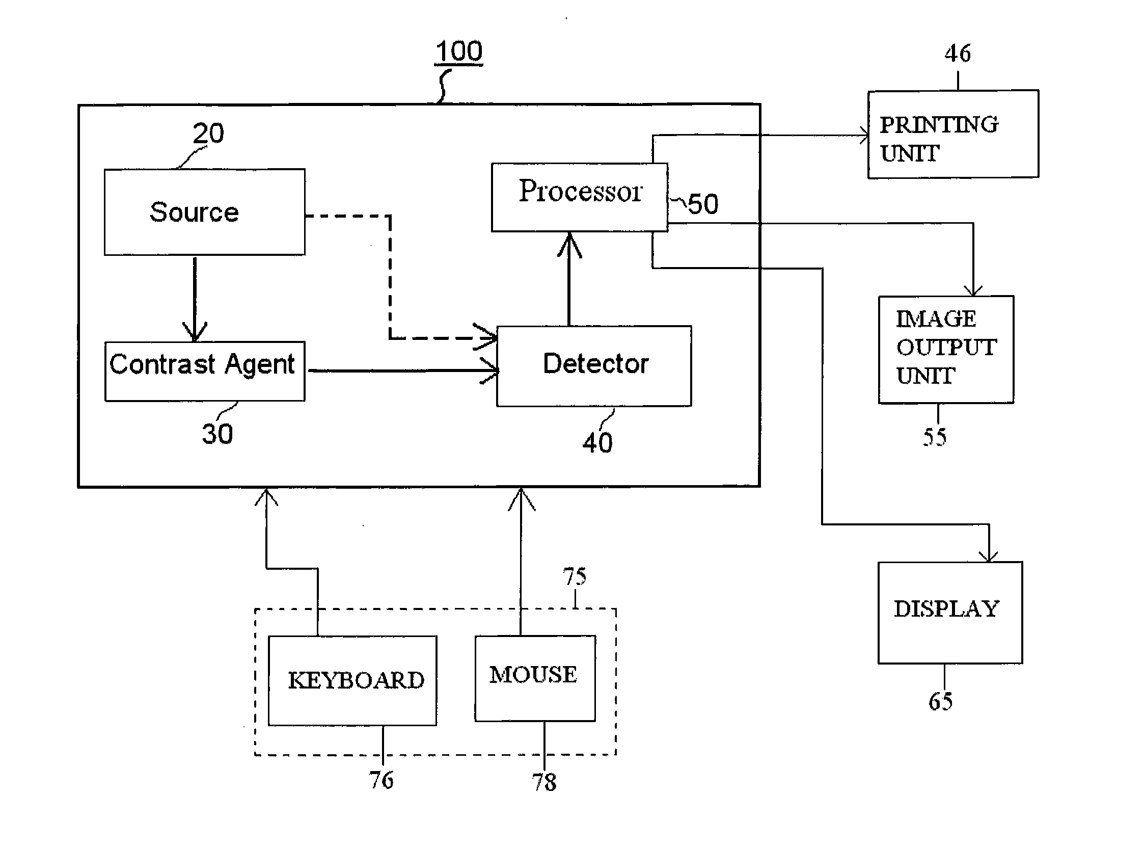
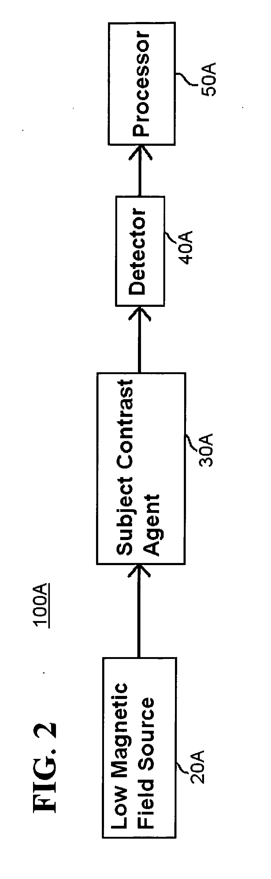
[0030]Aspects of the invention are more specifically set forth in the accompanying description with reference to the appended figures. FIG. 1 is a general block diagram of a Portable Bio-Magnetic Imager 100 according to an embodiment of the present invention. The system 100 illustrated in FIG. 1 includes the following components: a magnetic field source 20; a contrast agent 30; a magnetic field detector 40 and processor 50. Operation of the system 100 in FIG. 1 will become apparent from the following discussion.

[0031]The magnetic field source 20 generates electromagnetic radiation or an electromagnetic field which is applied to a region containing a contrast agent 30. The source 20 may be a permanent magnet coil, or some other device which produces a magnetic field through an electrical, magnetic, mechanic or other type(s) / combinations of mechanism(s).

[0032]The contrast agent 30 is applied to a subject (for example, a human) for detection of a signature of a certain type of anatomic...


PUM


Abstract

Methods and apparatuses of the present invention perform imaging using a contrast agent and/or a metamaterials lens, together with a low magnetic field detector. The apparatus according to one embodiment comprises: a field source capable of generating a magnetic field directed to an area in a subject; a low magnetic field detector arranged downstream from the field source, the low magnetic field detector being capable of detecting a low magnetic field signature associated with the area in the subject; and a metamaterials lens arranged downstream from the field source, the metamaterials lens concentrating the magnetic field produced by the field source to the area in the subject, and/or concentrating back the magnetic signature from the area in the subject to the low magnetic field detector.

Description

CROSS REFERENCE TO RELATED APPLICATION[0001]This non-provisional application claims priority under 35 U.S.C. §119(e) to U.S. Provisional Application No. 61 / 213,624 filed on Jun. 25, 2009, the entire contents of which are hereby incorporated by reference.BACKGROUND OF THE INVENTION[0002]1. Field of the Invention[0003]The present invention relates to an imaging technique and imaging apparatus, and more particularly to a method and apparatus for bio-imaging to detect injury or trauma.[0004]2. Description of the Related Art[0005]Traumatic Brain Injury (TBI) is considered to be the “signature wound” of modern wars such as the Iraq war. With the prevalence of Improvised Explosive Devices (IEDs) used by adversaries on the battlefield, undiagnosed mild to moderate TBI is likely to remain a significant problem for soldiers returning from combat. A main reason for undiagnosed TBI is the inability to quantify the damage to a soldier's brain soon after blast exposure. Currently a qualitative ex...

Claims


Application Information

Patent Timeline
Patent Type & Authority Applications(United States)
IPC IPC(8): G01R33/00
CPCG01R33/10
Inventor DRAKE, CHRISTINABALEINE, CLARA R.
Owner LOCKHEED MARTIN CORP
Why Eureka
Try Eureka