Multi-dimensional cardiac imaging

a cardiac imaging and multi-dimensional technology, applied in the field of multi-dimensional cardiac imaging methods and systems, can solve the problems of “corrupt patient datasets” and inadequate current non-invasive diagnostic techniques, and achieve the effects of improving robustness to motion/flow effects, robustness to missing data points, and high scan acceleration

Active Publication Date: 2012-06-14
THE BOARD OF TRUSTEES OF THE LELAND STANFORD JUNIOR UNIV +1
View PDF5 Cites 33 Cited by
  • Summary
  • Abstract
  • Description
  • Claims
  • Application Information

AI Technical Summary

Benefits of technology

[0007]The present invention provides a 5-dimensional imaging method and system to acquire and display the effect of dynamic physiologic changes (either spontaneous or induced) on cardiac function of a patient's heart to elucidate their effects on diastolic myocardial function: In a patient free-breathing magnetic resonance imaging study of said patient's heart, a computer system is used to acquire 3-dimensional spatial information and concurrently acquires recordings of cardiac and respiratory cycles of the patient. The 3-dimensional spatial information is sampled with a non-Cartesian 3-dimensional k-space readout trajectory which is incorporated into a magnetic resonance imaging sequence. The magnetic resonance imaging sequence could utilize principles of free-induction decay, steady-state free precession, gradient echo, spin echo, magnetization preparation modules, or a combination thereof to prepare image contrast from the magnetic resonant spins. The non-Cartesian 3-dimensional k-space readout trajectory traverses data points along arbitrary trajectories instead of straight rectilinear line trajectories common in Cartesian sampling. Examples of traversing data points could be along intersecting diagonal line trajectories, spiraling trajectories, circular trajectories in 3D k-space, or a combination thereof. Compared to traditional rectilinear Cartesian sampling, the non-Cartesian sampling provides higher scan acceleration (e.g., 3- to 8-fold or higher), improves robustness to motion / flow effects (incoherent instead of coherent artifacts) and provides robustness to missing data points in k-space.

Problems solved by technology

Diastolic left ventricular (LV) dysfunction remains one of the most important cardiac diseases for which current noninvasive diagnostic techniques remain inadequate.
However, many patient datasets are “corrupted” by dynamic physiologic motion.

Method used


Image

  • Multi-dimensional cardiac imaging
  • Multi-dimensional cardiac imaging
  • Multi-dimensional cardiac imaging

Examples


example

1. General Time-resolved Volumetric Acquisition

Acquisition

[0034]a. Continuous steady-state acquisition.

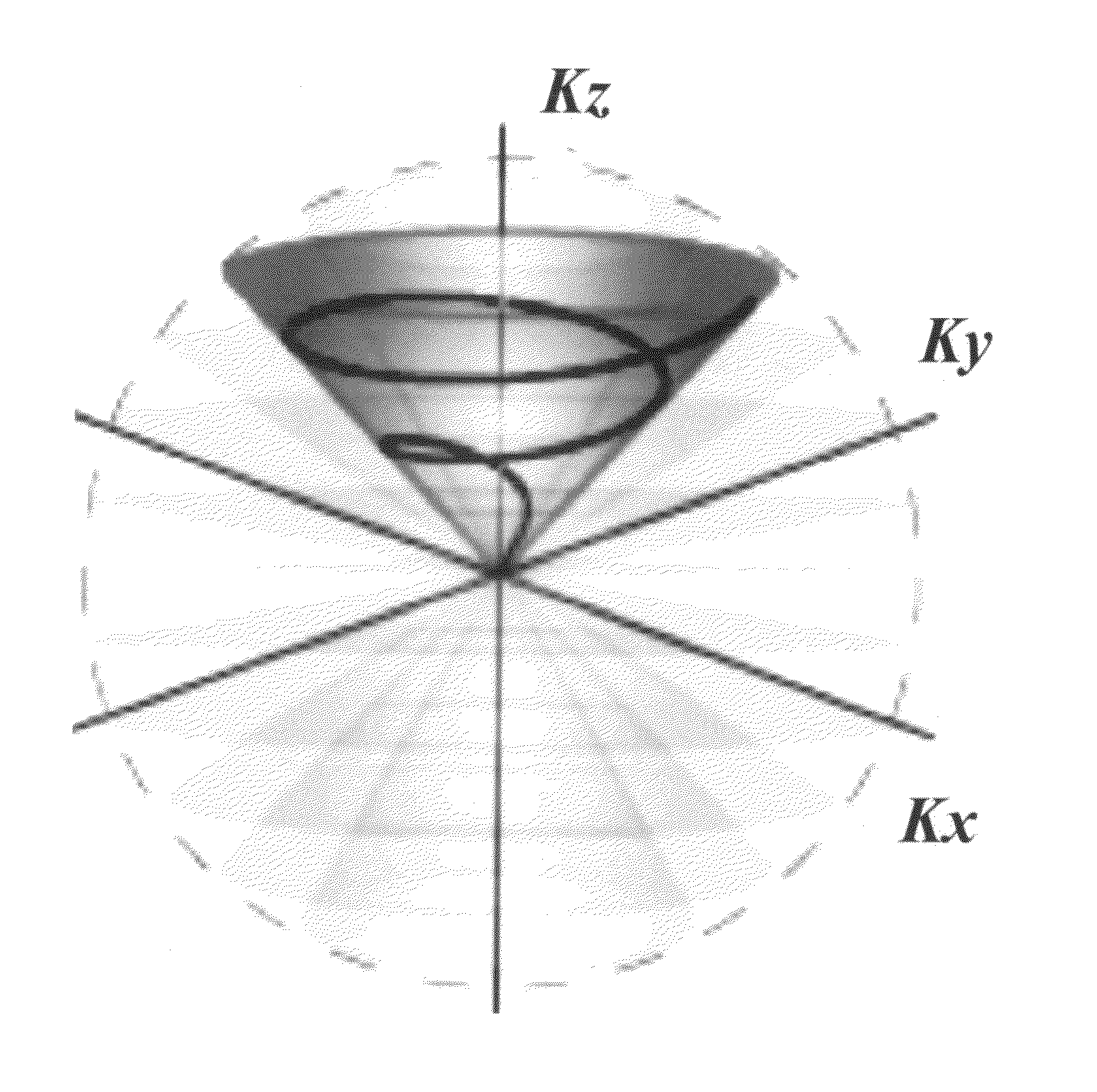
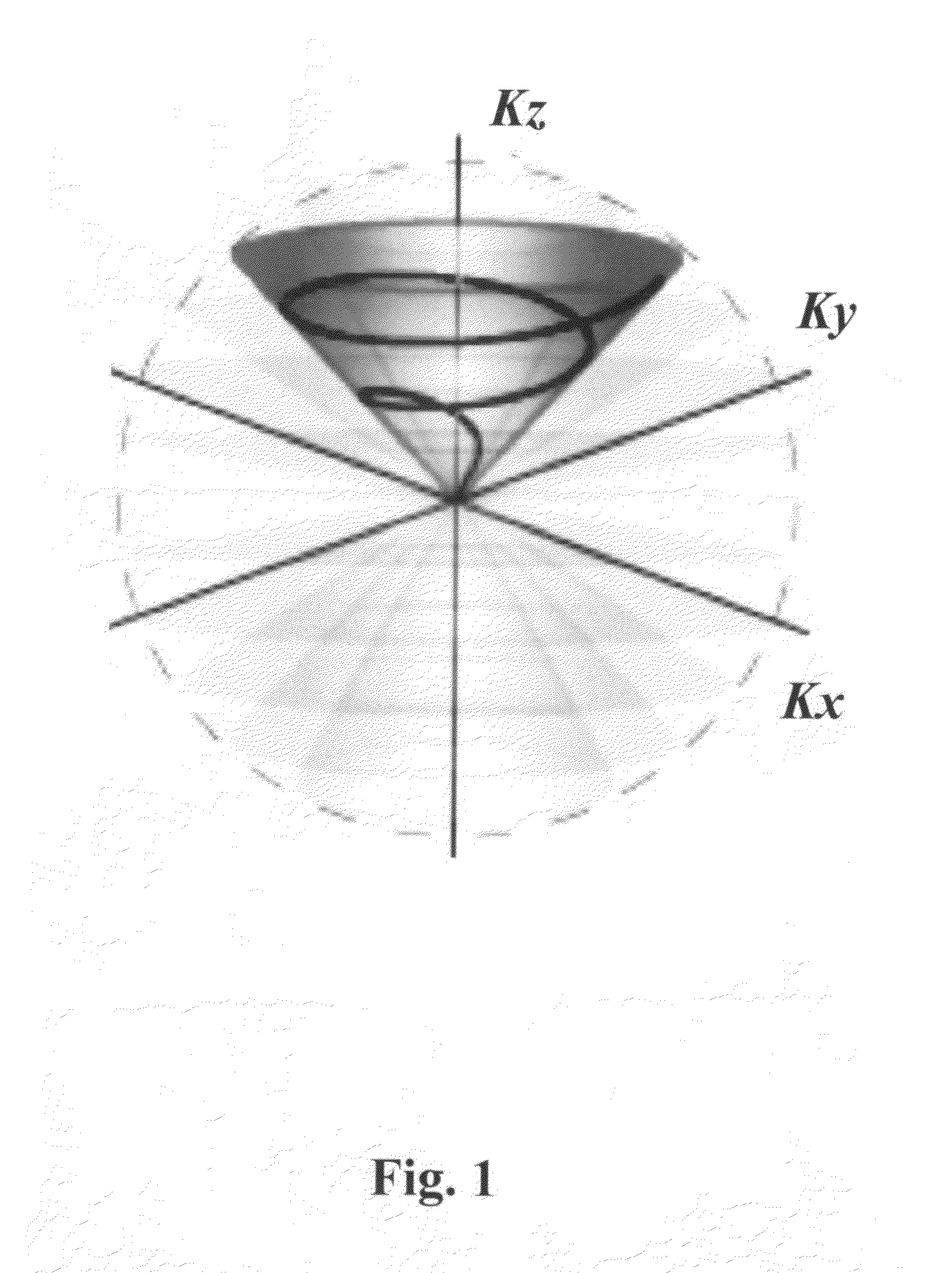
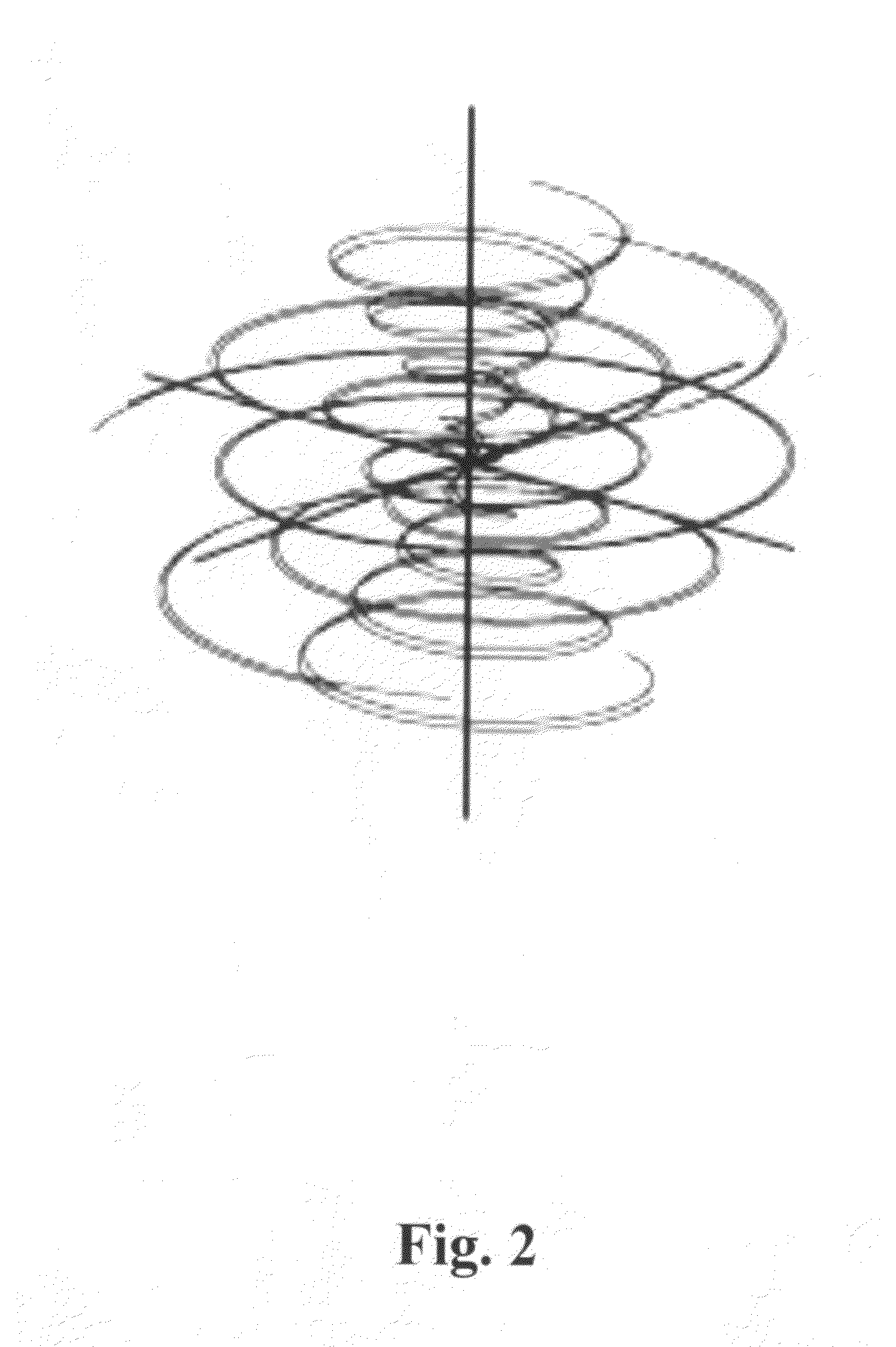
[0035]b. Use the 3D cones for spatial encoding.

[0036]c. Optional magnetization-preparation modules inserted for encoding of flow, diffusion, chemical shift, or other functional information.

[0037]d. Cardiac and respiratory information concurrently recorded during the scan from standard monitoring devices (ECG, pulse-ox, bellows) and / or MRI navigator signals.

[0038]e. Scan time reduction using multiple receiver elements.

Reconstruction

[0039]a. Retrospective synchronization of acquired anatomic / functional data to physiologic temporal phases (cardiac / respiratory phase).

[0040]b. Reconstruction of an entire volumetric and / or functional dataset for each desired temporal phase.

Processing and Display

[0041]a. Slice-by-slice or volume-rendered display of the 3D morphology as a temporal sequence with respect to cardiac motion at each stage of the respiratory cycle; a plot of the measured atrial / ...


PUM


Abstract

A 5-dimensional imaging method and system is provided to acquire and display the effect of dynamic physiologic changes (either spontaneous or induced) on cardiac function of a patient's heart to elucidate their effects on diastolic myocardial function. In a patient free-breathing magnetic resonance imaging study, 3-dimensional spatial information is encoded by a non-Cartesian 3-dimensional k-space readout trajectory and acquired concurrently with recordings of cardiac and respiratory cycles. The advantage of using non-Cartesian sampling in this invention compared to, for example, Cartesian sampling is higher scan acceleration, improved robustness to motion/flow effects (incoherent instead of coherent artifacts) and robustness to missing data points in k-space.

Description

CROSS-REFERENCE TO RELATED APPLICATIONS[0001]This application claims priority from U.S. Provisional Patent Application 61 / 459,238 filed Dec. 9, 2010, which is incorporated herein by reference in its entirety.STATEMENT OF GOVERNMENT SPONSORED SUPPORT[0002]This invention was made with Government support under contracts R01-HL039297 and NIH / NIBIB T32-EB009035 awarded by National Institutes of Health (NIH). The Government has certain rights in this invention.FIELD OF THE INVENTION[0003]The invention relates to multi-dimensional cardiac imaging methods and systems.BACKGROUND OF THE INVENTION[0004]Diastolic left ventricular (LV) dysfunction remains one of the most important cardiac diseases for which current noninvasive diagnostic techniques remain inadequate. For instance, more than 60% of women admitted for congestive heart failure have diastolic dysfunction as the cause of their symptoms. However, current noninvasive diagnostic criteria (primarily utilizing echocardiography) are able t...

Claims


Application Information

Patent Timeline
Patent Type & Authority Applications(United States)
IPC IPC(8): G01R33/48
CPCG01R33/4826G01R33/5602G01R33/5673G01R33/5635G01R33/56366G01R33/56341A61B5/05A61B6/503
Inventor WU, HOLDEN H.HU, BOB S.
Owner THE BOARD OF TRUSTEES OF THE LELAND STANFORD JUNIOR UNIV
Why Eureka
Try Eureka