Methods and systems for endobronchial diagnostics

a technology of endobronchial disease and diagnostics, applied in the field of lung disease diagnosis and treatment, can solve the problems of adjoining lung compartment collapse, affecting the patient's overall lung function and respiration, and not providing direct indication of imaging tests

Active Publication Date: 2012-06-14
PULMONX
View PDF2 Cites 10 Cited by
  • Summary
  • Abstract
  • Description
  • Claims
  • Application Information

AI Technical Summary

Problems solved by technology

However, imaging tests do not provide a direct indication of how the disease is affecting the patient's overall lung function and respiration.
One-way valves implanted in airways leading to a lung compartment restrict air flow in the inhalation direction and allow air to flow out of the lung compartment upon exhalation, thus causing the adjoining lung compartment to collapse over time.
Occluders block both inhalation and exhalation, also causing lung collapse over time.
If collateral airflow channels are present in a lung compartment, implanting a one-way valve or occluder might not be as effective, because the compartment might continue to fill with air from the collateral source and thus fail to collapse as intended.
Any one of these factors may extend the assessment time and compromise the assessment results.
Though promising, the accuracy and application of ventilation / perfusion scintigraphy has yet to be proven due to lackluster results in its application to lung volume reduction surgery.
Further, the dyes and separate scanning machinery necessary for scintigraphy means that this system is both complicated and costly to use.

Method used


Image

  • Methods and systems for endobronchial diagnostics
  • Methods and systems for endobronchial diagnostics
  • Methods and systems for endobronchial diagnostics

Examples


Embodiment Construction

[0033]Although the detailed description contains many specifics, these should not be construed as limiting the scope of the invention but merely as illustrating different examples and aspects of the invention. Various modifications, changes and variations may be made in the disclosed embodiments without departing from the spirit and scope of the invention.

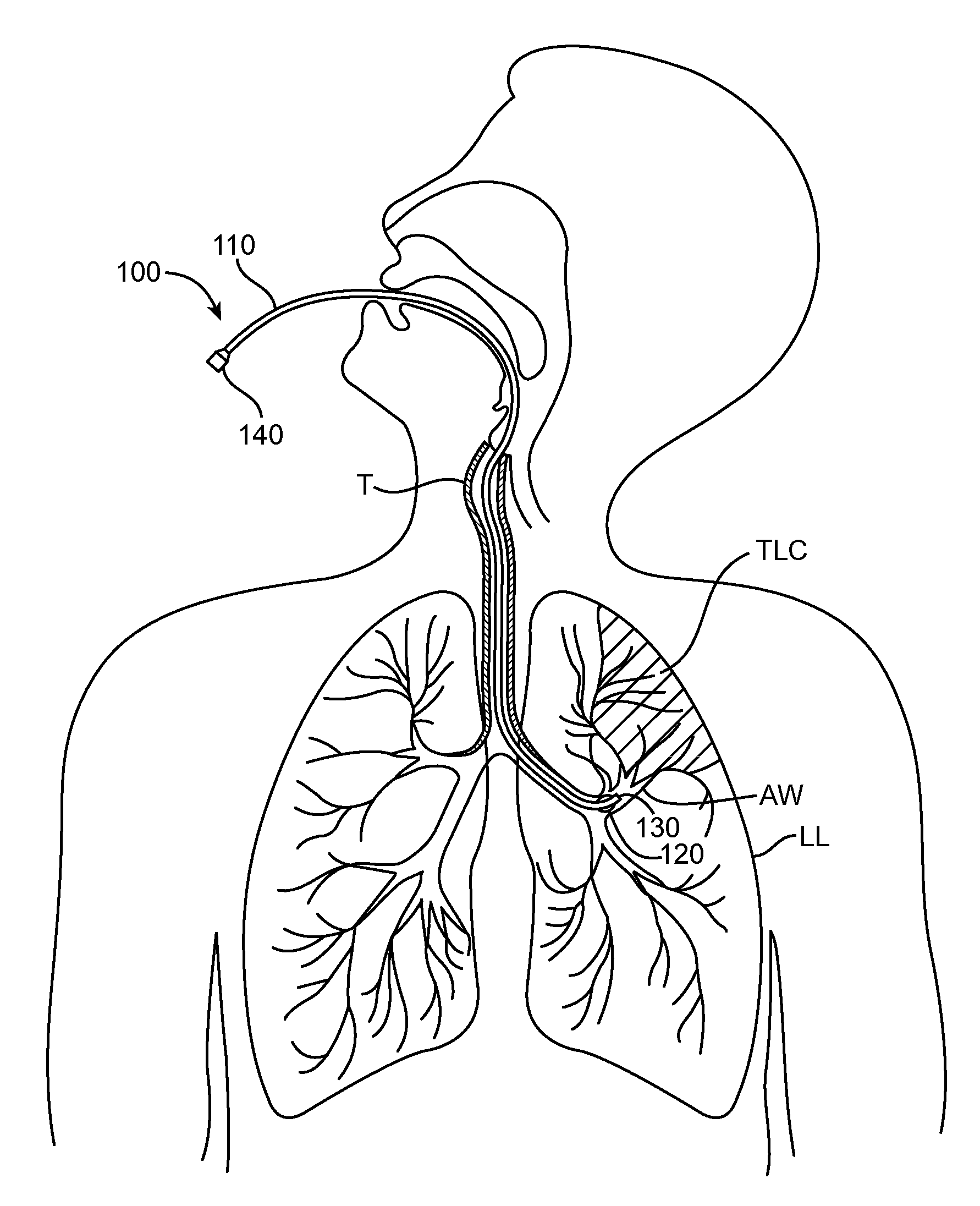
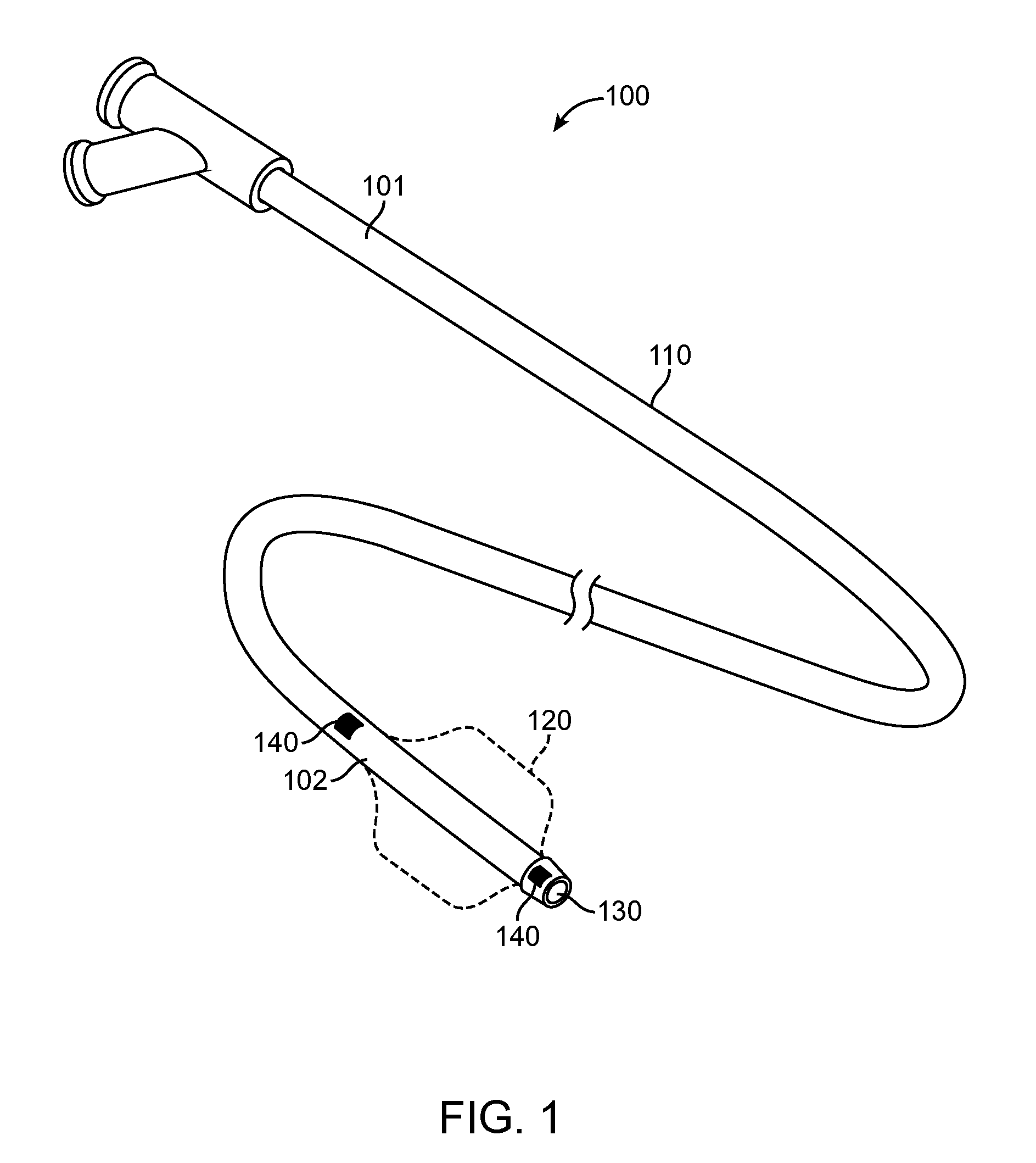
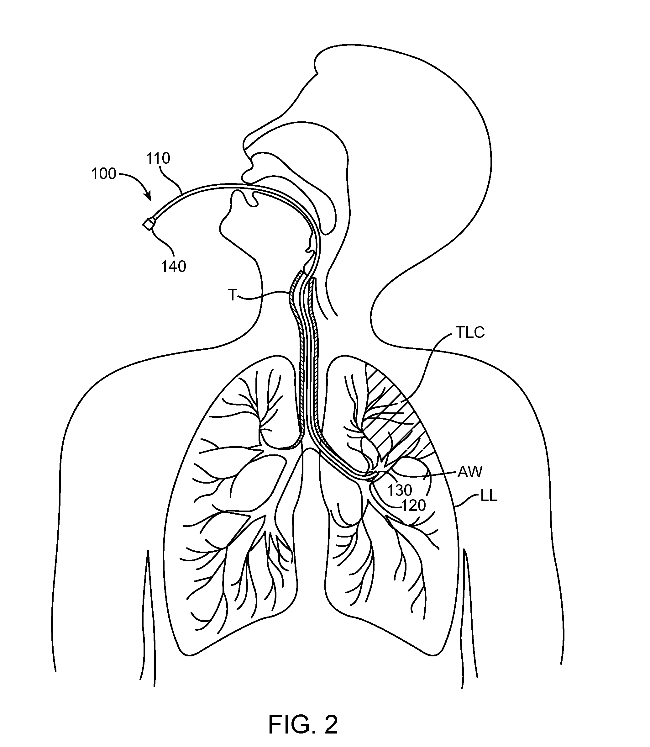
[0034]The present application provides methods and systems for targeting, accessing and diagnosing diseased lung compartments. Such compartments could be an entire lobe, a segment, a sub-segment or any such portion of the lung. Diagnosis is achieved in the disclosed embodiments by isolating a lung compartment to obtain various measurements to determine lung functionality. Though COPD is mentioned as an example, the applicability of these methods for treatment and diagnosis is not limited to COPD, but can be applicable to any lung disease.

[0035]The methods are minimally invasive in the sense that the required instruments are introdu...


PUM


Abstract

Methods and systems for targeting, accessing and diagnosing diseased lung compartments are disclosed. The method comprises introducing a diagnostic catheter with an occluding member at its distal end into a lung segment via an assisted ventilation device; inflating the occluding member to isolate the lung segment; and performing a diagnostic procedure with the catheter while the patient is ventilated. The proximal end of the diagnostic catheter is configured to be attached to a console. The method may also comprise introducing the diagnostic catheter into the lung segment; inflating the occluding member to isolate the lung segment; and monitoring blood oxygen saturation. The method may further comprise introducing the diagnostic catheter into the lung segment; determining tidal flow volume in the lung segment; determining total lung capacity of the patient; and determining a flow rank value based on the tidal flow volume of the lung segment and the total lung capacity.

Description

CROSS-REFERENCE TO RELATED APPLICATION[0001]This application claims priority under 35 U.S.C. §119(e) to U.S. Provisional Patent Application Ser. No. 61 / 360,811, entitled Methods and Systems for Endobronchial Diagnostics, filed Jul. 1, 2010, the full disclosure of which is hereby incorporated by reference.BACKGROUND OF THE INVENTION[0002]1. Field of the Invention[0003]This invention relates generally to methods for diagnosis and treatment of lung disease.[0004]2. Description of the Related Art[0005]Chronic obstructive pulmonary disease (COPD), including emphysema and chronic bronchitis, is a significant medical problem currently affecting around 16 million people in the U.S. alone (about 6% of the U.S. population). In general, two types of diagnostic tests are performed on a patient to determine the extent and severity of COPD: 1) imaging tests; and 2) functional tests. Imaging tests, such as chest x-rays, computerized tomography (CT) scans, Magnetic Resonance Imaging (MRI) images, p...

Claims


Application Information

Patent Timeline
Patent Type & Authority Applications(United States)
IPC IPC(8): A61B5/08A61B5/145A61B5/091A61M16/06A61B5/087A61B5/085
CPCA61B1/00082A61B1/2676A61B5/08A61B5/087A61B5/14542A61B5/6853A61M2016/0036A61M16/0404A61M16/0003A61B5/085A61B5/091A61B5/14552A61M2230/205A61M2016/102A61M2230/43
Inventor MANTRI, SURAGRADHAKRISHNAN, SRIKANTH
Owner PULMONX
Why Eureka
Try Eureka