Endoscopic diagnosis system

a diagnostic system and endoscope technology, applied in the field of endoscope diagnostic system, can solve the problems of high-quality autofluorescence, inability to obtain autofluorescence image, and inability to observe blood vessels

Active Publication Date: 2012-06-28
FUJIFILM CORP
View PDF7 Cites 23 Cited by
  • Summary
  • Abstract
  • Description
  • Claims
  • Application Information

AI Technical Summary

Benefits of technology

[0016]According to the present invention, a high-quality autofluorescence image can be obtained by increasing the emission amounts of the first and the second narrowband light (excitation light for autofluorescence observation) in an autofluorescence observation mode so as to be double those used in the narrowband light observation mode. Thus, using the first and the

Problems solved by technology

However, according to the method described in JP 2007-50106 A, whereby a narrowband light image and an autofluorescence image are acquired simultaneously, the excitation light has the same intensity for acquiring both images, but when the excitation light has the same intensity, a high-quality autofluorescence

Method used


Image

  • Endoscopic diagnosis system
  • Endoscopic diagnosis system
  • Endoscopic diagnosis system

Examples


Embodiment Construction

[0028]The endoscopic diagnosis system according to the present invention will be described in detail based on the preferred embodiments illustrated in the attached drawings.

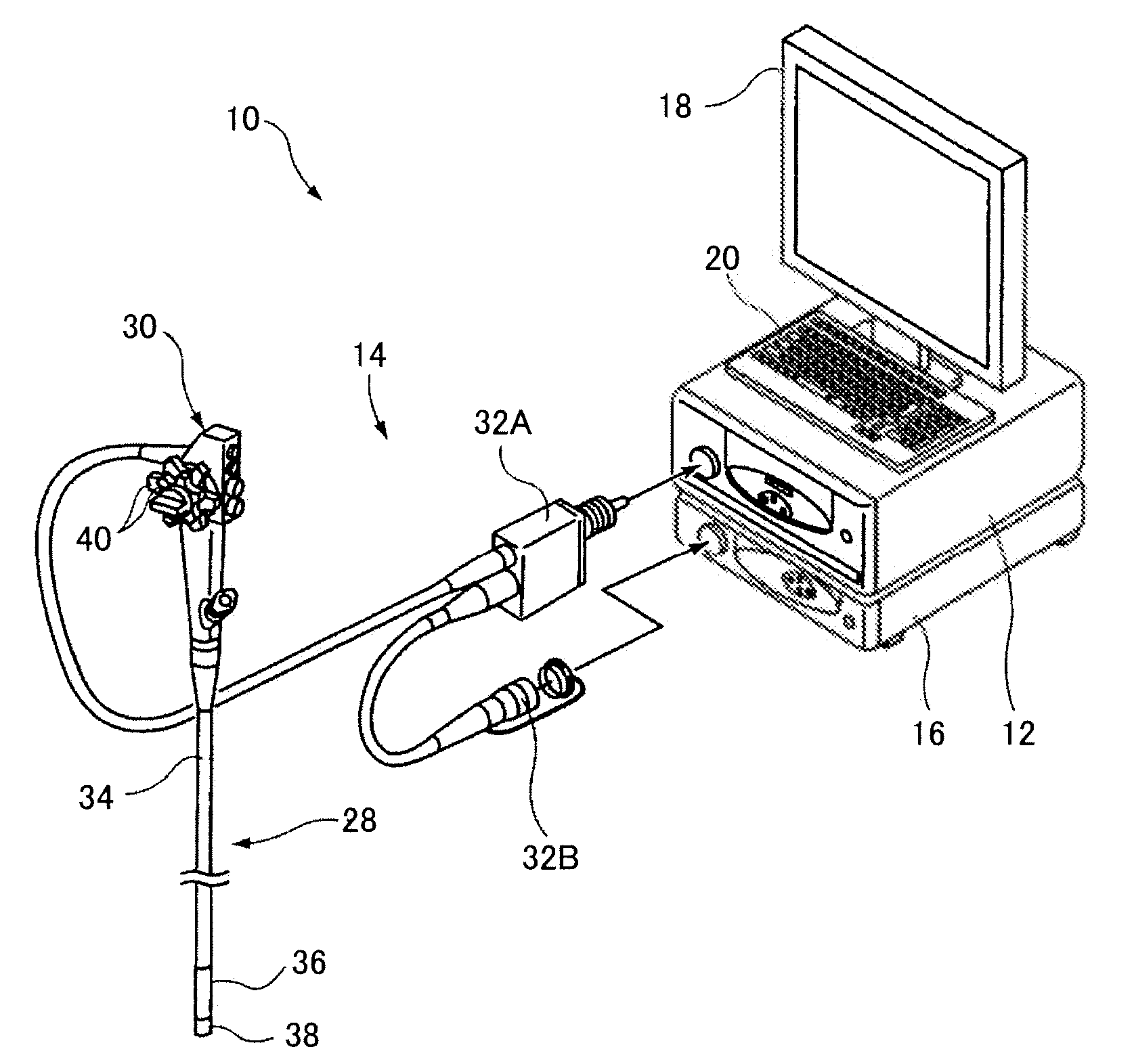
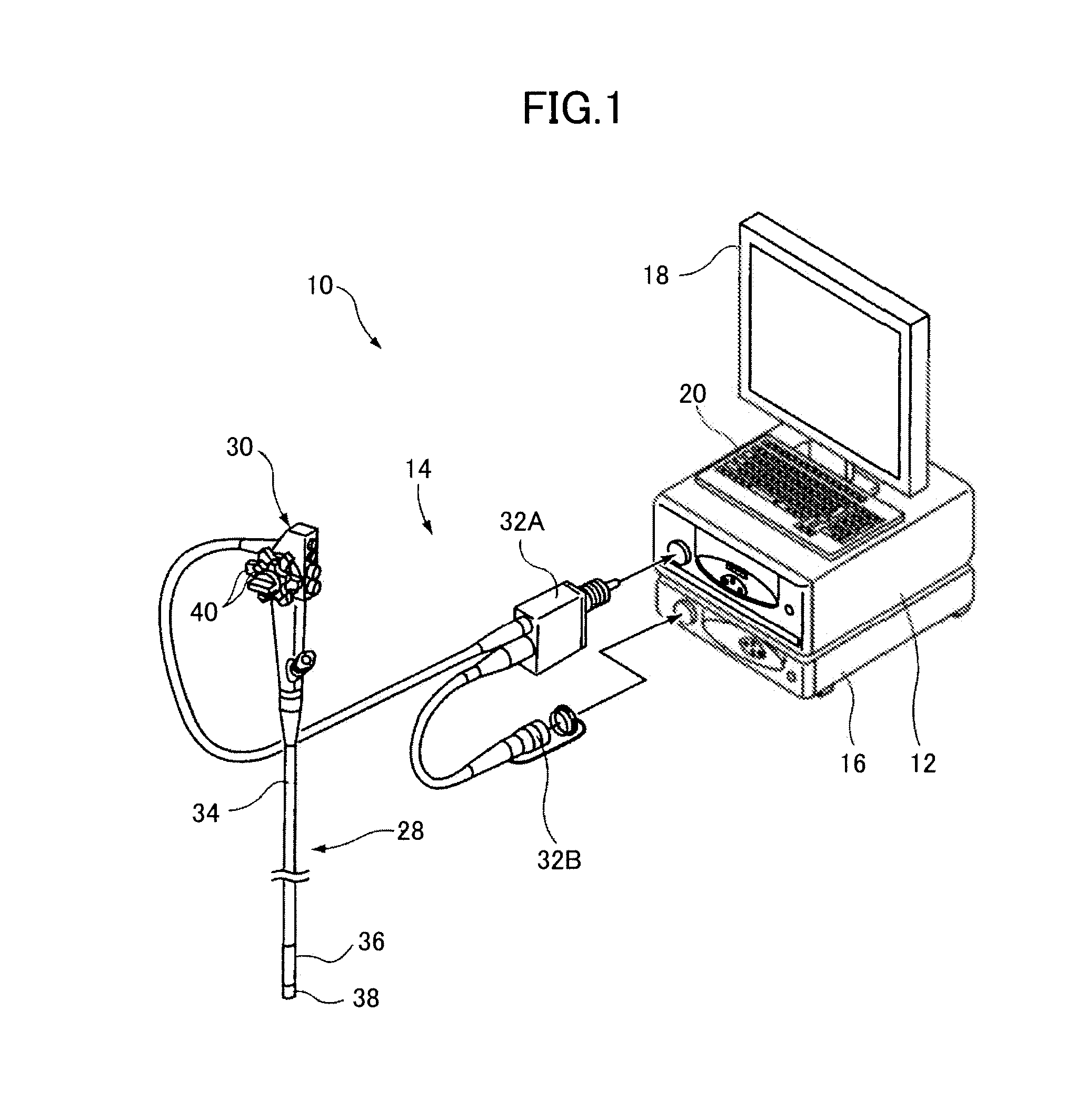
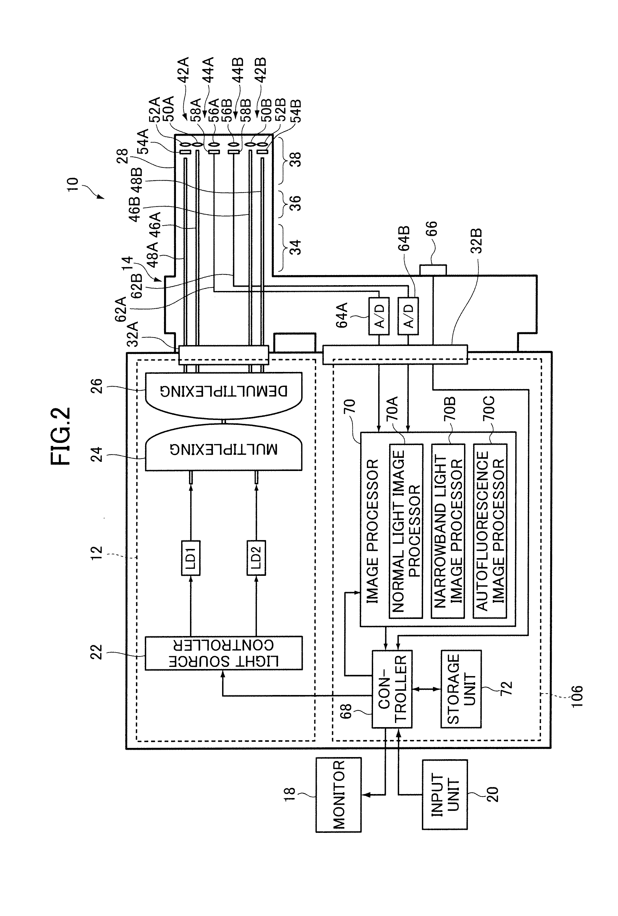
[0029]FIG. 1 is an external view of an embodiment illustrating a configuration of the endoscopic diagnosis system according to the invention; FIG. 2 is a block diagram illustrating an internal configuration thereof. A endoscopic diagnosis system 10 illustrated in these figures comprises a light source device 12 for emitting a plurality of light having different ranges of wavelength; an endoscope device 14 for guiding light emitted from the light source device 12 to illuminate a subject's region under observation with the illumination light and imaging the reflected light or an autofluorescence from the subject; a processor 16 for image-processing the acquired image acquired by the endoscope device 14 and outputting an endoscopic image; a monitor 18 for displaying an endoscopic image outputted from the processor 1...


PUM


Abstract

An endoscopic diagnosis system includes first and second narrowband light sources for emitting respectively first and second narrowband lights having a wavelength range different from each other, a first image sensor for receiving reflected light of the first narrowband light illuminating a subject to acquire a narrowband light image in a narrowband light observation mode, a second image sensor for receiving first autofluorescence emitted from the subject as the first narrowband light illuminates the subject to acquire a first autofluorescence image in a first autofluorescence observation mode and receiving second autofluorescence emitted from the subject as the second narrowband light illuminates the subject to acquire a second autofluorescence image in a second autofluorescence observation mode, and a light source controller for increasing emission amounts of the first and the second narrowband light in the first and the second autofluorescence observation mode.

Description

BACKGROUND OF THE INVENTION[0001]The present invention relates to an endoscopic diagnosis system for performing narrowband light observation and autofluorescence observation as special light observation.[0002]There is conventionally used an endoscope device wherein white light (normal light) emitted from a light source device is guided to the tip of an endoscope to illuminate a region under observation of a subject, and the reflected light is imaged to acquire a normal light image (white light image) in order to perform normal light observation (white light observation). In recent years, there is used an endoscope device wherein a region under observation of a subject is illuminated by narrowband light (special light) having a given wavelength range, and the reflected light and autofluorescence are imaged to acquire a special light image in order to perform special light observation in addition to normal light observation. See JP 2007-50106 A.[0003]An endoscope device capable of spe...

Claims


Application Information

Patent Timeline
IPC IPC(8): A61B1/06A61B5/1459
CPCA61B1/043A61B1/0638A61B1/0646A61B1/07A61B5/14551A61B1/063A61B1/0653A61B1/0655
Inventor YAMAMOTO, HIROAKI
Owner FUJIFILM CORP
Why Eureka
Try Eureka