DEK as a urine based biomarker for bladder cancer
a biomarker and bladder cancer technology, applied in the field of urine sample detection method, can solve the problems of poor patient compliance, invasiveness, unpleasantness, and high cost, and achieve the effect of reducing the risk of cancer, and improving the patient's complian
- Summary
- Abstract
- Description
- Claims
- Application Information
AI Technical Summary
Benefits of technology
Problems solved by technology
Method used
Image
Examples
example 1
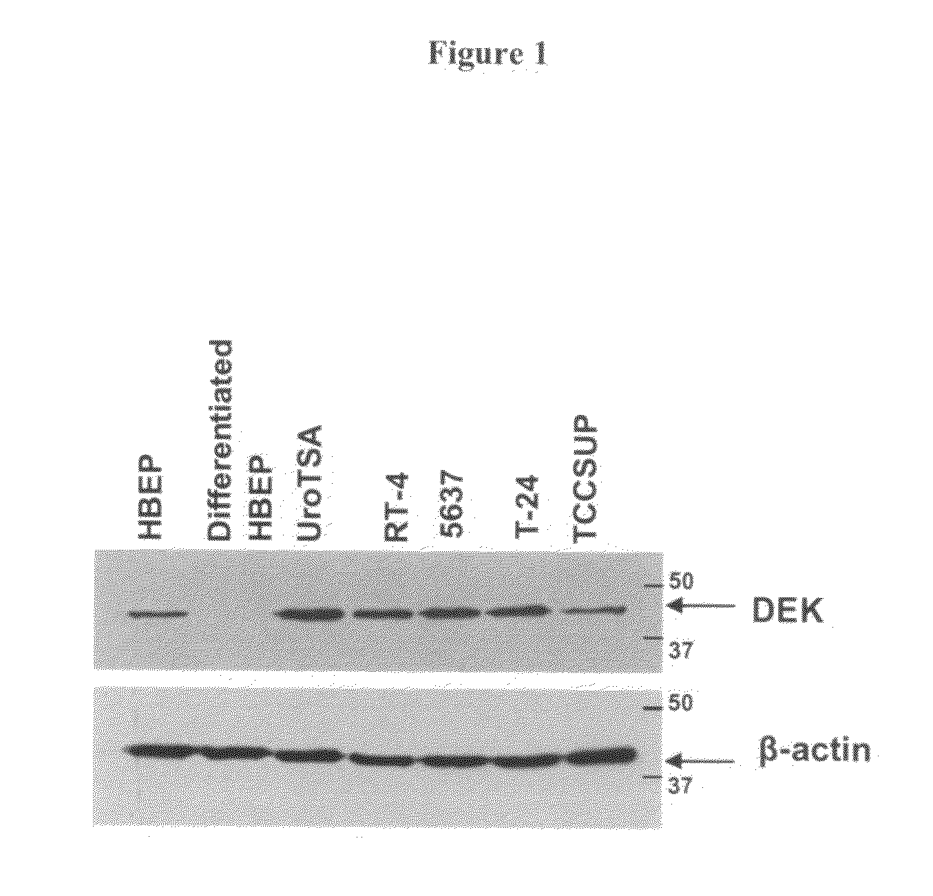
Western Blot Detection of DEK Protein in Bladder Cell Lines
[0100]In this series of studies, we optimized the detection of DEK protein in a biological sample (e.g., urine) using Western blot assay. We tested cell lysate extracts obtained from four (4) bladder cancer cell lines (i.e., RT-4, 5637, T-24 and TCCSUP) and examined their DEK protein expression. These cells were chosen to represent different stages of bladder cancer. In addition, we tested cell extracts from bladder epithelial cells transformed with SV-40 T-antigen (i.e., UroTSA), progenitor human epithelial cells (i.e., HBEP cells), and differentiated HBEP cells for their DEK protein expression. Note that HBEP cells were treated with 1 mM calcium chloride to prevent cycling in growth media.
[0101]Cell extracts from 1×10⁷ cells were obtained using RIPA buffer (i.e., 25 mM Tris-HCl (pH 7.6), 150 mM NaCl, 1% NP-40, 1% sodium deoxycholate, and 0.1% SDS). Protein was quantified using a BCA assay kit (Pierce, Thermo Fisher Scienti...
example 2
Western Blot Fails to Detect DEK Protein in Cultured Media
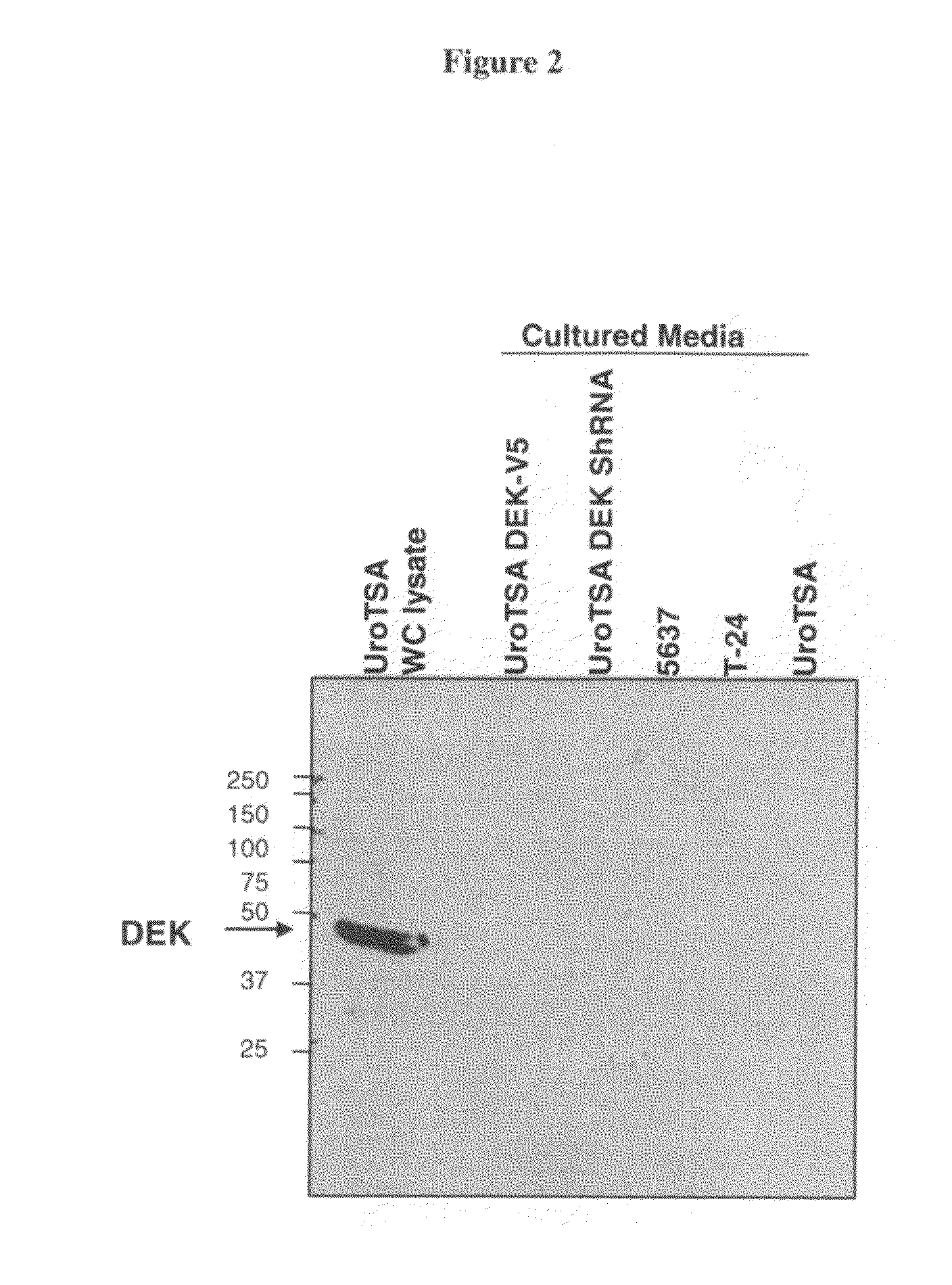
[0104]In this study, we examined if DEK protein can be released from bladder cancer cells (i.e., secreted from cells). To do so, we first collected cultured media from two (2) bladder cancer cell lines (i.e., T-24 and 5637) and then examined DEK protein expression in these cultured media. One (1) ml of cultured media was collected and briefly centrifuged (3,000 rpm, 5 min) to remove any cellular debris. The cultured media was subsequently concentrated to 50-fold (i.e., from 500 μl to 10 μl) using a Microcon® 3K filter (Millipore, Billerica, Mass.). The entire 10 μl of the concentrated cultured media sample was loaded in a Western blot assay. DEK protein was examined using a polyclonal anti-DEK antibody (cat. no. A-301-335A) (Bethyl Labs., Montgomery, Tex.). UroTSA (10 μg) whole cell lysate was used as a control.
[0105]FIG. 2 shows that DEK protein was not detectable in the cultured media of the two (2) bladder cancer cell line...
example 3
Western Blot Detection of DEK Protein in Bladder Tissues
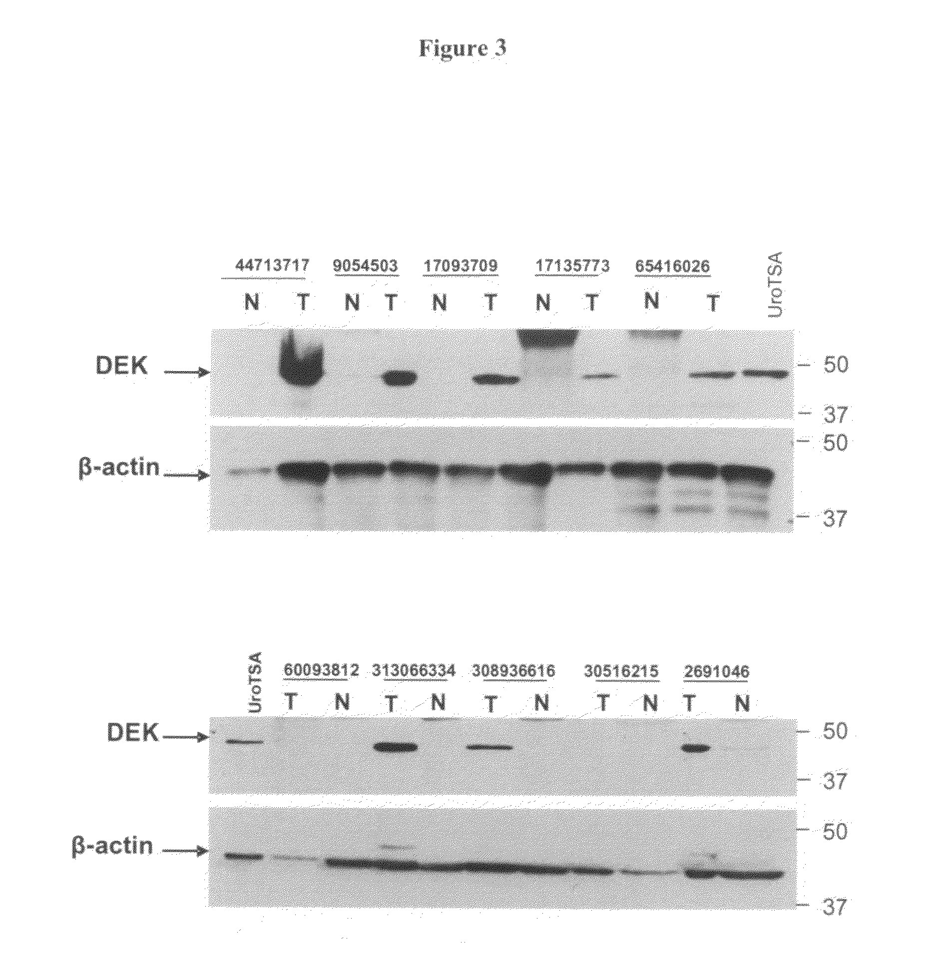
[0106]In this study, we examined if our Western blot assay (see Example 1) could detect DEK protein in bladder tissues obtained from human subjects (e.g., bladder cancer patients or healthy individuals).
[0107]Twenty-seven (27) bladder tumor tissue samples were obtained from patients who suffered from low and high grade transitional cell carcinoma (TCC). For comparison, twenty-seven (27) normal bladder tissue samples were obtained from the adjacent sites of the same individuals. Tissue lysate extracts were prepared using RIPA buffer (as described above) and the tissue extracts were prepared to a protein concentration of 2-10 μg / μl. Protein concentration of the tissue lysates extracts was determined using a BCA assay kit (Pierce, Thermo Fisher Scientific, Rockford, Ill.). ˜50 μg of the tissue lysates extracts from each sample was analyzed for their DEK protein expression in our Western blot assay, using a monoclonal anti-DEK anti...
PUM
 Abstract
Description
Claims
Application Information
 - R&D
- Intellectual Property
- Life Sciences
- Materials
- Tech Scout
- Unparalleled Data Quality
- Higher Quality Content
- 60% Fewer Hallucinations
Browse by: Latest US Patents, China's latest patents, Technical Efficacy Thesaurus, Application Domain, Technology Topic, Popular Technical Reports.
© 2025 PatSnap. All rights reserved.Legal|Privacy policy|Modern Slavery Act Transparency Statement|Sitemap|About US| Contact US: help@patsnap.com


