Display Orientation Adjustment Device And Adjustment Program For Medical Three-Dimensional Image

a three-dimensional image and display orientation technology, applied in the field of three-dimensional image display orientation adjustment devices and program, can solve the problems of difficult to reflect a region, low clinical reproducibility, low validity, etc., and achieve low clinical reproducibility, high reproducibility, and low validity of ct image.

Inactive Publication Date: 2012-09-20
IMAGNOSIS
View PDF11 Cites 17 Cited by
  • Summary
  • Abstract
  • Description
  • Claims
  • Application Information

AI Technical Summary

Benefits of technology

[0008]In recent years, it has been tried to apply a CT (Computed Tomography) image three-dimensionally constructed based on three-dimensional CT data to frontal face evaluation. However, the CT is not standardized photography, and when evaluating the bilateral symmetry of a face by using a CT image, first, instead of using the coordinate system with low clinical reproducibility and low validity of the CT image adopted when photographing as it is, the coordinate system must be re-positioned in a frontal orientation preferable for morphological evaluation. As a positioning means for re-positioning the coordinate system, a positioning means that can reflect a large number of anatomical maxillofacial feature points (hereinafter, referred to as “landmarks”) and can set various frontal orientations is required. The inventor of the present invention has already developed a method for setting a frontal face orientation by setting an anatomical or morphological reference surface based on landmarks on a CT image and a method for correcting a set frontal face orientation by further using desired landmarks as reference points, and applied for a patent (refer to Patent Documents 2 and 3 listed above). These methods enable setting of various orientations with high reproducibility and high validity required for utilizing CT images for morphological diagnosis.
[0011]An object of the present invention is to further improve diagnostic and treatment correctness by further improving variation, accuracy, and operability of positioning of a medical three-dimensional image.
[0016]According to the present invention, a reference image to be used as a reference for adjustment of the display orientation of a medical three-dimensional image is displayed, so that the display orientation of the medical three-dimensional image is easily adjusted. For example, when adjusting the frontal face orientation of a head image, by displaying a plane, cells, or a face symmetric model outline orthogonal to an image reference axis (Yo axis) extending in the front-back direction of the face, contour lines or a moire projected in the image reference axis direction, etc., as a reference image, the frontal face orientation is easily adjusted based on the bilateral symmetry of the face.
[0017]In addition, according to the present invention, the display orientation of the medical three-dimensional image can be adjusted from a plurality of observation directions, so that the display orientation can be adjusted more accurately. Further, according to the present invention, according to rotative movement of the medical three-dimensional image by the display orientation changing means, the medical three-dimensional image data according to the three-dimensional reference coordinate system is corrected, so that it is not necessary to specify landmarks that are hard to accurately specify.
[0020]In the third aspect of the present invention, when a parallel movement command is input, the medical three-dimensional image displayed by the display means is parallel-moved relative to the specified image reference axis (second image reference axis) by the parallel moving means according to the input parallel movement command. Then, according to parallel movement by the parallel moving means, medical three-dimensional image data on the three-dimensional reference coordinate system is corrected. For example, when the second image reference axis is the Yo axis, an origin that the Yo axis passes through can be moved relative to the medical three-dimensional image on the Xo-Zo plane. Similarly, when the second image reference axis is the Zo axis, an origin that the Zo axis passes through can be moved relative to the medical three-dimensional image on the Xo-Yo plane, and when the second image reference axis is the Xo axis, an origin that the Xo axis passes through can be moved relative to the medical three-dimensional image on the Yo-Zo plane. According to the present invention, by parallel-moving the medical three-dimensional image with respect to a specified image reference axis, the display orientation can be adjusted.

Problems solved by technology

However, the CT is not standardized photography, and when evaluating the bilateral symmetry of a face by using a CT image, first, instead of using the coordinate system with low clinical reproducibility and low validity of the CT image adopted when photographing as it is, the coordinate system must be re-positioned in a frontal orientation preferable for morphological evaluation.
However, many landmarks are defined for morphological measurement on a radiophotograph, so that when three-dimensionally identifying the two-dimensionally defined landmarks, an error occurs, and for example, it is difficult to reflect a region such as the buccal region of a face in which landmarks cannot be set morphologically due to its shape in positioning.
In addition, the length of a side of a minimum constituent unit (voxel) of CT data is approximately 0.5 mm at minimum, so that it is impossible to specify landmarks with high accuracy on the CT data.

Method used


Image

  • Display Orientation Adjustment Device And Adjustment Program For Medical Three-Dimensional Image
  • Display Orientation Adjustment Device And Adjustment Program For Medical Three-Dimensional Image
  • Display Orientation Adjustment Device And Adjustment Program For Medical Three-Dimensional Image

Examples


Embodiment Construction

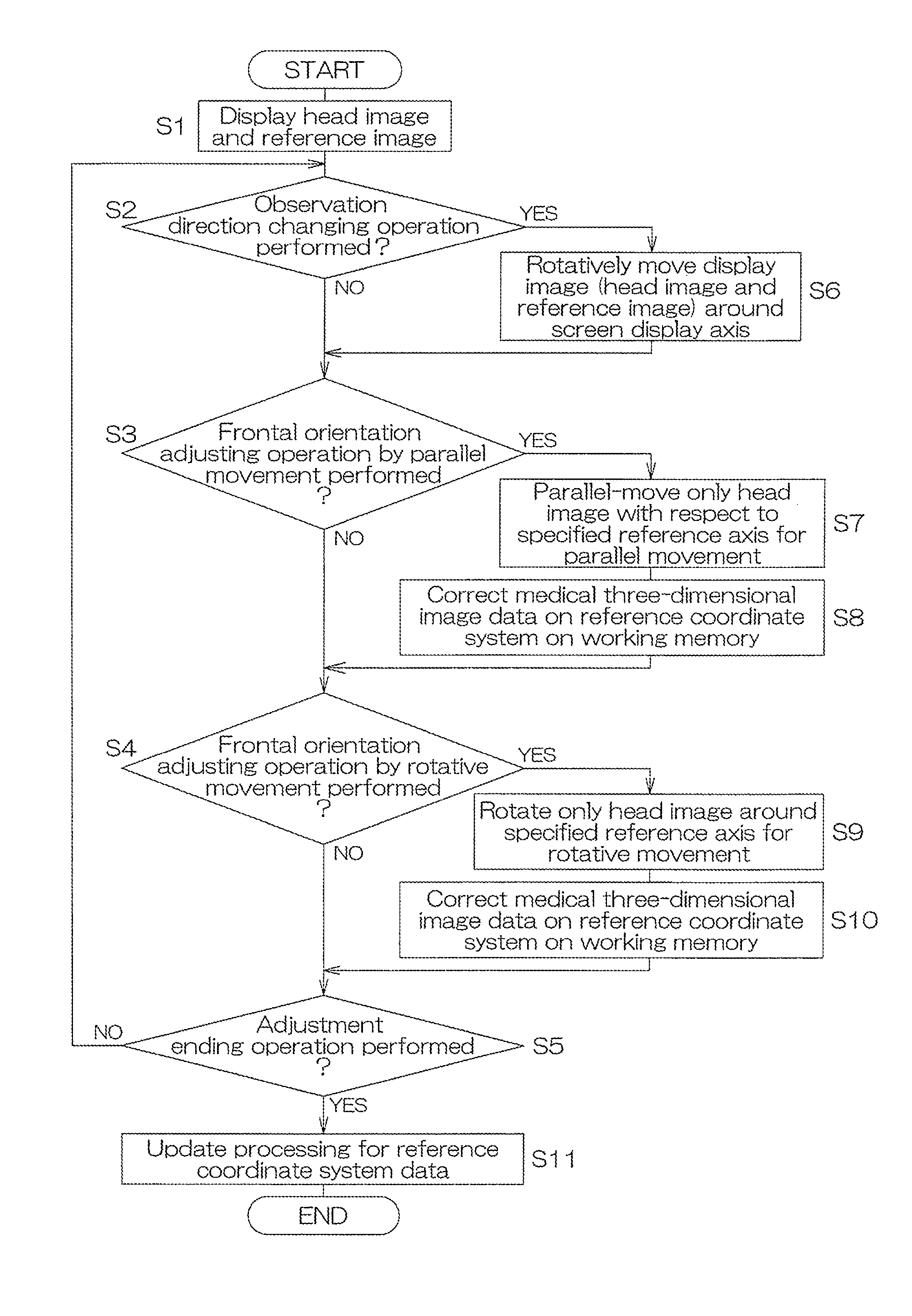
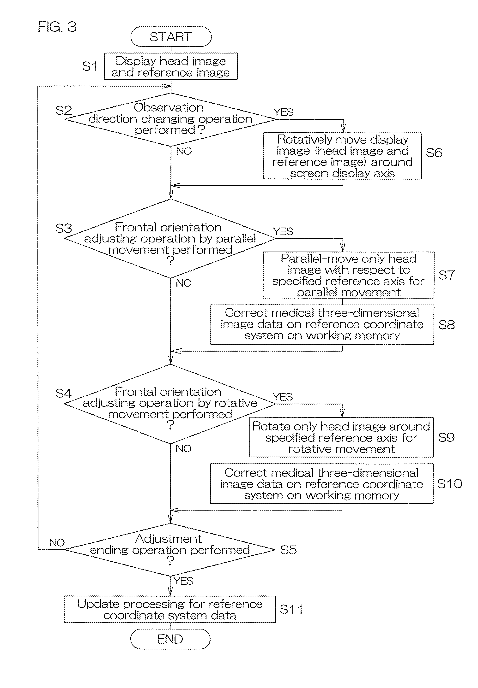
[0035]Hereinafter, preferred embodiments of the present invention are described with reference to the drawings.

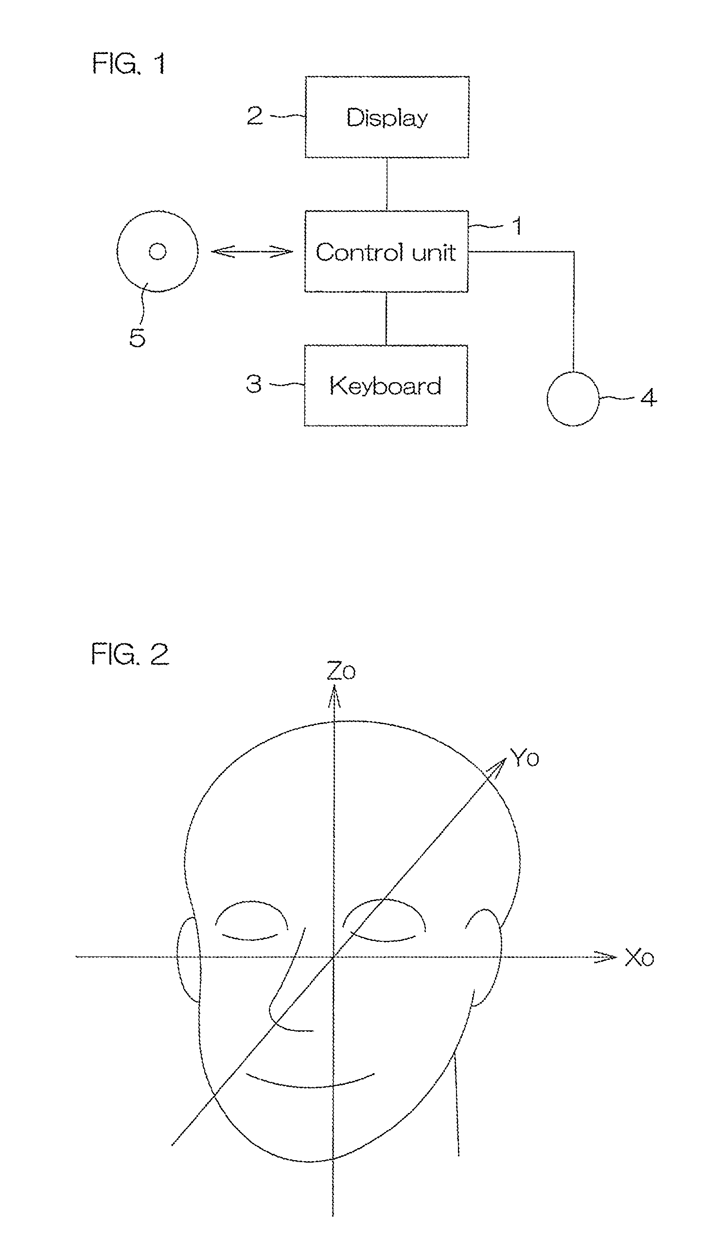
[0036]FIG. 1 shows a configuration of an image processing device according to a preferred embodiment of the present invention.

[0037]The image processing device is realized by, for example, a computer such as a personal computer (PC). The image processing device includes a control unit 1 equipped with a CPU, a ROM, a RAM, and a hard disk, etc. To the control unit 1, a display (monitor) 2, a keyboard 3, a mouse 4, etc., are connected. On the hard disk of the control unit 1, a display orientation adjustment program according to a preferred embodiment of the present invention is installed from, for example, a storage medium 5, etc., storing the display orientation adjustment program.

[0038]The hard disk stores medical three-dimensional image data according to a predetermined three-dimensional reference coordinate system (body coordinate system: coordinate system having a referen...


PUM


Abstract

A display orientation adjustment device for a medical three-dimensional image, capable of adjusting a display orientation of a medical three-dimensional image. When an observation direction changing operation is performed by a user, a control unit rotatively moves the whole display image by a predetermined amount in a direction corresponding to the observation direction changing operation around a specified screen display axis. When a frontal orientation adjusting operation by rotative movement is performed, the control unit rotatively moves a head image by a predetermined amount in a direction corresponding to the adjusting operation around a specified reference axis (specified reference axis for rotative movement). The control unit corrects medical three-dimensional image data on a reference coordinate system on a working memory based on the specified reference axis and specified rotative movement direction and rotative movement amount.

Description

CROSS-REFERENCE TO RELATED APPLICATIONS[0001]The present application is a continuation of pending International Patent Application PCT / JP2010 / 069837, filed on Nov. 8, 2010, which designates the United States and claims priority from Japanese Patent Application 2009-268620, filed on Nov. 26, 2009, and Japanese Patent Application 2009-260077, filed on Nov. 13, 2009. The content of all prior applications is incorporated herein by reference.FIELD OF THE INVENTION[0002]The present Application relates to a device and a program for adjusting a display orientation of a medical three-dimensional image.BACKGROUND OF THE INVENTION[0003]When treating jaw deformity involving jaw distortion in the dental or plastic surgery field, for grasping functional and aesthetic maxillofacial problems, diagnosis of the maxillofacial morphology is important. Conventionally, a cephalometric radiograph is key information for diagnosis of maxillofacial morphology, however, image magnification and distortion occu...

Claims


Application Information

Patent Timeline
Patent Type & Authority Applications(United States)
IPC IPC(8): G06T15/00
CPCA61B6/032A61B6/14A61B6/463A61B6/501A61B6/466A61B6/467A61B6/465A61B6/5247A61B5/0064A61B5/0077A61B5/0035G06T19/20G06T2219/2016A61B6/03
Inventor KIM, HAN-JOON
Owner IMAGNOSIS
Why Eureka
Try Eureka