

Method and device for retinal image analysis
a retinal image and image technology, applied in the field can solve the problems of increasingaffecting the accuracy of retinal image analysis, and affecting so as to achieve the effect of reducing the risk of stroke, and reducing the accuracy of image analysis
- Summary
- Abstract
- Description
- Claims
- Application Information
AI Technical Summary
Benefits of technology
Problems solved by technology
Method used
Image

Examples
example 1
Method
Case: Stroke Patients.
[0188]Inclusion criteria: clinical diagnosed stroke patients.
[0189]Diagnostic criteria of stroke105:
[0190]1. Stroke symptoms: numbness, paralysis, slurring of speech, blurring of vision, etc. and the diagnosis was confirmed by experienced neurologists;
[0191]2. Brain MRI or CT manifestation: ischemic or hemorrhage change of the brain.
[0192]Patients were diagnosed as stroke case with the 1st criteria with or without the examination of brain MRI or CT. There were totally 122 stroke cases in this study. The cases of stroke were from two sources: 64 of them were from diabetic retinopathy screening program. The screening program was started in January 2008 and diabetic patients followed in the Prince of Wales Hospital were invited to be screened for diabetic retinopathy. There were 64 patients with prevalent stroke have their color retina image record in this screening program. Another 58 stroke cases were acute stroke patients from Acute Stroke U...
example 2
Automated Computer Methodology
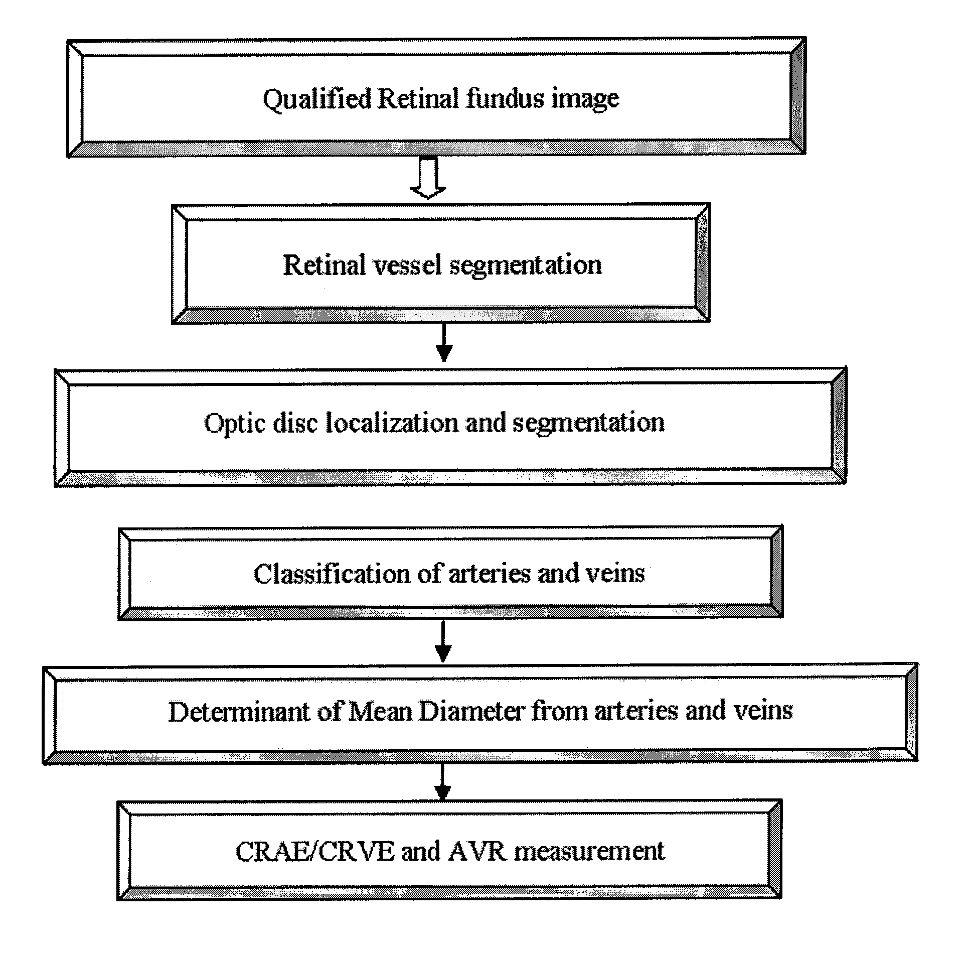
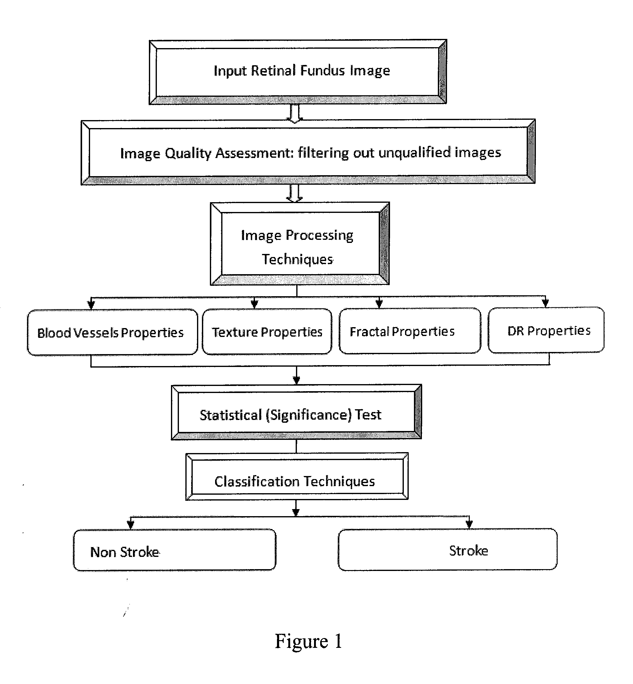
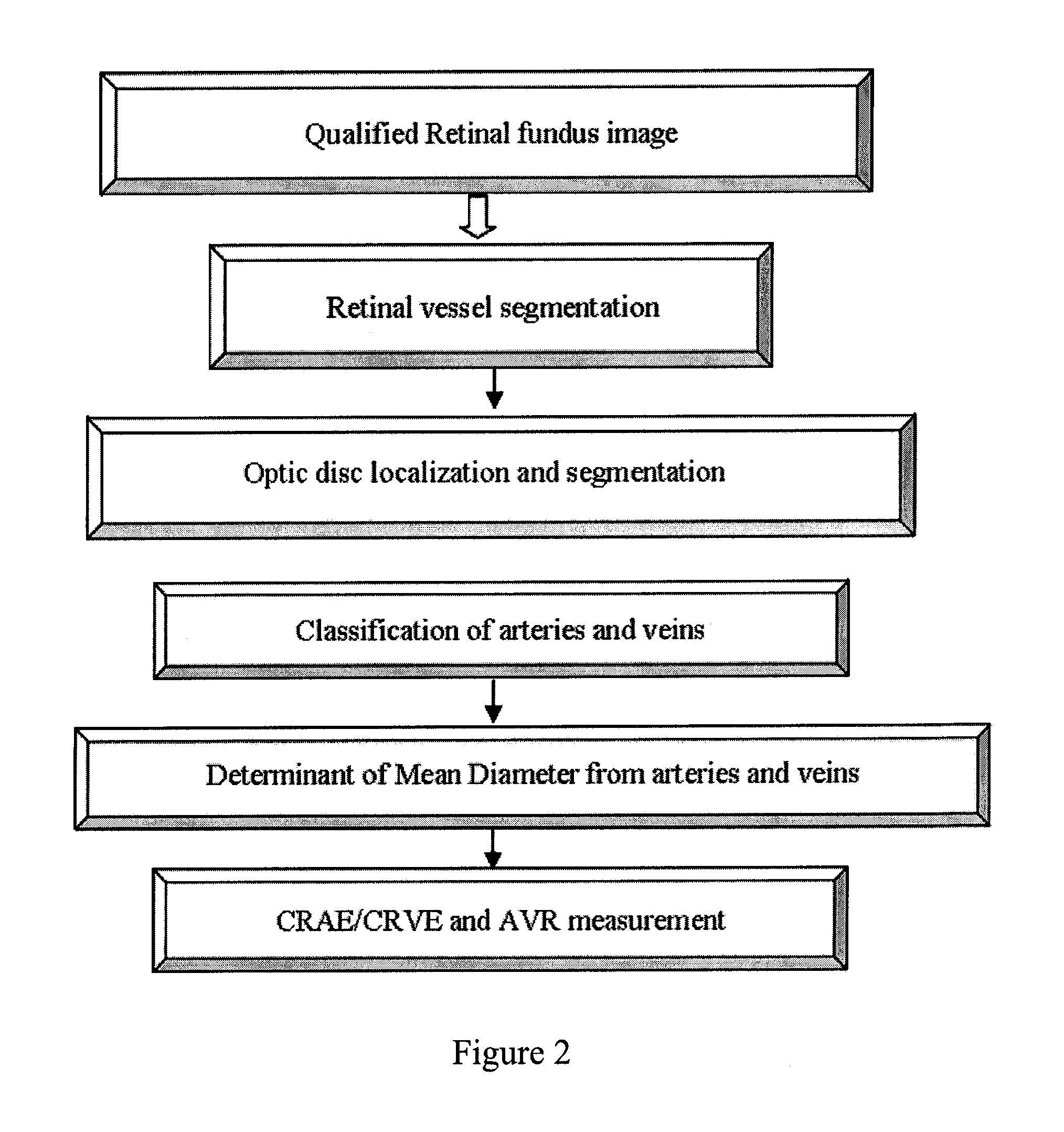
[0248]FIG. 1 shows the overall system used for stroke epidemiology prevention and risk assessment tools. As can be seen from FIG. 20, there are total five steps that need to be addressed for the system to carry out diagnosis for stroke.
[0249]2.1 Automated and fast measurement of the image quality
[0250]2.2.1 Blood vessels features: vessels width and the Arteriolar-to-Venular diameter Ratio (AVR), we may also consider part e) as one of the blood vessel feature.
[0251]2.2.3 Texture properties related to stroke: mainly from the spectrum concerning.
[0252]2.2.4 Fractal Analysis—Other features including retinal arteriolar narrowing, arteriole-venule nicking and vessel tortuosity, which any irregular shape related to stroke (may be too complicate measured / extracted from image). We use fractal analysis to deal with such kind of features.
[0253]2.2.5 Other stroke features that related to DR retinopathy: Hemorrhages
[0254]The detailed information for above five steps...
example 3
Screening or Grading of Diabetic Retinopathy
Materials and Methods
[0328]Public dataset “DIARETDB0” (Kauppi, T., et. al, DIARETDB0: Evaluation Database and Methodology for Diabetic Retinopathy Algorithms. Technical report) is used to train the system of the application. This database consists of 130 color retina images of which 20 are normal and 110 contain signs of the diabetic retinopathy. Characteristics of the dataset are summarized in Table 18. “redsmalldots”, “hemorrhages”, “hardexudates”, “softexudates” and “neovascularisation” represent “microaneurysms”, “hemorrhages”, “hard exudates”, “cotton wool spots” and “new vessels” respectively.
TABLE 18CHARACTERISTIC SUMMARIES OF DATASET “DIARETDB0”CharacteristicsredsmalldotshemorrhagesHardexudatessoftexudatesNeovascularisationNumber10680734120
[0329]In the system utilized in the Example, the first module is digital fundus camera integrated to other modules, the second module is configured to conduct color histogram and dual tree comple...
PUM
 Abstract
Description
Claims
Application Information