Apparatus, method, and computer-accessible medium for b1-insensitive high resolution 2d t1 mapping in magnetic resonance imaging

a magnetic resonance imaging and t1 mapping technology, applied in the field of medical imaging apparatus, methods, and computer accessible medium for longitudinal relaxation time (t1) mapping using fast spin echo, can solve the problems of mr imaging of these structures in three orthogonal planes susceptible to partial volume effects, cartilage that appears morphologically normal in routine mri may already be irreversibly compromised in early oa, etc., to achieve high spatial resolution, increase snr facilitated by 3 tes

Inactive Publication Date: 2012-10-25
NEW YORK UNIV
View PDF6 Cites 11 Cited by
  • Summary
  • Abstract
  • Description
  • Claims
  • Application Information

AI Technical Summary

Benefits of technology

[0011]For example, according to certain exemplary embodiments of the present disclosure, it is possible to provide apparatus, methods, and computer-accessible medium for obtaining high spatial resolution 2D T1 mapping. For example, an increased SNR facilitated by 3 Tesla imaging can be exploited by performing high spatial resolution 2D T1 mapping in radial imaging planes to take advantage of the geometry of the hip joint (see, e.g., References 4 and 12). According to certain exemplary embodiments of the present disclosure, a B1-insensitive 2D T1 mapping pulse sequence with high in-plane resolution for dGEMRIC in radial planes of the hip can be provided. Exemplary embodiments can, for example, image the hip using an exemplary fast spin-echo (FSE) pulse sequence at 3 Tesla to achieve high spatial resolution with adequate SNR and employ a B1-insensitive saturation pulse to perform uniform T1 weighting. The scan time of the proposed pulse sequence can be, for example, about 1 minute and 20 second per 21) slice. Compared with the previously reported 3D dGEMRIC pulse sequence, the exemplary pulse sequence can be relatively less sensitive to patient motion. Further, according to certain exemplary embodiments of the present disclosure, the exemplary results can be validated, for example, against a rigorous multi-point saturation recovery (SR) pulse sequence at 3 Tesla, by comparing measured T1 in a phantom and in the hip cartilage of FAI patients. Additionally, the accuracy and SNR efficiency of the exemplary pulse sequence against the 3D F2T1 pulse sequence can be compared in phantom experiments.

Problems solved by technology

The acetabular cartilage's and labrum's position and orientation within the pelvis make MR imaging of these structures in three orthogonal planes susceptible to partial volume effects.
Cartilage that appears morphologically normal in routine MRI may already be irreversibly compromised in early OA.

Method used


Image

  • Apparatus, method, and computer-accessible medium for b1-insensitive high resolution 2d t1 mapping in magnetic resonance imaging
  • Apparatus, method, and computer-accessible medium for b1-insensitive high resolution 2d t1 mapping in magnetic resonance imaging
  • Apparatus, method, and computer-accessible medium for b1-insensitive high resolution 2d t1 mapping in magnetic resonance imaging

Examples


Embodiment Construction

[0009]These and other objects, features and advantages of the present disclosure will become apparent upon reading the following detailed description of exemplary embodiments of the present disclosure, when taken in conjunction with the appended drawings and claims.

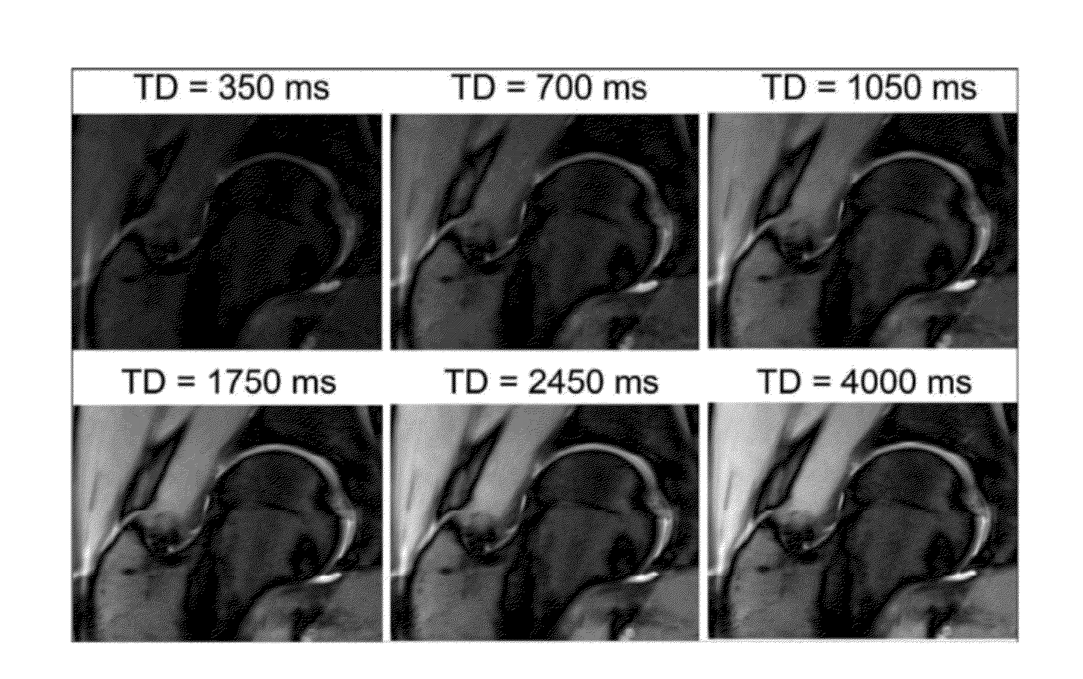
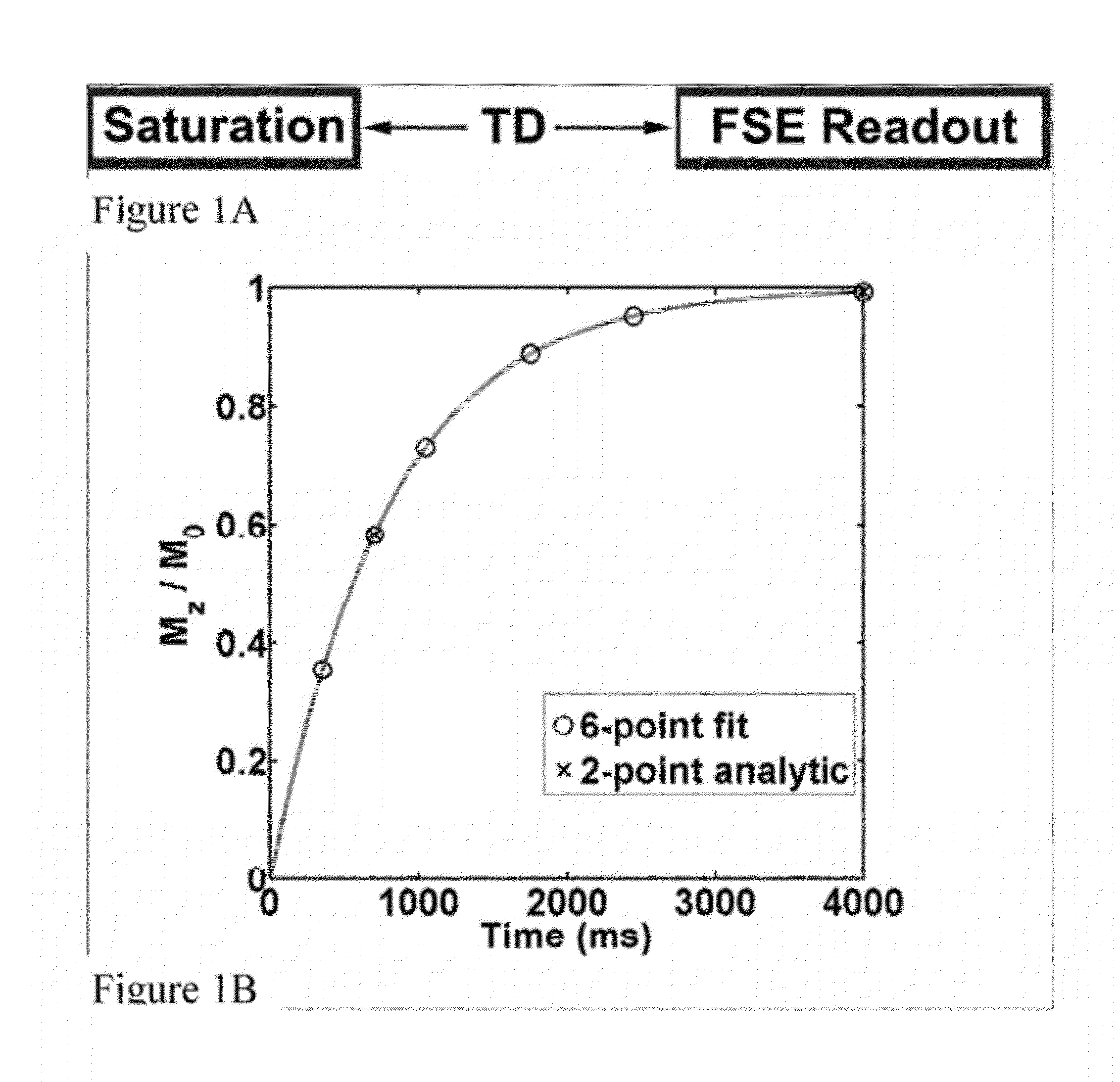
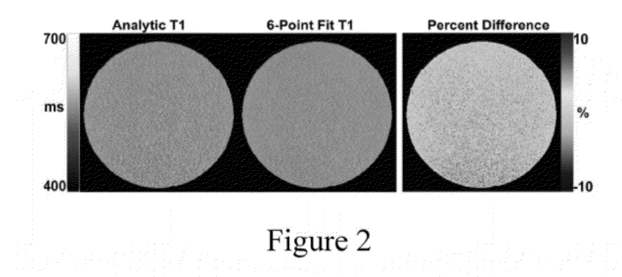
[0010]According to exemplary embodiments of the present disclosure, apparatus, methods, and computer-accessible medium for generating a high-resolution 2D T1 mapping sequence suitable for dGEMRIC in radial planes of the hip at 3 Tesla can be provided. The T1 measurements can be accurate, repeatable and reproducible. An exemplary technique implemented by the exemplary apparatus, systems, methods, and computer-accessible medium can be applied to measure cartilage T1 in other joints (e.g., knee, etc.) and T1 of other tissues, and it can be suitable for applications at 3 Tesla, because it can be insensitive to B1+ inhomogeneities.

[0011]For example, according to certain exemplary embodiments of the present disclosure, it is po...


PUM


Abstract

Exemplary systems, methods and computer-accessible mediums can be provided for imaging at least one anatomical structure. For example, it is possible to direct a saturation-recovery (SR) pulse sequence having fast spin echo (FSE) to or at the anatomical structure(s). At least one T1 image of the at least one anatomical structure can be generated based on the SR pulse sequence. In one example, the anatomical structure(s) can include a hip. According to another example, T1 image(s) can include a plurality of T1 images generated or provided in a plurality of rotating radial planes.

Description

CROSS-REFERENCE TO RELATED APPLICATION(S)[0001]The present application relates to and claims priority from U.S. Provisional Patent Application No. 61 / 478,271 filed Apr. 22, 2011, the entire disclosure of which is incorporated herewith by reference.FIELD OF THE DISCLOSURE[0002]The present disclosure relates to exemplary embodiments of apparatus, methods, and computer accessible-medium for medical imaging, and more particularly, to exemplary embodiments of apparatus, methods, and computer accessible-medium for longitudinal relaxation time (T1) mapping using fast spin echo.BACKGROUND INFORMATION[0003]It has been recognized that femoroacetabular impingement (FAI), a condition in which structural abnormalities of the femoral headneck junction and / or acetabulum cause mechanical blockage in the terminal range of hip motion, can lead to osteoarthritis (OA) of the hip (see, e.g., Ganz R, Parvizi J, Beck M, Leunig M, Notzli H, Siebenrock K A. Femoroacetabular impingement: a cause for osteoart...

Claims


Application Information

Patent Timeline
Patent Type & Authority Applications(United States)
IPC IPC(8): A61B5/055
CPCA61B5/055G01R33/5617G01R33/50
Inventor KIM, DANIELLATTANZI, RICCARDOGLASER, CHRISTIANRECHT, MICHAEL
Owner NEW YORK UNIV
Why Eureka
Try Eureka