

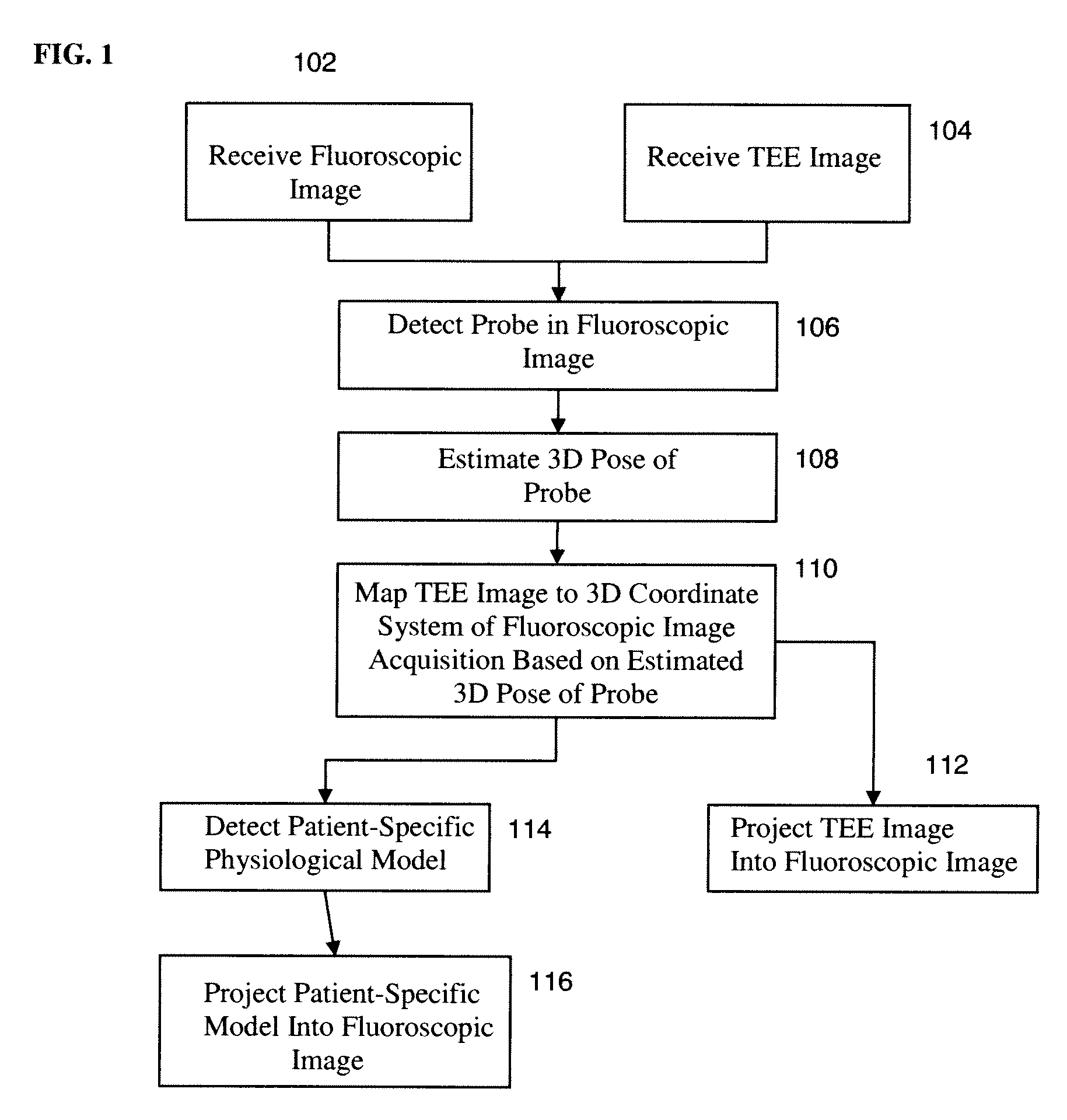
Method and System for Registration of Ultrasound and Physiological Models to X-ray Fluoroscopic Images
a fluoroscopic image and ultrasound technology, applied in the field of multimodal medical image registration, can solve the problems of difficult catheter navigation inside the vessel of a patient, and the imaging modality does not capture the soft tissue structure of the patien
- Summary
- Abstract
- Description
- Claims
- Application Information
AI Technical Summary
Benefits of technology
Problems solved by technology
Method used
Image

Examples
Embodiment Construction
[0019]The present invention relates to registration of ultrasound and physiological models to x-ray fluoroscopic images. Embodiments of the present invention are described herein to give a visual understanding of the model-based image fusion method. A digital image is often composed of digital representations of one or more objects (or shapes). The digital representation of an object is often described herein in terms of identifying and manipulating the objects. Such manipulations are virtual manipulations accomplished in the memory or other circuitry / hardware of a computer system. Accordingly, is to be understood that embodiments of the present invention may be performed within a computer system using data stored within the computer system.
[0020]Methods that attempt fusion of ultrasound and x-ray fluoroscopy can be broadly categorized as either hardware based or image based. Hardware based approaches typically attach additional devices to an ultrasound probe (used to acquire Transe...
PUM
 Abstract
Description
Claims
Application Information