
Detecting complement activation

a technology of complement activation and detection field, which is applied in the direction of antibody medical ingredients, instruments, drug compositions, etc., can solve the problems of significant deterioration of a patient's condition, difficult accurate measurement of complement activation, etc., and achieve the effect of minimizing spontaneous complement activation

Active Publication Date: 2012-12-13
KYPHA
View PDF2 Cites 21 Cited by
  • Summary
  • Abstract
  • Description
  • Claims
  • Application Information

AI Technical Summary

Benefits of technology

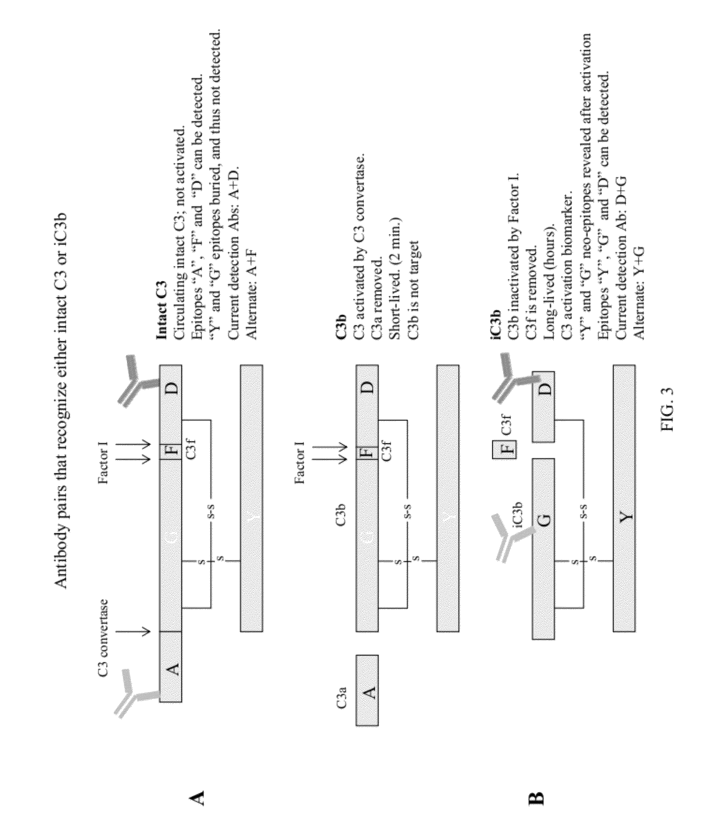
[0010]By carefully selecting, capturing and detecting antibody pairs that avoid interfering cross-talk, the inventors have surprisingly found that it is possible to detect and quantify biomarkers of complement activation including native or intact C3 and / or iC3b, while avoiding the false positive results that have plagued more conventional testing methods for these analytes. This invention may be applied in a variety of formats; the inventors have further found that a lateral flow assay type format, and particularly a lateral flow immunoassay format, provides particular advantages and surprising features.
[0012]The present invention provides technologies that address these previously unidentified sources of problems. For example, in some embodiments, the present invention provides methods in which relevant steps are all performed within a restricted time period. According to the present invention, such methods provide advantages including minimizing spontaneous complement activation and, alternatively or additionally, providing clinically relevant data within a time period, measured from initiation of sample collection from a subject, that is materially reduced as compared with standard methodologies. In some embodiments, provided assays are completed within a time period, measured from initiation of sample collection from a subject, that is less than about 60 minutes or fewer, about 50 minutes or fewer, about 40 minutes or fewer, about 30 minutes or fewer, about 20 minutes or fewer, about 10 minutes or fewer, or about 5 minutes or fewer. By contrast, many prior art assays do not provide data for hours. Such a difference in time can make a life or death difference to a patient.

Problems solved by technology

While the complement response is an important defense system against disease, several features make accurate measurement of complement activation difficult, particularly in a clinically relevant time period.
During this time, significant deterioration of a patient's condition may be occurring that does not visibly manifest until significant, or even irreversible, damage has occurred.

Method used


Image

  • Detecting complement activation
  • Detecting complement activation
  • Detecting complement activation

Examples


example 1

Patient Triage

[0160]Before the first test sample is assayed, a standard curve is performed using 10 ng / ml, 30 ng / ml, 100 ng / ml, 300 ng / ml, and 1000 ng / ml of intact C3 and iC3b standards. Lateral flow immunoassay cassettes are read with an electronic reader after 20 minutes.

[0161]The test is used to gauge injury severity within 15, 30, or 60 minutes of injury. It is most useful for patients who may have suffered injuries not obvious by visual inspection. A drop of blood is collected either from an arterial line (A-line) or finger stick. A 10 ul sample is drawn up using a fixed volume pipet. The sample then mixed with 990 ul of sample buffer. The blood and sample buffer are mixed. Using a fixed volume pipet bulb, 100 ul is drawn up and pipetted onto the lateral flow immunoassay cassette containing integrated intact C3 and iC3b test strips. Alternatively, 100 ul can be applied to separate intact C3 and iC3b lateral flow assay cassettes. After 10 minutes but before 40 minutes, the casse...

example 2

Trajectory Monitoring of a Trauma Patient

[0162]At the beginning of the shift, ICU staff performs a standard curve using 10 ng / ml, 30 ng / ml, 100 ng / ml, 300 ng / ml, and 1000 ng / ml of intact C3 and iC3b standards. Lateral flow immunoassay cassettes are read with an electronic reader after 20 minutes.

[0163]The objective of trajectory monitoring is to detect changes in inflammatory and immune status of patients that have been stabilized after severe trauma. In this example, respiratory distress caused by either pneumonia or inflammatory dysfunction is to be detected. The expected patient profile is one who has an injury severity score (ISS) equal or greater to 16 and who requires ventilator assistance for breathing.

[0164]The patient receives a complement test at frequent intervals, which aligns with the time points for testing glucose levels in blood. This interval between testing is usually about two hours. Blood is collected using the same method as for glucose testing, either by A-line...

example 3

Determining Disease Severity and Effectiveness of Treatment in a Patient with Systemic Lupus Erythematosus (SLE)

[0166]Before the first test sample is assayed, a standard curve is performed using 10 ng / ml 30 ng / ml, 100 ng / ml, 300 ng / ml, and 1000 ng / ml of intact C3 and iC3b standards. Lateral flow immunoassay cassettes are read with an electronic reader after 20 minutes.

[0167]The test is used to gauge the initial severity of disease as well as the effectiveness of therapy. One of the standard diagnostics performed on SLE patients is measurement of total C3 levels. C3 levels are normally depressed in SLE patients and return to normal (>1 mg / ml) following successful treatment. However, it is not known generally whether the C3 activation has been abrogated or only slowed enough to allow normal replenishment mechanisms to restore C3 levels to normal.

[0168]At each doctor visit, a patient's blood is collected for total C3, intact C3, and iC3b tests. Only one drop is required for the combine...


PUM

PropertyMeasurementUnit
concentrationaaaaaaaaaa
volumeaaaaaaaaaa
volumeaaaaaaaaaa

Abstract

Methods of detecting complement activation including steps of detecting in a sample from a subject a level of iC3b wherein the detecting involves specific interaction between the iC3b and a non-cross-reactive antibody thereto, comparing the detected level with a reference level, which reference level is within a range of about 10 ng / ml to about 5,000 ng / ml, wherein determination that the detected level is above the reference level indicates that the subject is suffering from or susceptible to undesirable and / or pathologic complement activation, and administering treatment to treat undesired complement activation if the detected level is above the reference level. Other methods of detecting complement activation with or without measuring iC3b are also provided.

Description

RELATED APPLICATIONS[0001]This application is a continuation-in-part of U.S. Non-Provisional application Ser. No. 13 / 287,432, filed Nov. 2, 2011, which claims priority under 35 U.S.C. §119(e) to U.S. Provisional Application Ser. No. 61 / 409,297, filed Nov. 2, 2010, the contents of which are incorporated by reference herein in their entirety.BACKGROUND[0002]Inflammation is the physiological response of vascularized tissue to injury, infection, and certain diseases. The inflammatory process is a biological requirement for wound healing after traumatic injury and for the clearance of infection. However, inflammation can also damage self-tissue. For this reason, inflammation is often considered a double-edged sword.[0003]Complement is the most ancient arm of the immune system and is deeply rooted in the inflammatory process. The complement protein cascade is a first line of defense against invading microbes and a critical player in the wound healing process. The complement cascade involv...

Claims


Application Information

Patent Timeline
Patent Type & Authority Applications(United States)
IPC IPC(8): G01N33/566A61P29/00A61P31/00A61P27/02A61P25/00A61P13/12A61K39/395A61P9/00
CPCG01N33/564G01N2333/4716G01N33/5306G01N2800/26G01N2800/125A61P9/00A61P13/12A61P25/00A61P27/02A61P29/00A61P31/00G01N2800/104G01N2800/52
Inventor OLSON, PAULMOSS, DON W.STATEN, NICHOLAS
Owner KYPHA
Learn More