Physics based image processing and evaluation process of perfusion images from radiology imaging

a radiology imaging and image processing technology, applied in the field of physics based image processing and evaluation process of perfusion images from radiology imaging, can solve the problems of difficult visual evaluation without quantification, the opportunity to distinguish between wall-thickening function and perfusion, and the cost of invasive diagnostic examination every year, so as to achieve accurate detection

Active Publication Date: 2012-12-20
CARNEGIE MELLON UNIV +1
View PDF2 Cites 53 Cited by
  • Summary
  • Abstract
  • Description
  • Claims
  • Application Information

AI Technical Summary

Benefits of technology

[0017]In an embodiment, the process and system provide for a method of data representation of left ventricular myocardium of risk from perfusion data as a polar plot. The polar plot may assist the clinician in his or her diagnosis of the patient by performing tasks which include the following: (a) a classification task is to generate a map (i.e., a perfusion characteristics map) of probability of b

Problems solved by technology

However, the use of coronary angiograms expose over 2.7 million Medicare/Medicaid reimbursed Americans to the risks and result in costs of invasive diagnostic examination every year, whereas only about 61% of these patients require clinical intervention.
While DSE primarily assesses wall motion, SPECT allows simultaneous determination of myocardial perfusion and wall motion at relatively low resolution, although it exposes th

Method used


Image

  • Physics based image processing and evaluation process of perfusion images from radiology imaging
  • Physics based image processing and evaluation process of perfusion images from radiology imaging
  • Physics based image processing and evaluation process of perfusion images from radiology imaging

Examples


Embodiment Construction

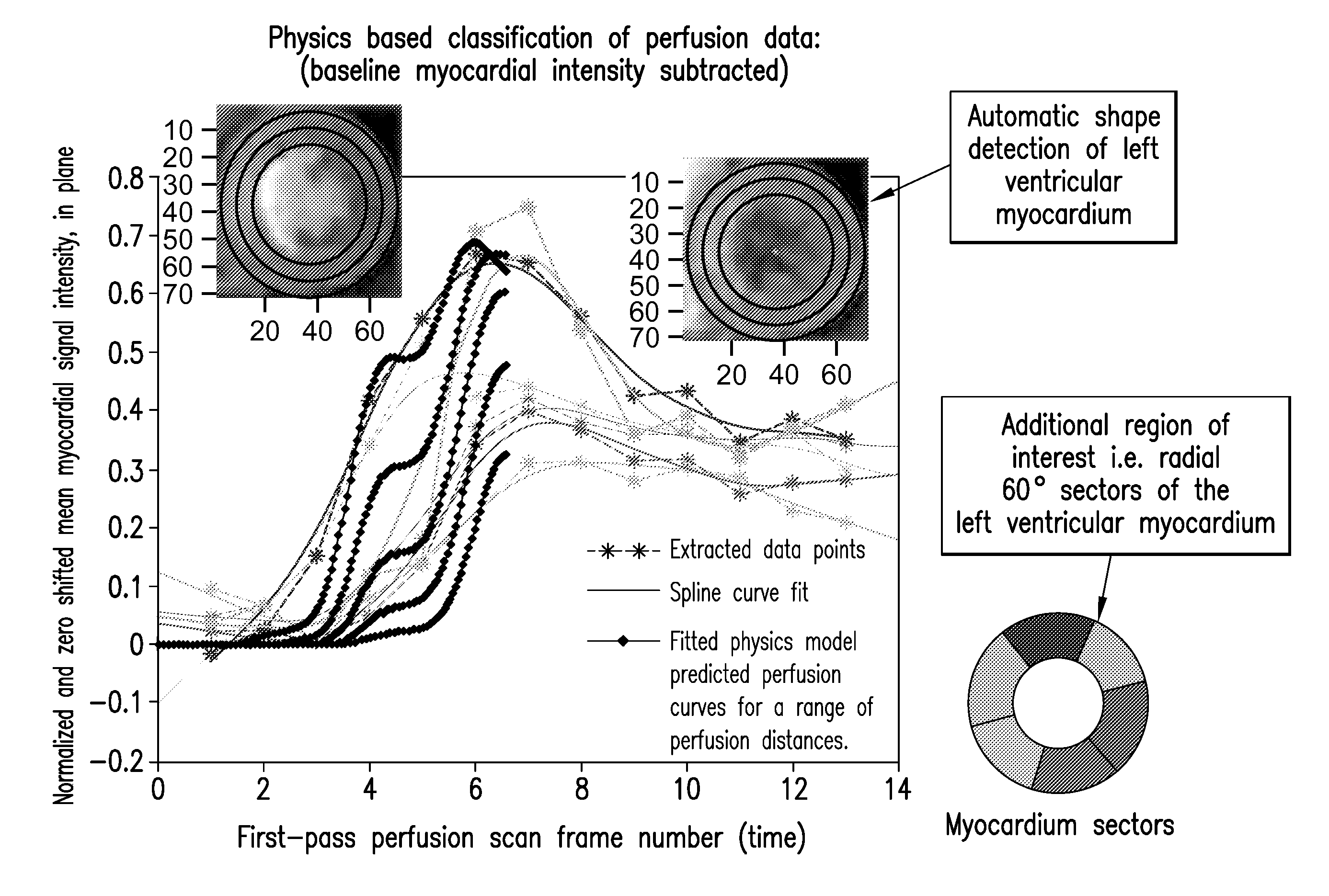
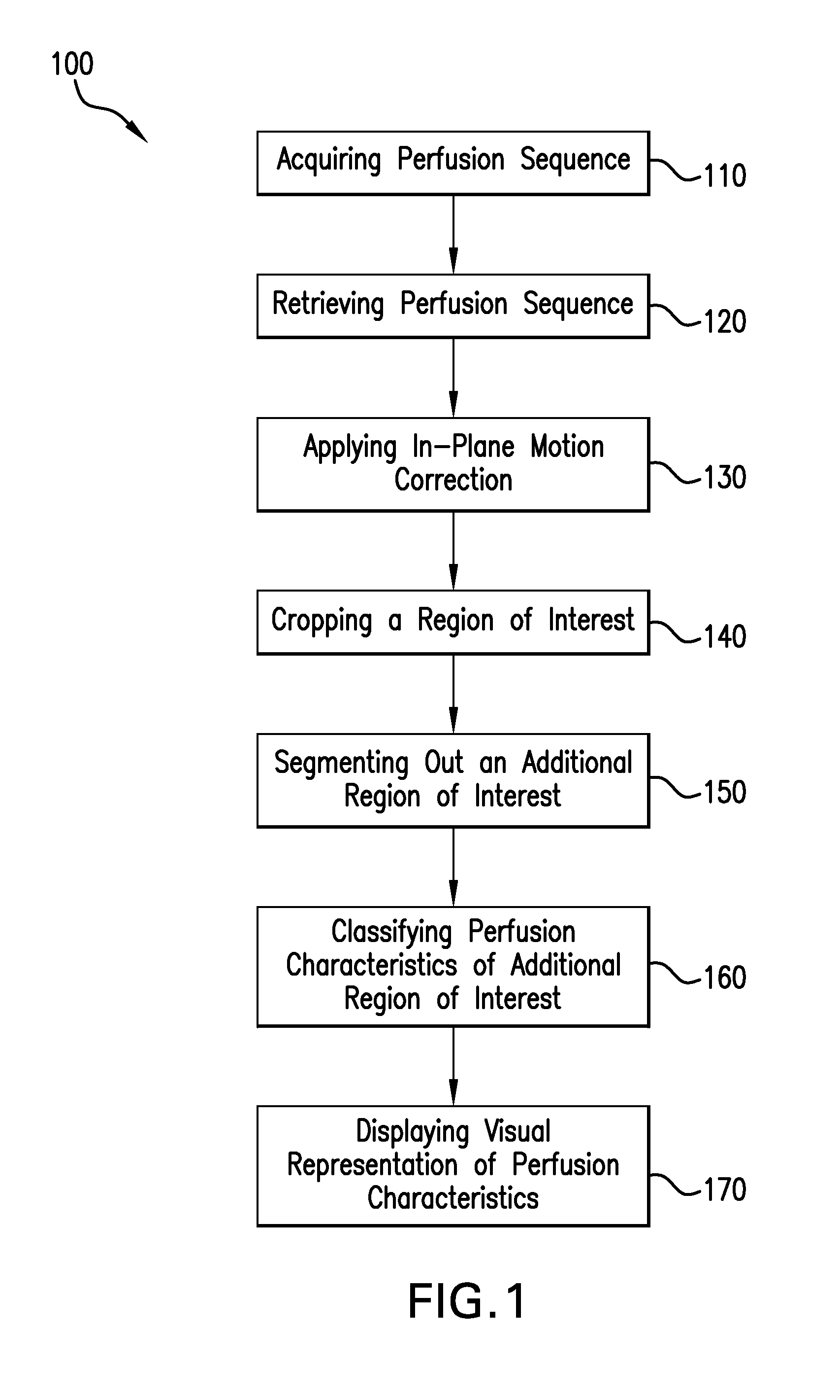
[0027]A physics-based image processing technique and an evaluation process for diagnosis of perfusion abnormalities in non-invasively imaged tissue, for example in left ventricular myocardial ischemia, using radiology imaging is disclosed. Patient specific time series images of a tissue undergoing perfusion, for example first-pass perfusion CMR images, are analyzed for perfusion characteristics and displayed as perfusion characteristic maps of tissue at risk of impending death. In the case of cardiac tissue, patient-specific perfusion information may be correlated against myocardial function (i.e., wall motion and wall thickening) information in order to confirm suspicion of myocardial infarction or myocardial ischemia.

[0028]As described herein, this quantification technology may be used to extend a cardiologist's clinical diagnosis capabilities by assisting him or her with the identification of early signs of heart disease long before a patient experiences a myocardial infarction o...


PUM


Abstract

A process and a system for assisting a clinician in identification and evaluation of perfusion characteristics of an organ are disclosed. The process and system may be used to extend clinical diagnosis capabilities by augmenting the ability to analyze perfusion images acquired from a radiology imaging modality through the use of the quantification technology as described. Classification of perfusion characteristics of a region of interest is facilitated through the use of a quantitative spatial perfusion characteristics map. The map is generated by a computing device which applies at least one physics-based image processing technique. This map is displayed on a clinician device for review by the clinician to assist in the spatial identification of perfusion abnormalities.

Description

CROSS-REFERENCE TO RELATED APPLICATIONS[0001]This application claims the benefit of an earlier-filed U.S. Provisional Application, entitled “Physics based image processing and evaluation techniques applied to diagnosis of left ventricular ischemia using cardiac MRI first-pass contrast perfusion imaging” having Ser. No. 61 / 520,922, filed Jun. 17, 2011 and an earlier-filed International Application entitled “Physics Based Image Processing And Evaluation Process Of Perfusion Images From Radiology Imaging” having Serial No. PCT / US12 / 42834, filed Jun. 15, 2012.FIELD OF THE INVENTION[0002]The present invention relates to a process and system for quantification of tissue perfusion using radiology images. More particularly, the present invention is concerned with augmenting the ability to analyze perfusion images acquired from a radiology imaging modality through the use of the physics based perfusion quantification technology.BACKGROUND OF THE INVENTION[0003]Heart disease is the leading ca...

Claims


Application Information

Patent Timeline
IPC IPC(8): A61B6/02
CPCA61B6/463A61B6/481A61B6/507A61B6/5217A61B6/5229A61B8/06A61B8/065A61B8/463A61B8/5223G16H50/30
Inventor MENON GOPALAKRISHNA, PRAHLADDOYLE, MARK
Owner CARNEGIE MELLON UNIV
Why Eureka
Try Eureka