Diagnostic and therapeutic methods for corneal ectasia following refractive surgery, keratoconus or pellucid degeneration

a technology of corneal ectasia and refractive surgery, applied in the direction of biological material analysis, sense disorder, drug composition, etc., can solve problems such as difficult diagnosis

Inactive Publication Date: 2013-03-07
THE UNIV OF SYDNEY
View PDF0 Cites 5 Cited by
  • Summary
  • Abstract
  • Description
  • Claims
  • Application Information

AI Technical Summary

Benefits of technology

[0009]The Wnt signalling pathway describes a complex network of proteins involved in a cascade that controls many physiological processes in mammals, including a role in apoptosis. The canonical Wnt signalling pathway involves Wnt proteins binding to cell-surface receptors of the Frizzled family, causing receptors to activate Dishevelled proteins, eventually regulating β-catenin movement to the nucleus and subsequent gene expression. Since apoptosis is one pathological feature of keratoconus, the present inventors examined whether one or more defective elements in the Wnt pathway in the corneal stroma or epithelium may be involved in pathological processes of this condition.

Problems solved by technology

Diagnosis may be difficult if the disease affects one eye only which is common in the early stages, and accordingly in some subjects the time of diagnosis can occur relatively late in the progression of the disease.

Method used


Image

  • Diagnostic and therapeutic methods for corneal ectasia following refractive surgery, keratoconus or pellucid degeneration
  • Diagnostic and therapeutic methods for corneal ectasia following refractive surgery, keratoconus or pellucid degeneration
  • Diagnostic and therapeutic methods for corneal ectasia following refractive surgery, keratoconus or pellucid degeneration

Examples


example 1

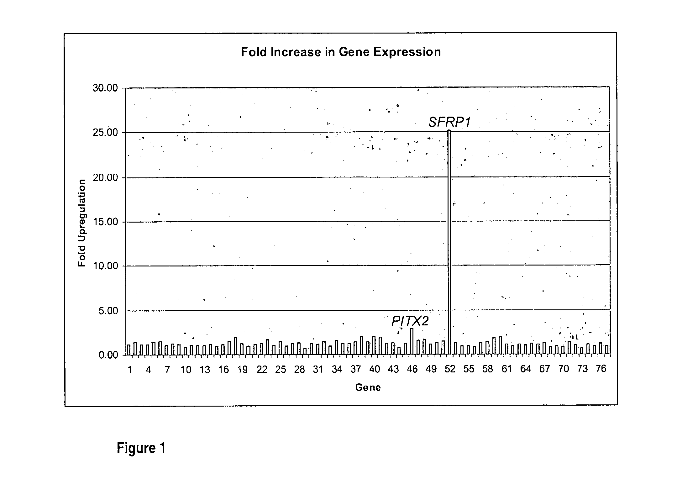
Quantification of the change of Gene Expression in the Corneal Epithelium of Keratoconus Subjects

[0130]Samples of corneal epithelium were collected from KC patients undergoing corneal transplantation and 6 age-matched controls undergoing photorefractive keratectomy (PRK). The clinical characteristics of the KC patients including age, best spectacle corrected visual acuity (BSCVA), mean keratometry and rate of corneal steepening were documented at the time of surgery and are set out in Table 2.

[0131]All keratoconus subjects exhibited poor vision which was not correctable by contact lenses. The rate of progression of keratoconus in the subjects was assessed by quantifying the amount of corneal steepening over time between assessments.

[0132]Tetracaine 1% (Minims) and Chloramphenicol 0.5% (Minims) eye drops were administered every ten minutes for 30 minutes prior to surgery. An 8 mm trephine was used to mark the epithelial surface and an 8 mm diameter of central epithelium was removed w...

example 2

Diagnosis of Keratoconus Using expression of SFRP1

[0158]Subjects for diagnosis may be selected on the basis of previous clinical diagnosis, for example by corneal topography, where the methods described herein may provide a confirmatory diagnosis and may identify whether the condition will progress rapidly or slowly. In addition, subjects intending to undergo refractive surgery, such as LASIK, may be screened. This diagnostic test may also be used for screening donor corneas prior to corneal transplantation procedures. This diagnostic test may also be carried out on subjects following refractive surgery in which there is suspected corneal ectasia.

[0159]A sample of full thickness epithelium of approximately 1 mm2 area from one or both corneas is taken under local anaesthetic using a sterile blade or scraper. Corneal epithelial samples are taken from the region overlying any corneal topographical anomaly, or from the corneal periphery. A similarly sized sample of conjunctival epitheli...

example 3

Treatment of Keratoconus, of Pellucid Marginal Degeneration or of Corneal Ectasia Following refractive Surgery

[0163]Subjects are diagnosed with keratoconus, pellucid marginal degeneration or corneal ectasia following refractive surgery using the diagnostic methods described herein and / or using standard diagnostic criteria. For example, a keratoconic subject with loss of best spectacle corrected vision of at least 80% would be eligible for the treatment.

[0164]In certain methods, subjects are topically administered a sterile aqueous solution of an agent which comprises neutralising antibodies to SFRP1 polypeptide to the corneal surface. In other methods, the agent comprises antisense molecules to SFRP1 or RNAi to SFRP1 and is administered to the eye using the general methods described in WO 2005 / 053600.

[0165]This administration may be formulated as aqueous eye drops, gels or creams, or may be provided in a depot such as a hydrogel lens which substantially covers the corneal surface or...


PUM

PropertyMeasurementUnit
timeaaaaaaaaaa
timeaaaaaaaaaa
diameteraaaaaaaaaa

Abstract

The present invention relates to methods of diagnosis and treatment of corneal ectasia following refractive surgery, keratoconus or pellucid marginal degeneration in a subject by determining or modulating the level of expression of molecules associated with the Wnt signalling pathway. Marker molecules of the present invention include SFRP1, PITX2, LEF1, WNT16 and WNT5A.

Description

RELATED APPLICATION[0001]This application claims benefit from Australian provisional patent application number 2009905883, filed 2 Dec. 2009, entitled “Diagnostic and Therapeutic Methods”, the entire disclosure of which is incorporated herein by reference.TECHNICAL FIELD[0002]The present invention relates to methods of diagnosis and treatment of corneal ectasia following refractive surgery or keratoconus or pellucid marginal degeneration in a subject.BACKGROUND OF THE INVENTION[0003]Keratoconus is a bilateral progressive, non-inflammatory but degenerative ectasia of the cornea which usually presents in the second decade of life and progresses into the third and fourth decade with a decreasing visual function. It is rare for progressive thinning and protrusion of the cornea to continue beyond that period. Keratoconus causes loss of visual function due to corneal thinning, irregular astigmatism and progressive myopia. The alteration to corneal topography occurs very early in the disea...

Claims


Application Information

Patent Timeline
Patent Type & Authority Applications(United States)
IPC IPC(8): C40B30/00A61K31/7052A61P27/02A61K33/00A61K31/5377A61K39/395A61K31/437
CPCA61K31/437A61K31/5377A61K33/00G01N2800/16C12Q2600/158G01N33/5044C12Q2600/112C12Q1/6883A61P27/02
Inventor SUTTON, GERARDMCAVOY, JOHNMADIGAN, MICHELE
Owner THE UNIV OF SYDNEY
Why Eureka
Try Eureka