

Diagnostic autoantibody profiles for the detection and diagnosis of neurodegenerative diseases
a technology for diagnosing and diagnosing autoantibodies, applied in the field of diagnosing autoantibodies for the detection and diagnosis of neurodegenerative diseases, can solve the problems of brain-reactive antibodies in mothers of autistic children eliciting behavioral abnormalities, autoantibodies, inflammation and damage,
- Summary
- Abstract
- Description
- Claims
- Application Information
AI Technical Summary
Benefits of technology
Problems solved by technology
Method used
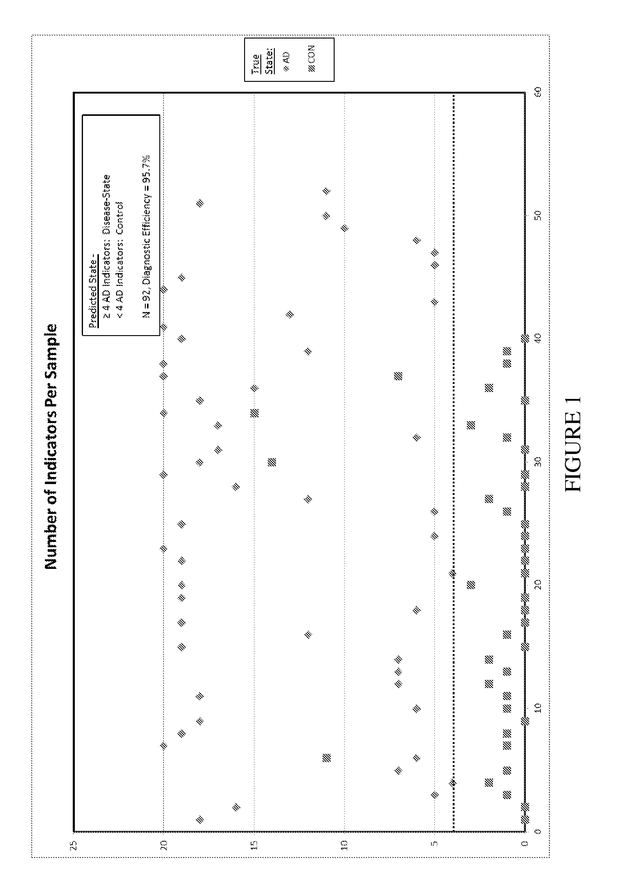
Image

Examples
example 1
Materials and methods
Animals
[0070]Swiss-Webster mice were obtained from Taconic Farms (Hudson, N.Y.) and used for experiments at 3-6 months of age. Sprague-Dawley rats were also obtained from Taconic Farms and used at 7-9 weeks of age. Both were maintained on ad libitum food and water with 12-hour light / dark cycle in an AALAC-accredited vivarium. Animals use was reviewed and approved by the UMDNJ IACUC.
Human Brain Tissue
[0071]Brain tissue from patients with sporadic AD (n=23, age range=71-88) and age-matched, neurologically normal individuals (n=14, age range=69-83) were obtained from the Harvard Brain Tissue Resource Center (Belmont, Mass.), the Cooperative Human Tissue Network (Philadelphia, Pa.), the UCLA Tissue Resource Center (Los Angeles, Calif.) and Slidomics (Cherry Hill, N.J.). Post-mortem intervals were J Neuropathol Exp Neurol 56, 1095-7). Formalin-fixed tissues were processed for routine paraffin embedding and sectioning according to established protocols. Control tissue...
example 2
Brain Reactive Autoantibodies in Human Sera
[0079]Sera from AD patients (n=52, age range 61-97 years), age-matched, non-demented control subjects (n=28, age range 51-86 years) and younger healthy individuals (n=28, age range 19-30 years) were tested for the presence of brain-reactive autoantibodies. For western analyses, individual sera were tested for the presence of brain-reactive autoantibodies by probing proteins obtained from whole cell homogenate derived from adult rat brain. Results confirmed the presence of brain-reactive autoantibodies in all sera from the three groups tested. The number of immunoreactive protein bands generated by each serum sample was similar for all three subject groups: mean=5.1+ / −3.1 for AD sera (n=52); 7.4+ / −4.0 for age-matched control sera (n=28); and 6.0+ / −3.8 for younger healthy control sera (n=28). Comparable results were obtained when human sera were used to probe mouse and human brain proteins. Based on apparent molecular weights in western blots...
example 3
IgG-Positive Neurons in Brain Regions Exhibiting AD Pathology
[0080]Ig-positive neurons in postmortem AD brains have been reported (Bouras et al. (2005) Brain Res Brain Res Rev. 48, 477-87; Clifford et al. (2007) Brain Res. 1142, 223-36; Deane and Zlokovic (2007) Curr Alzheimer Res. 4, 191-7; Franceschi et al. (1989) J. Gerontol. 44, M128-30; Kalaria (1999) Ann NY Acad. Sci. 893, 113-125; Kulmala et al. (1987) Exp Aging Res 13:67-72; Loeffler et al. (1997) Neurochem Res. 22, 209-14; Mooradian (1988) Neurobiol Aging. 9, 31-9; Nandy et al. (1975) J. Gerontol. 30, 269-74; Stein et al. (2002) J Neuropathol Exp Neurol. 61, 1100-8). In this example, immunohistochemistry using anti-human IgG antibodies was employed to test for the presence of IgG-immunopositive brain components in 23 AD and 14 age-matched control brains. IgG-positive neurons with immunolabeled cell bodies and dendrite trunks were found in all brains that were examined. IgG-positive neurons were far more abundant, widespread...
PUM
| Property | Measurement | Unit |
|---|---|---|
| pH | aaaaa | aaaaa |
| thick | aaaaa | aaaaa |
| excitation wavelength | aaaaa | aaaaa |
Abstract
Description
Claims
Application Information