Cell penetrating nucleolytic antibody based cancer therapy
- Summary
- Abstract
- Description
- Claims
- Application Information
AI Technical Summary
Benefits of technology
Problems solved by technology
Method used
Image
Examples
example 1
5C6 is a Lupus Autoantibody that Penetrates into Cell Nuclei
[0164]Materials and Methods
[0165]Hybridomas and Cell Lines
[0166]A panel of hybridomas, including the 3E10, 5C5, and 5C6 hybridomas was previously generated from the MRLmpj / lpr lupus mouse model and DNA binding activity was evaluated (Zack, et al., J. Immunol. 154:1987-1994 (1995); Gu, et al., J. Immunol., 161:6999-7006 (1998)). The hybridomas were maintained in serum free hybridoma media (BD Cell MAb medium; BD Biosciences, San Jose, Calif.) supplemented with 2 mM L-glutamine (Life Technologies, Carlsbad, Calif.). Antibodies were harvested in hybridoma supernatants and exchange-dialyzed into appropriate media. BRCA2 proficient (BRCA2(−) and deficient (BRCA2(−)) DLD1 colon cancer cells (Horizon Discovery, Ltd., Cambridge, UK) were grown in RPMI 1640 (Life Technologies) supplemented with 10% fetal bovine serum (FBS; Sigma-Aldrich, Saint Louis, Mo.). The identities of the BRCA2(+) and BRCA2(−) DLD1 colon cancer cells were conf...
example 2
5C6 Induces γH2AX in BRCA2—Cancer Cells
[0172]Materials and Methods
[0173]γH2AX Assays
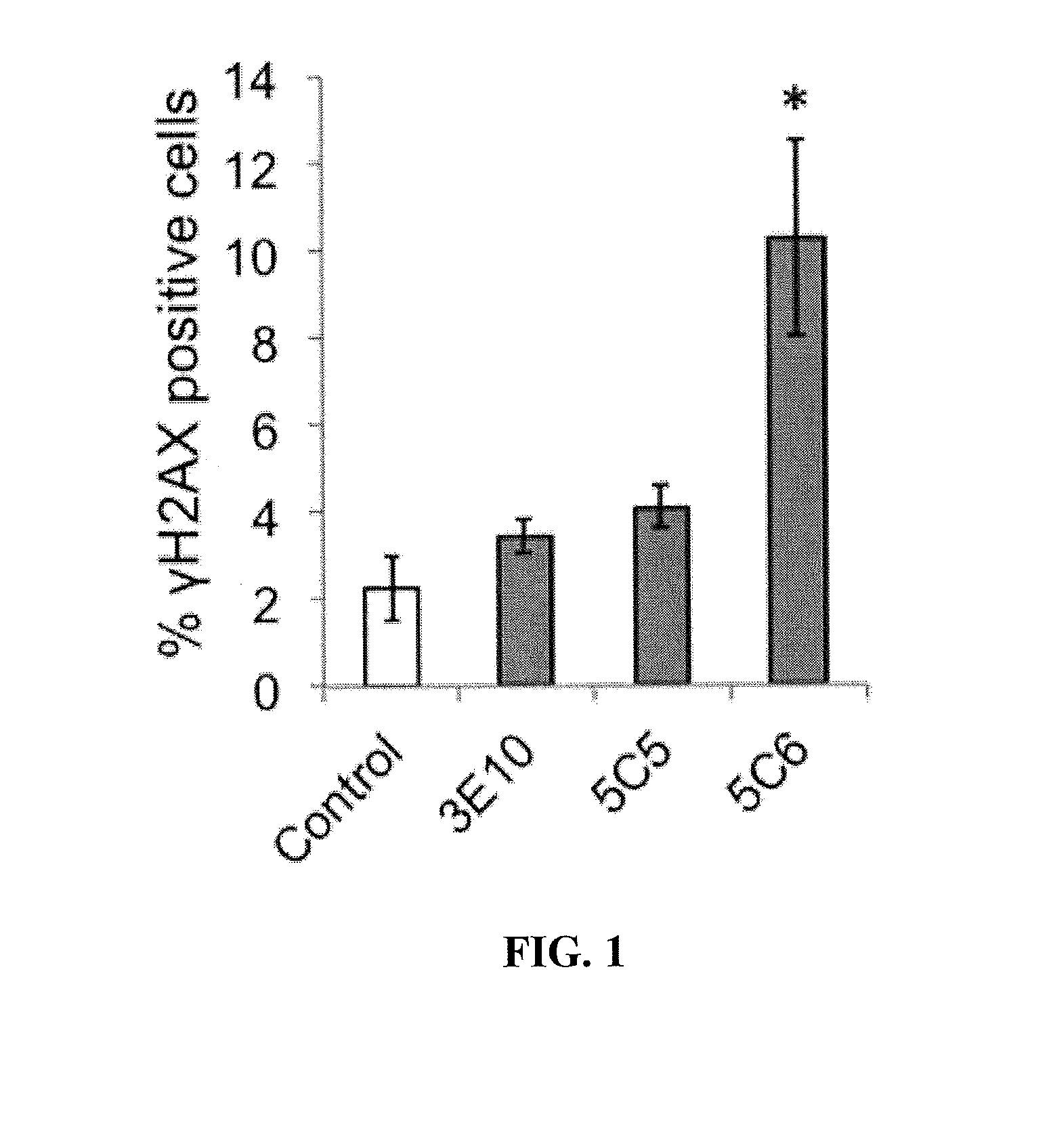
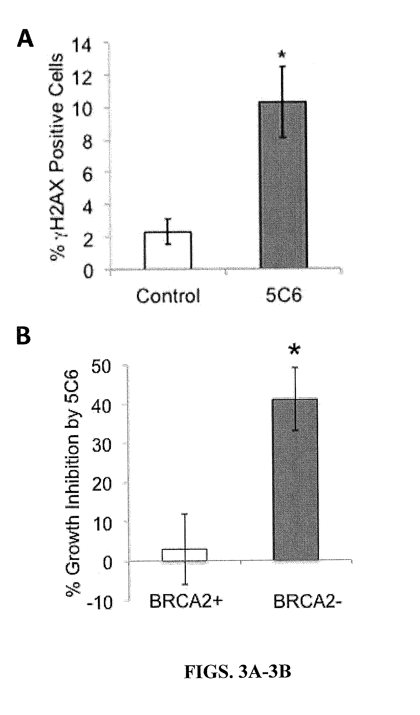
[0174]BRCA2(+) and BRCA2(−) DLD1 cells were treated with control media or media containing 10 μM 3E10, 5C5, or 5C6 for one hour, followed by evaluation of the percentage of cells positive for γH2AX (a marker of DNA double strand breaks) by immunofluorescence. Cells were washed with PBS, fixed with chilled 100% ethanol for 5 minutes, washed again with PBS, blocked with 1% BSA / PBS for 45 minutes and then probed with Alexa488-conjugated anti-γH2AX antibody overnight at 4° C. (Cell Signaling). Fluorescent signal corresponding to γH2AX (a marker of DNA double-strand breaks) was then imaged using an EVOS fl digital fluorescence microscope using a GFP filter.
[0175]Results
[0176]The antibodies were screened for the ability to induce DNA double-strand breaks in the DLD1 cells. None of the antibodies induced γH2AX in the BRCA2(+) cells. Similarly, neither 3E10 nor 5C5 caused any significant increase in the perc...
example 3
5C6 Catalyzes Degradation of Single-Stranded DNA
[0177]Materials and Methods
[0178]Catalytic Assays—Single-Stranded Plasmid DNA Assays
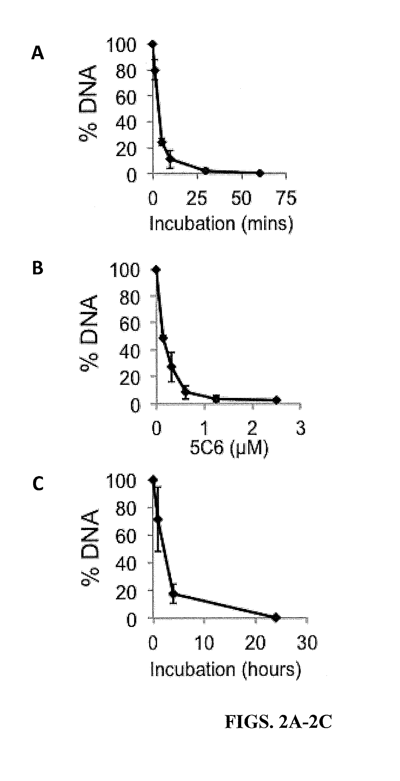
[0179]Single-stranded M13mp18 DNA (New England Biolabs, Ipswich, Mass.) was incubated with 0-2.5 μM 5C6 for 0-60 minutes in binding buffer (50 mM Tris-HCl, 100 mM NaCl, 10 mM MgCl2). The reaction was terminated by the addition of 1% SDS. Samples were boiled and then run on 1% agarose gels and stained with SYBR Gold (Life Technologies) for 30 minutes. The proportion of DNA remaining after treatment relative to untreated M13mp18 DNA was then calculated by band densitometry using Image J (National Institutes of Health, Bethesda, Mass.). Single-stranded M13mp18 circular DNA was incubated with buffer containing 2.5 μM 5C6 for 0-60 minutes, followed by visualization of DNA on an agarose gel.
[0180]The percentage of M13mp18 DNA remaining after incubation with 5C6 was quantified relative to untreated M13mp18 DNA. 5C6 degrades single-stranded DNA in a dose-depend...
PUM
| Property | Measurement | Unit |
|---|---|---|
| Fraction | aaaaa | aaaaa |
| Dimensionless property | aaaaa | aaaaa |
| Composition | aaaaa | aaaaa |
Abstract
Description
Claims
Application Information
 - R&D
- Intellectual Property
- Life Sciences
- Materials
- Tech Scout
- Unparalleled Data Quality
- Higher Quality Content
- 60% Fewer Hallucinations
Browse by: Latest US Patents, China's latest patents, Technical Efficacy Thesaurus, Application Domain, Technology Topic, Popular Technical Reports.
© 2025 PatSnap. All rights reserved.Legal|Privacy policy|Modern Slavery Act Transparency Statement|Sitemap|About US| Contact US: help@patsnap.com


