Diagnostic Methods for Glaucoma

a glaucoma and glaucoma technology, applied in the field of medical diagnostics, can solve the problems of false negative, limited early detection methods for glaucoma, and no standard diagnostic tests to identify which persons have an elevated intraocular pressure, so as to prevent the cell death of retinal ganglion cells and stop the progression of glaucoma diseas

Active Publication Date: 2013-07-04
M LAB GMBH
View PDF3 Cites 7 Cited by
  • Summary
  • Abstract
  • Description
  • Claims
  • Application Information

AI Technical Summary

Benefits of technology

[0062]In preferred embodiments of using ocular antigens for a pharmaceutical composition to treat glaucoma, they are used as blocking agents binding to autoantibodies present at increased levels in a glaucoma patient. In particular, it has been found that many of the elevated autoimmune reactivities in glaucoma patients are directed against cytoskeletal proteins, for example actin and actin binding proteins such as annexin V, alpha fodrin, myelin binding protein, HSP 27. It is known that autoantibodies against cytoseleteal structures such as the f-actin meshwork when taken up by retinal ganglion cells disrupt the actin meshwork and induce cell apoptosis. Thus, according to this aspect of the invention, a pharmaceutical composition is provided, to prevent cell death of retinal ganglion cells. The composition comprising at least one of the ocular antigens of the group 2 stops the progression of glaucoma disease by ocular antigens, which bind to the autoantibodies and block their destructive effects on retinal ganglion cells in glaucoma patients.
[0063]While the first diagnostic method for glaucoma, which is independent of an elevated intraocular pressure, relies on a biomarker profile of identified ocular antigens, a second such diagnostic method which is independent of an elevated intraocular pressure relies on the effect of body fluids on a cell culture in an in vitro assay.

Problems solved by technology

So far there are no standard diagnostic tests to identify which persons with an elevated intraocular pressure develop glaucoma.
Early detection methods for Glaucoma are still limited.
Measuring the intraocular pressure can detect some of the patients but fluctuant pressure levels can also give a false negative result.
The traditional detection method measuring an elevated intraocular pressure fails with these patients.
Furthermore, it could be shown that the direct application of anti-HSP antibodies results in an apoptosis of retinal ganglion cells in a cell culture approach (Tezel G, et al.
However, studies in the diagnostic field concerning other diseases such as cancer have revealed, that diagnosis based on autoimmune reactivity against biomarkers of unknown identity is often unreliable.

Method used


Image

  • Diagnostic Methods for Glaucoma
  • Diagnostic Methods for Glaucoma
  • Diagnostic Methods for Glaucoma

Examples


example 1

Antigen Microarrays Comparing Autoimmunoreactivty in Sera and Aqueous Humor, with Characteristic Differences in Glaucoma Patients and Healthy Individuals

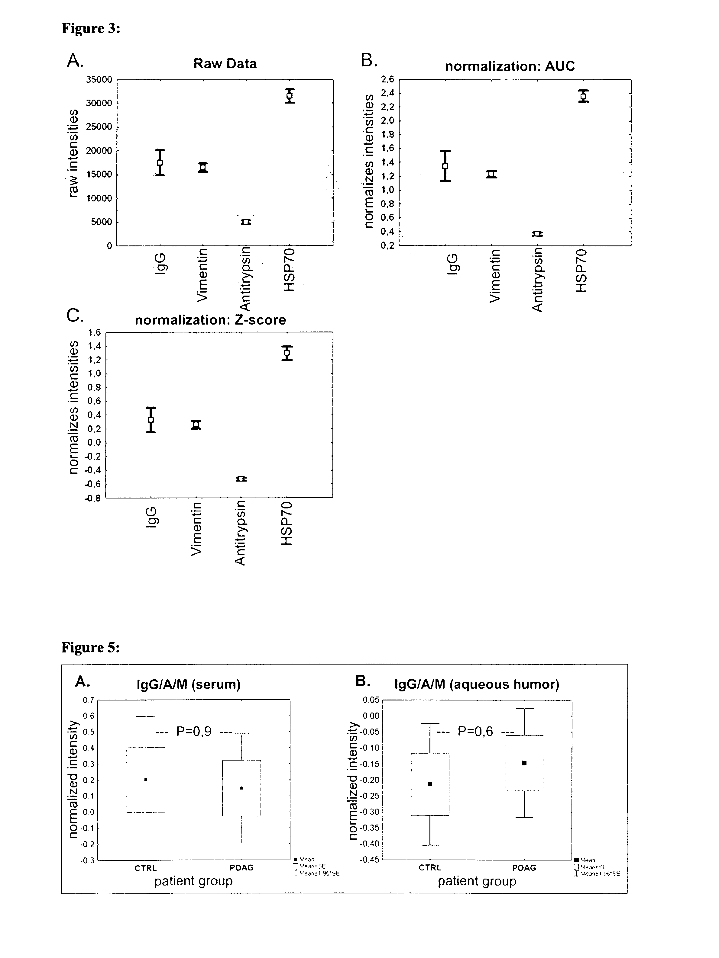
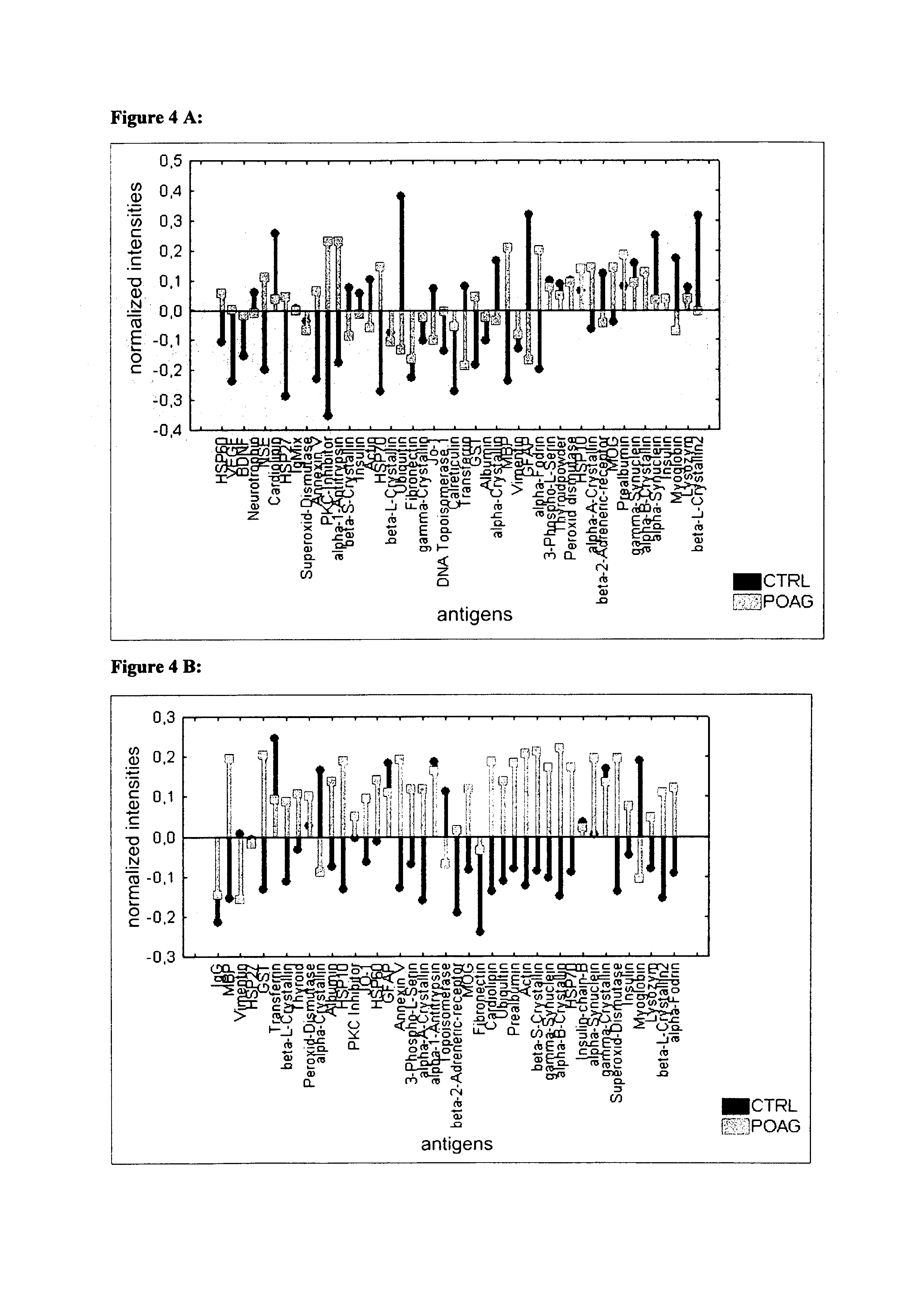
[0118]Sera and aqueous humor of patients with primary open-angle glaucoma (POAG; n=13) and healthy controls (CTRL; n=13) were used for antibody analysis. The protein arrays were prepared by spotting 40-100 different purified antigens (known biomarkers) onto nitrocellulose-coated slides. The arrays were incubated with sera (1:250) and aqueous humor (1:20) respectively. For visualization of the antibody-antigen-reactions arrays were treated with a fluorescence labeled anti-human IgG antibody, followed by fluorescence scanning. The signals emitted from secondary antibodies were digitized and the spot intensities were compared using multivariate statistical techniques.

[0119]Results: The intraindividual comparison revealed congruencies but also differences between antibody patterns of sera and aqueous humor. In both, aqueous humor and se...

example 2

Procurement of Sera and Aqueous Humor Samples

[0120]Procurement of samples was performed in accordance with the Declaration of Helsinki on biomedical research involving human subjects. Blood and aqueous humor was collected from all volunteers giving their informed consent. The blood samples were centrifuged at 1000 g and the serum was stored at −80° C. for subsequent analysis. Aqueous humor samples were stored at −80° C. directly after sampling. All participants were subject of a full ophthalmologic examination, including Goldmann Applanation, Tonometry, optical coherence tomography (OCT) and Heidelberg retina Tomography (HRT), at the Department of Ophthalmology (University of Mainz, Germany) and they were classified in accordance with the guidelines of the European Glaucoma Society. 31 patients, undergoing cataract surgery, with a mean age of 73 (SD±10) and 37 primary open-angle glaucoma patients (POAG; mean age: 67, SD±10) were included in this study. Cataract patients with no clin...

example 3

Preparation of Microarrays

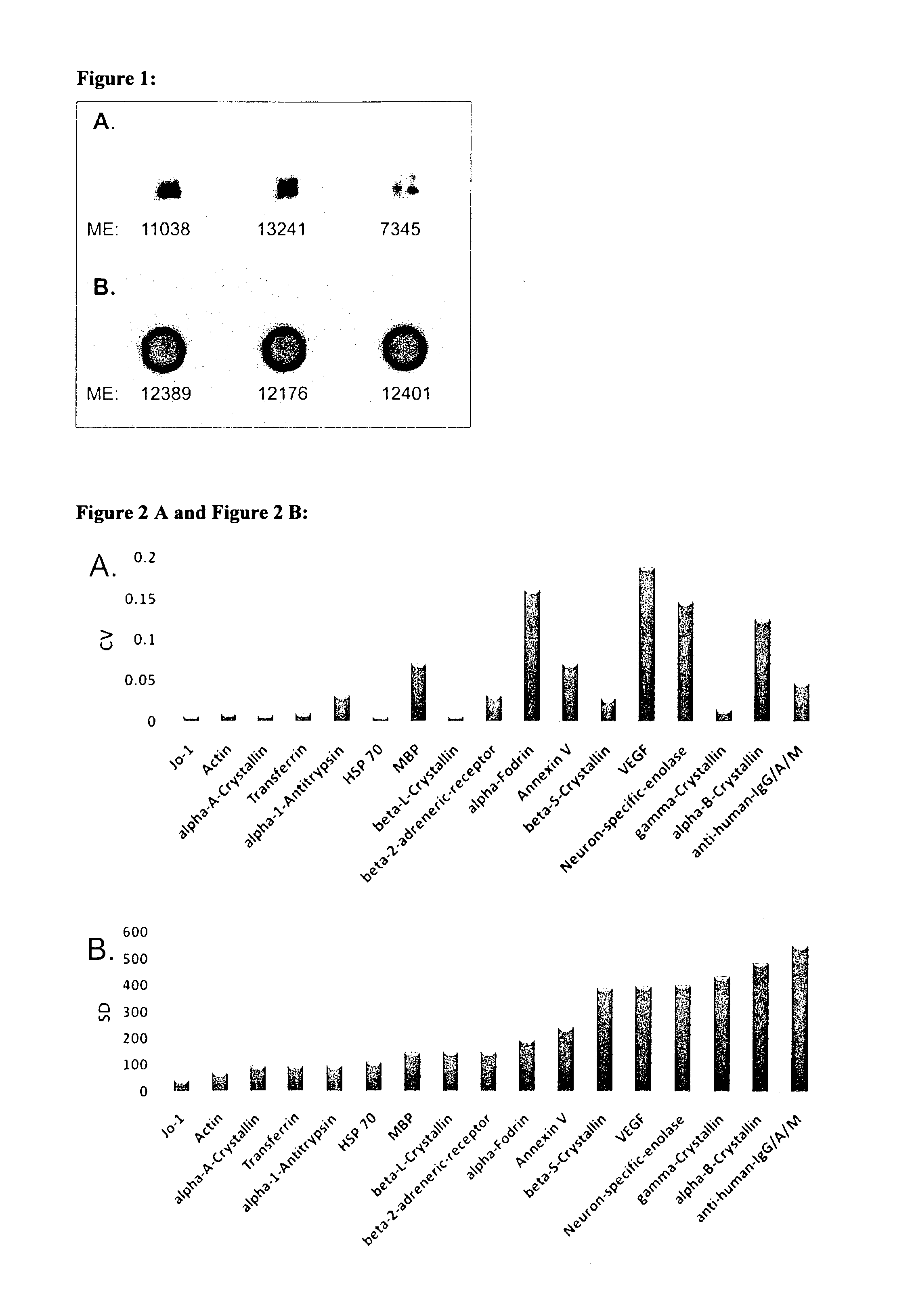
[0121]We used highly purified proteins, purchased at Sigma-Aldrich (Germany) and BioMol (Hamburg, Germany), as antigens. Antigens were diluted to 1 μg / μl with PBS buffer containing 1.5% Trehalose for optimal printing conditions. The spotting of antigens was performed with both a non contact printing technology (sciFLEXARRAYER S3, Scienion, Berlin, Germany), based on piezo dispensing, and the commonly used pin based contact printing technique (OmniGrid100, Digilab Genomic Solutions, Ann Arbor, USA). Results were comparatively evaluated for spot morphology and spot to spot variability. For printing of the whole set of study microarrays the piezo based spotting technique was used. Each antigen was spotted in triplicate onto nitrocellulose-slides (Oncyte, nitrocellulose 16 multi-pad slides, Grace Bio-Labs, Bend, USA). As a positive and negative control we used mouse anti human IgG / A / M (10 μg / μl) and spotting buffer. The spotting process was performed at RT and ...


PUM

PropertyMeasurementUnit
temperatureaaaaaaaaaa
pressureaaaaaaaaaa
molecular weightsaaaaaaaaaa

Abstract

The invention concerns a first diagnostic method for glaucoma based on an analysis of autoimmune reactivity in body fluids against at least one sample of at least partially purified ocular antigens, wherein the autoimmune reactivity against individual antigens is measured and transformed into a glaucoma score to determine the diagnostic result. Further aspects of the invention include antigen carrying elements carrying at least one sample of the at least partially purified ocular antigens and kits for diagnosis of glaucoma. Further aspects include methods of collecting a body fluid such as tears for the use in the diagnostic method for glaucoma. Yet further aspects include ocular antigens serving as diagnostic markers and/or for preparing pharmaceutical compositions for treatment of glaucoma. The invention further concerns a second diagnostic method for glaucoma comprising the steps of a) providing an in vitro culture of cells; b) incubating a body fluid from a test individual with the in vitro culture of cells or incubating components, which are fractionated from the body fluid or from a body specimen of the test individual with the in vitro culture of cells; c) analyzing protein expression of the cells and/or analyzing the viability of the cells after treatment according to step b); and d) comparing the results of the analysis in step c) with standard data to determine a diagnostic result.

Description

FIELD OF THE INVENTION[0001]The invention lies in the field of medical diagnostics and relates in particular to methods of diagnosing glaucoma based on analysis of autoimmune reactivity. This invention comprises two diagnostic methods for glaucoma. Both the first and the second diagnostic method rely on the autoimmune reactivities in body fluids of glaucoma patients. A further aspect of the invention relates to therapeutic methods modulating the autoimmune reactivity of glaucoma patients.[0002]A first diagnostic method for glaucoma is based on an analysis of autoimmune reactivity of bodily fluids against ocular antigens which are at least partially purified. A second diagnostic method for glaucoma is based on the analysis of the effect of autoantibodies in body fluids, on the protein expression of in vitro cultured retinal ganglion cells (RGC).BACKGROUND OF THE INVENTION[0003]Glaucoma is a group of ocular disorders characterized by progressive loss of retinal ganglion cells and thei...

Claims


Application Information

Patent Timeline
Patent Type & Authority Applications(United States)
IPC IPC(8): G01N33/68
CPCG01N2800/168G01N33/6893A61P27/02A61P27/06G01N33/564G01N33/53G01N33/68G01N33/48
Inventor GRUS, FRANZBOEHM, NILSPFEIFFER, NORBERTBELL, KATHARINA
Owner M LAB GMBH
Why Eureka
Try Eureka