Composition and method for clusterin-mediated stem cell therapy for treatment of atherosclerosis and heart failure
a clusterin and stem cell technology, applied in the field of cellular therapy and tissue engineering for treating atherosclerosis and heart failure, can solve the problems of cell death by apoptosis, little information concerning the differentiation of implanted fetal myocytes in the heart, etc., and achieve the effect of reducing the risk of atherosclerosis, preventing apoptosis cell death, and reducing the risk
- Summary
- Abstract
- Description
- Claims
- Application Information
AI Technical Summary
Benefits of technology
Problems solved by technology
Method used
Image
Examples
example 1
Establishment of a Murine Infarct Model
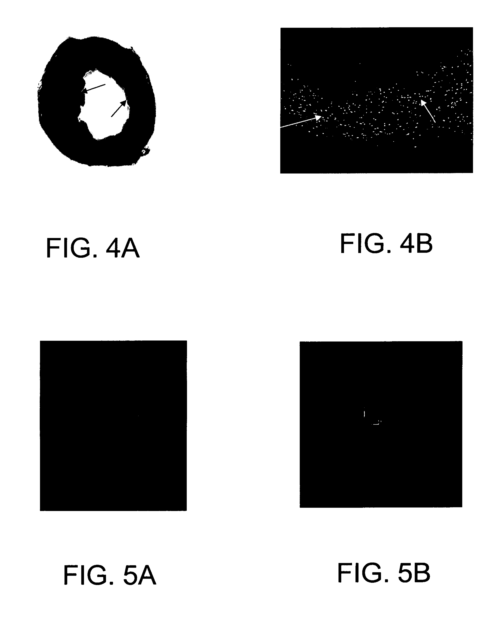
[0075] In order to test the apoptosis and development of stem cells in the heart with ischemic injury, a murine model was established for cell transplantation. Open chest surgery was performed on C57BL / 6J mice under anesthesia. After coronary ligation for one hour, the hearts were reperfused for 30 min and then sacrificed 2 weeks later. The hearts were perfused-fixed in formalin. Cell death by apoptosis was determined by in situ labeling (TUNEL) of DNA fragments. Typical infarcts were observed in the myocardium injured by ischemia-reperfusion (FIG. 4A-B). Many TUNEL positive nuclei exist in the lesions. FIG. 4A, left ventricular section, TTC / blue dye staining showing an infarct in the heart (indicated by arrows); and FIG. 4B, numerous TUNEL positive (green fluorescent) nuclei in infarcted and surrounding areas, indicated by white arrows.
example 2
Apoptosis of Cardiac Stem Cells and Clusterin Treatment
[0076] The effect of clusterin on apoptosis of cardiac stem cells induced by serum starvation was tested. as described above in the General Methods and Materials. Cardiac stem cells were prepared from the newborn murine heart by gradient centrifugation. In the presence of clusterin-containing DMEM medium (FIG. 5A), the cardiac stem cells showed no sign of apoptosis while many cells died without clusterin. The dead cells exhibited a morphology characteristic of apoptosis including cell shrinkage, chromatin condensation, and nuclear fragmentation (FIG. 5B). In FIGS. 5A,B, images of murine cardiac stem cells in the presence (FIG. 5A) or absence (FIG. 5B) of clusterin. Cells were stained with acridine orange and ethidium bromide, and images were taken under an Olympus fluorescence microscope with ×40 objective.
example 3
GFP Expression by cDNA Transfection in Fetal Cardiac Myoblasts
[0077] A cDNA transfection method was established by which cardiac myoblasts or stem cells can be labeled with GFP. A plasmid was constructed with a neomycin-resistant gene. The transfected cells were selected using G418. GFP labeled cells can differentiate into mature myocytes in the same way as their parent cells. FIGS. 6A,B are representative images of GFP-transfected fetal cardiac myoblasts, illustrating GFP cDNA transfection leading to expression of the green fluorescent protein (GFP) in fetal cardiac myoblasts. Fetal cardiac myoblasts cultured in DMEM medium were transfected with a plasmid containing an insert coding for GFP. FIG. 6A is a phase-contrast image; and FIG. 6B is a fluorescent image showing GFP positive cells 48 hours after transfection
PUM
| Property | Measurement | Unit |
|---|---|---|
| concentrations | aaaaa | aaaaa |
| concentrations | aaaaa | aaaaa |
| emission wavelength | aaaaa | aaaaa |
Abstract
Description
Claims
Application Information
 - R&D
- Intellectual Property
- Life Sciences
- Materials
- Tech Scout
- Unparalleled Data Quality
- Higher Quality Content
- 60% Fewer Hallucinations
Browse by: Latest US Patents, China's latest patents, Technical Efficacy Thesaurus, Application Domain, Technology Topic, Popular Technical Reports.
© 2025 PatSnap. All rights reserved.Legal|Privacy policy|Modern Slavery Act Transparency Statement|Sitemap|About US| Contact US: help@patsnap.com


