
System and method for molecular breast imaging energy spectrum imaging and analysis

a technology of energy spectrum imaging and molecular breast, which is applied in the field of system and method for molecular breast imaging energy spectrum imaging and analysis, can solve the problems of reducing the usefulness of this procedure, not being widely used as a screening technique, and mri is currently approximately 20 times more expensive, so as to increase the range of photon energy and reduce the dose

Active Publication Date: 2013-08-22
MAYO FOUND FOR MEDICAL EDUCATION & RES
View PDF9 Cites 21 Cited by
  • Summary
  • Abstract
  • Description
  • Claims
  • Application Information

AI Technical Summary

Benefits of technology

The patent describes a way to make MRIs using less radiation by using a system to collect data with a wider range of energy levels. This approach reduces the amount of radioactive material needed for the MRI.

Problems solved by technology

However, since bilateral breast MRI is currently approximately 20 times more expensive than mammography, it is not in widespread use as a screening technique.
The PEM technology offers excellent resolution; however, the currently available radiotracer (F-18 Fluoro deoxyglucose) requires that a patient fast overnight, the patient must have low blood levels (this is often a problem for diabetics), and after injection, the patient must wait 1-2 hours for optimum uptake of F-18FDG in the tumor.
The high cost of these PET procedures coupled with the long patient preparation time reduces the usefulness of this procedure and makes it difficult to employ for routine breast evaluation.
However, these results only hold for large tumors and several studies have shown that the sensitivity falls significantly with tumor size.
This limitation is particularly important in light of the finding that up to a third of breast cancers detected by screening mammography are smaller than 10 mm.
The failure of conventional scintimammography to meet this limit led to its abandonment as a useful technique in the United States.
Despite this improved percentage of success, the failure to identify lesions of any size can have significant consequences.
Because MBI involves intravenous injections of the radiotracer, it poses a very different type of radiation risk than x-ray mammography.
However simply reducing the administered dose of Tc-99m sestamibi is not possible as the increased noise level in the resulting images significantly degrades image quality.
In addition, studies have shown that MBI detected 3 times as many cancers as digital and analog mammography in asymptomatic women at increased risk of breast cancer.

Method used


Image

  • System and method for molecular breast imaging energy spectrum imaging and analysis
  • System and method for molecular breast imaging energy spectrum imaging and analysis
  • System and method for molecular breast imaging energy spectrum imaging and analysis

Examples


Embodiment Construction

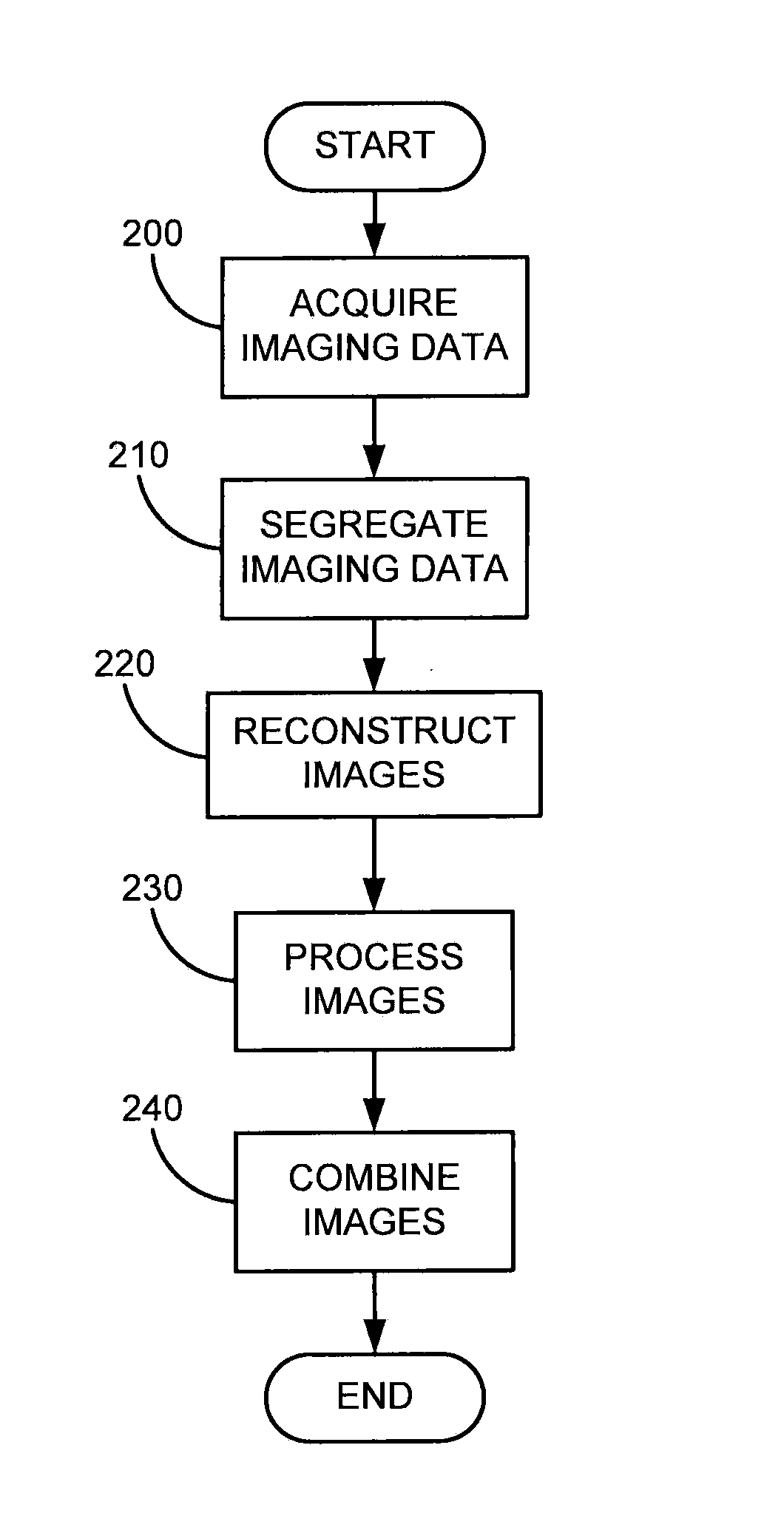
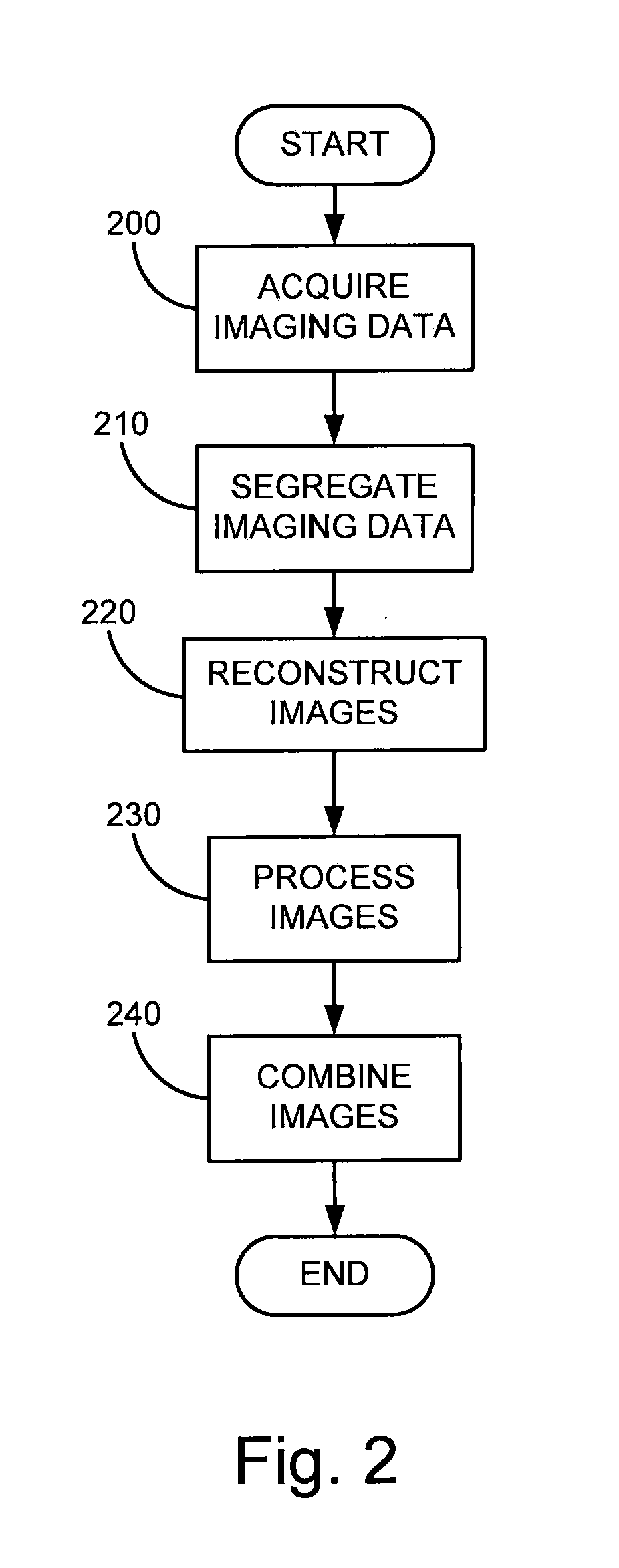
[0026]Conventionally, nuclear medicine gamma cameras employ energy windows centered on the primary gamma ray energy emitted by the radioisotope being detected. For example, with Tc-99m, a 20 percent energy window (from 126-154 keV) is centered on the 140 keV gamma rays. At lower energies, scatter within the patient and the gamma camera degrade image quality and these events are not considered useful.

[0027]In molecular breast imaging, the present invention recognizes that useful clinical images can be obtained at energies below the primary gamma ray energy. With this recognition in place, the present invention provides a method for extracting useful clinical information and images at these lower energies. By doing so the present invention improves image quality and can use this additional information to reduce the scan time or reduce patient radiation dose.

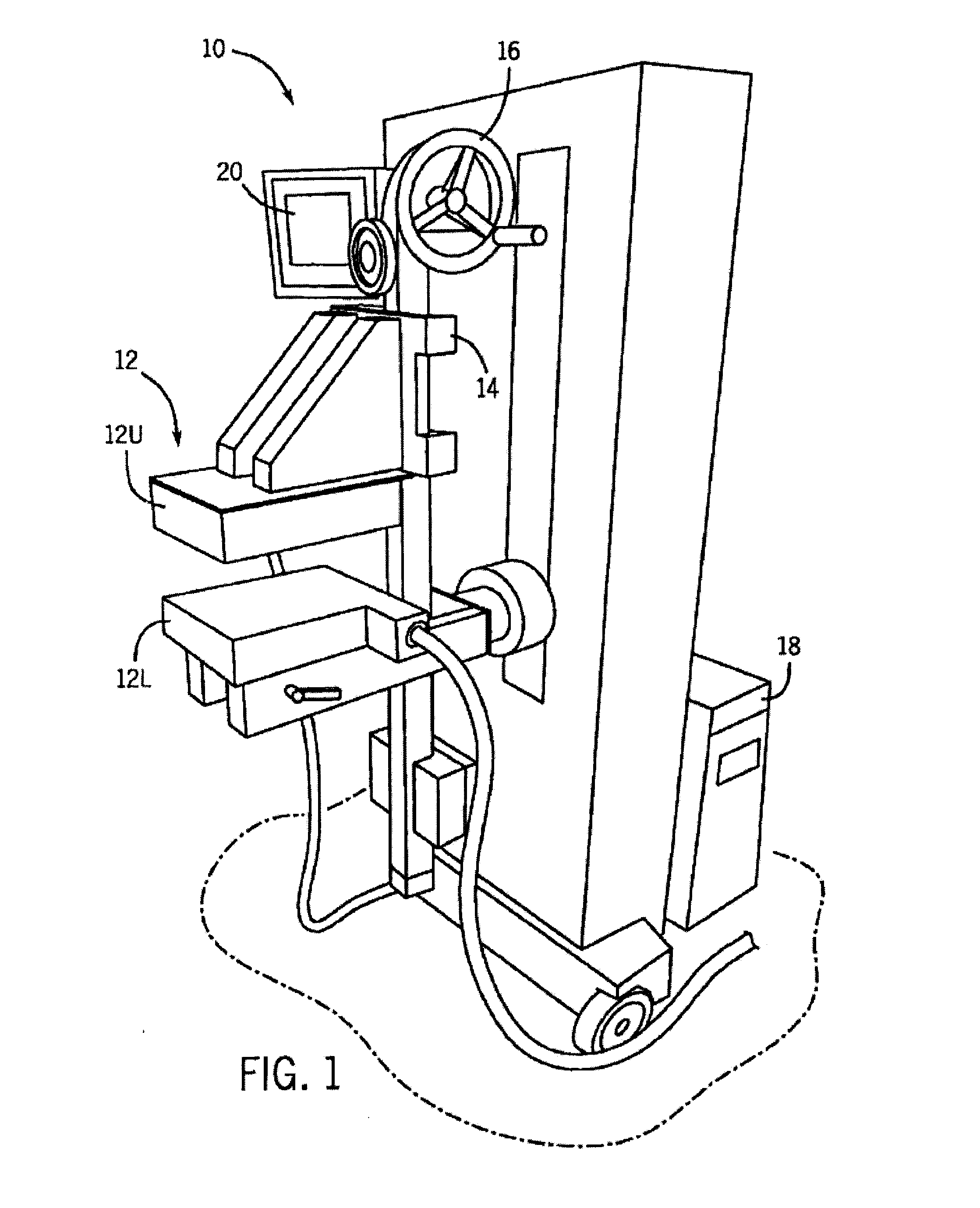
[0028]Referring to FIG. 1, a molecular breast imaging (MBI) system 10 includes two opposing CZT detectors (detector heads) 12. In...


PUM


Abstract

A system and method is provided for imaging a region of interest (“ROI”) including a breast having received a reduced dose of radionuclide using an imaging system including at least two gamma detectors. The acquired imaging data is analyzed respect to an energy characteristic and divided into subsets of data based on the analysis. A plurality of images is generated using the subsets of data, wherein the plurality of images include a primary image corresponding to subsets of data having energy including the energy characteristic and multiple secondary images corresponding to subsets of data having energy not including the energy characteristic. Each of the plurality of images is processed using energy information associated with each of the plurality of images and the processed plurality of images is combined to form a composite image of the ROI.

Description

CROSS-REFERENCE TO RELATED APPLICATIONS[0001]This application is based on U.S. Provisional Patent Application Ser. No. 61 / 408,301 filed on Oct. 29, 2010 and entitled “System and Method For Molecular Breast Imaging Energy Spectrum Imaging and Analysis.”STATEMENT REGARDING FEDERALLY SPONSORED RESEARCH[0002]No funding informationFIELD OF THE INVENTION[0003]The present invention relates to a method for imaging a region of interest using an imaging system including at least two gamma detectors. Specifically, the present invention relates to a system method for imaging a breast using a molecular breast imaging system and an increased spectrum analysis over traditional systems and methods.BACKGROUND OF THE INVENTION[0004]Screening mammography has been the gold standard for breast cancer detection for over 30 years. However, the sensitivity of screening mammography varies considerably. The most important factor in the failure of mammography to detect breast cancer is radiographic breast den...

Claims


Application Information

Patent Timeline
IPC IPC(8): G06T11/60
CPCA61B6/482A61B6/502A61B6/4258G06T11/60A61B6/5235
Inventor O'CONNOR, MICHAEL K.
Owner MAYO FOUND FOR MEDICAL EDUCATION & RES
Learn More