Scaffold for growing neuronal cells and tissue
a technology for neuronal cells and scaffolds, applied in the field of scaffolds for growing neuronal cells and tissue, can solve the problems of lack of interconnection channels, inability to form new tissues, and inability to separate cells, and achieve the effects of reducing the number of scaffolds
- Summary
- Abstract
- Description
- Claims
- Application Information
AI Technical Summary
Benefits of technology
Problems solved by technology
Method used
Image
Examples
example 1
Seeding OB Primary Cultures on PLLA / PLGA Scaffolds
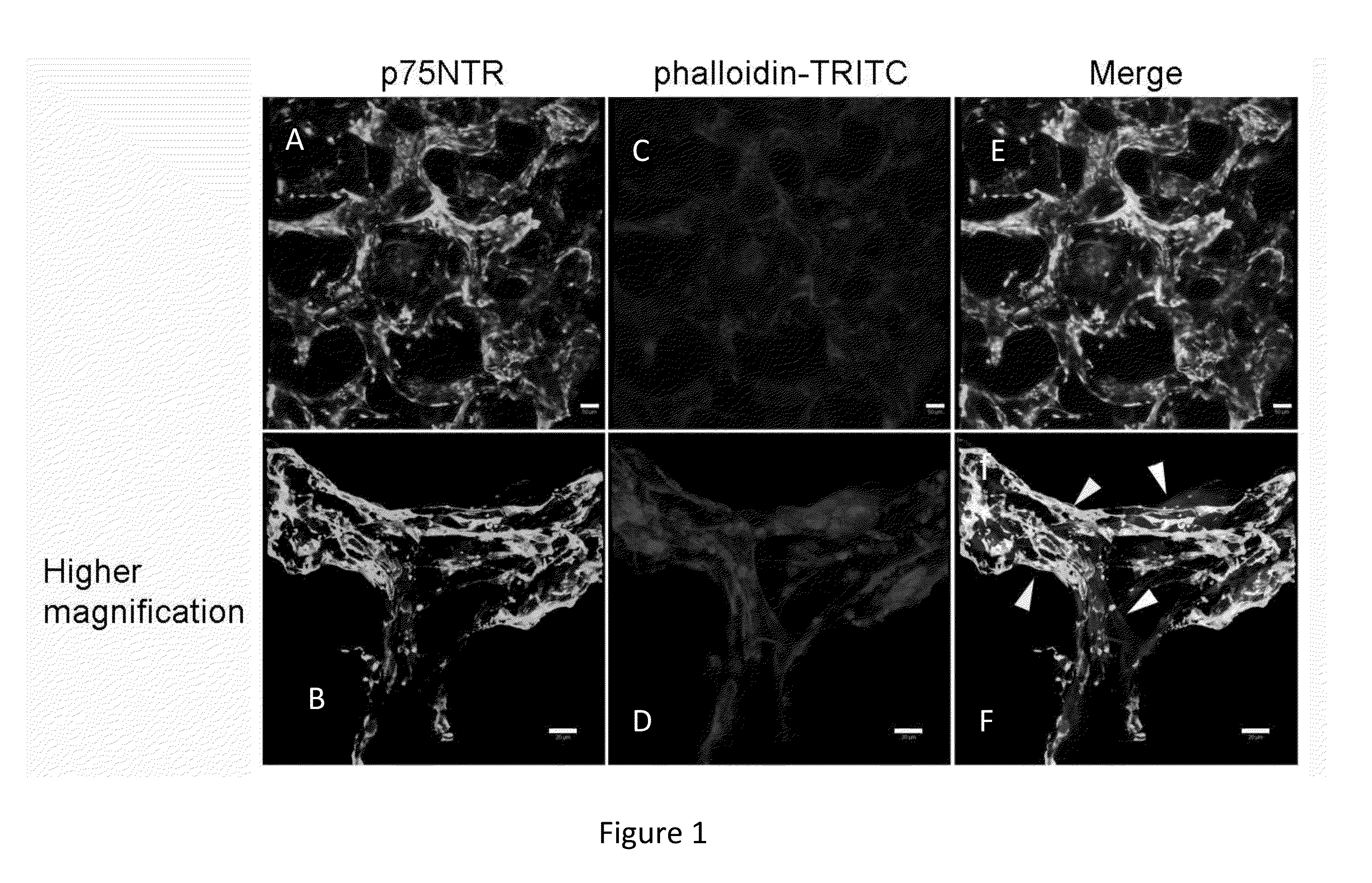
[0079]Olfactory bulbs from P7 mice were dissected and used for the preparation of primary cultures. These cultures were initially grown on 2D plates and were then seeded on PLLA / PLGA porous scaffolds. Cells were grown for a period of 28 days and were then fixed and stained with phalloidin-TRITC and p75NTR specific antibody (FIG. 1). By using low magnification microscopy, cell organization around the scaffold's pores was visualized, with few cells residing in the center of the pores (FIG. 1, upper panel). Higher magnification images (lower panel) revealed the organization and interaction between OECs (positively stained for p75NTR) and phalloidin stained cells in the culture.
[0080]The results indicated that following multicellular culturing on scaffolds, the OEC marker p75NTR was detected around the scaffold pores (FIG. 1). This cell distribution is unique to the present scaffold. Moreover, the scaffold of the present invention unexpe...
example 2
Cells Grown on Scaffolds Remain Viable and Proliferative
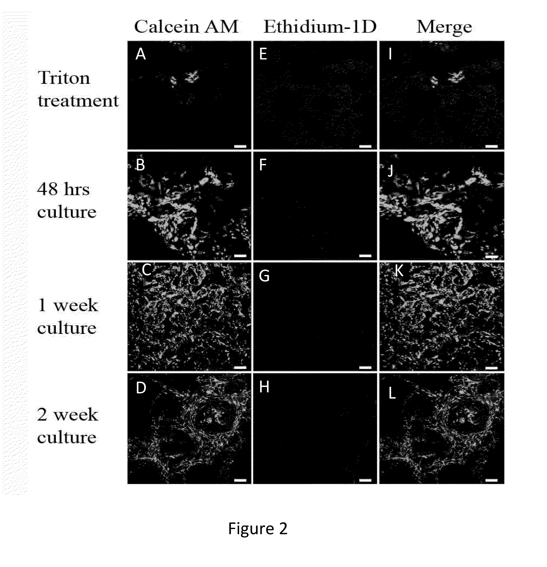
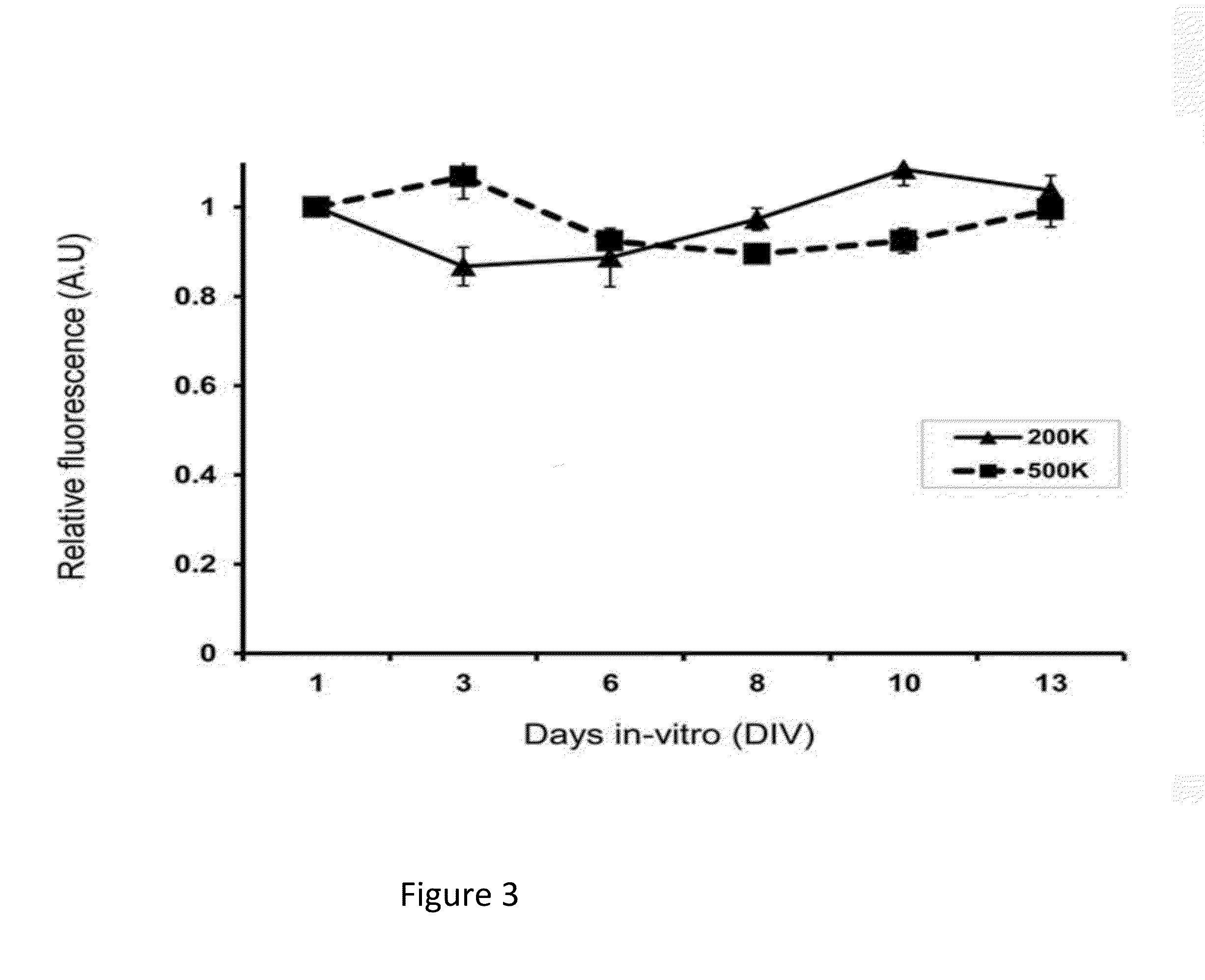
[0081]In order to analyze the viability of the cells and cell numbers, a live / dead assay and an Alamar Blue assay, were used (FIGS. 2 and 3, respectively). In the live / dead assay, the majority of cells were still viable at two weeks post-seeding (FIG. 2). As a negative control, the cells on the scaffold were treated with Triton X-100, prior to the analysis. In this case, the cells appeared dead, as they were positive for ethidium homodimer (Et-1D) staining. For the quantitative measurement of cell numbers, we used Alamar Blue analysis (FIG. 3). This analysis revealed that the cell numbers initially decreases but then returns to base level after 14-days in culture. Thus the cell viability and proliferation data of the present invention provides that 14 days culturing in-vitro is the minimal culturing period for the OB-derived cell constructs. This period is necessary in order to reach baseline proliferation rates.
example 3
OB-Derived Cells Grown on Scaffolds Secrete NGF And Induce Neuronal Differentiation of Pc12 Cells
[0082]It has been previously shown that OECs express and secrete various neurotrophic factors, including NGF, BDNF, and GDNF. To examine whether OEC-containing OB-derived 3D culture affectively secrete NGF, these cells were co-seeded on PLLA / PLGA scaffolds with pheochromocytoma (PC12 ) cells. PC12 cell line differentiates to a neuronal lineage in response to NGF stimulation, manifested by neuronal phenotype and neuronal genes expression. In this study, differentiation of PC12 cells served as an index for NGF secretion by olfactory bulb cells. To identify PC12 and OECs, the co-culture was stained with P75NTR (expressed by both PC12 and OEC) and βIII-tubulin (mark only PC12 ), as shown in FIG. 3N. In the absence of OB-derived cells, PC12 cells appeared round, without processes and expressed both p75NTR and βIII-tubulin (FIG. 4A-D). However, when seeded together with OB-derived cells (FIG. ...
PUM
| Property | Measurement | Unit |
|---|---|---|
| diameter | aaaaa | aaaaa |
| porosity | aaaaa | aaaaa |
| porosity | aaaaa | aaaaa |
Abstract
Description
Claims
Application Information
 - R&D
- Intellectual Property
- Life Sciences
- Materials
- Tech Scout
- Unparalleled Data Quality
- Higher Quality Content
- 60% Fewer Hallucinations
Browse by: Latest US Patents, China's latest patents, Technical Efficacy Thesaurus, Application Domain, Technology Topic, Popular Technical Reports.
© 2025 PatSnap. All rights reserved.Legal|Privacy policy|Modern Slavery Act Transparency Statement|Sitemap|About US| Contact US: help@patsnap.com


