X-ray CT system

a ct system and x-ray technology, applied in the field of x-ray ct system, can solve problems such as excessive radiation exposure, and achieve the effect of preventing excessive radiation exposur

Active Publication Date: 2013-10-24
TOSHIBA MEDICAL SYST CORP
View PDF4 Cites 10 Cited by
  • Summary
  • Abstract
  • Description
  • Claims
  • Application Information

AI Technical Summary

Benefits of technology

[0018]This embodiment is for solving said problem with the intention of providing an X-ray CT system that can prevent excessive radiation exposure caused by human error by determining whether or not input imaging conditions are appropriate based on patient attributes measured by the apparatus itself.

Problems solved by technology

There was a problem in that excessive exposure to radiation may occur with these conventional technologies; however, because they may not alert to prevent excess exposure to radiation when appropriate imaging conditions are not input (human error) due to factors such as lack of knowledge and insufficient attention by the technologist as well as if there is no opportunity to correct the imaging conditions.

Method used


Image

  • X-ray CT system
  • X-ray CT system
  • X-ray CT system

Examples


Embodiment Construction

[0025]The embodiment of the X-ray CT system will be explained with reference to each figure.

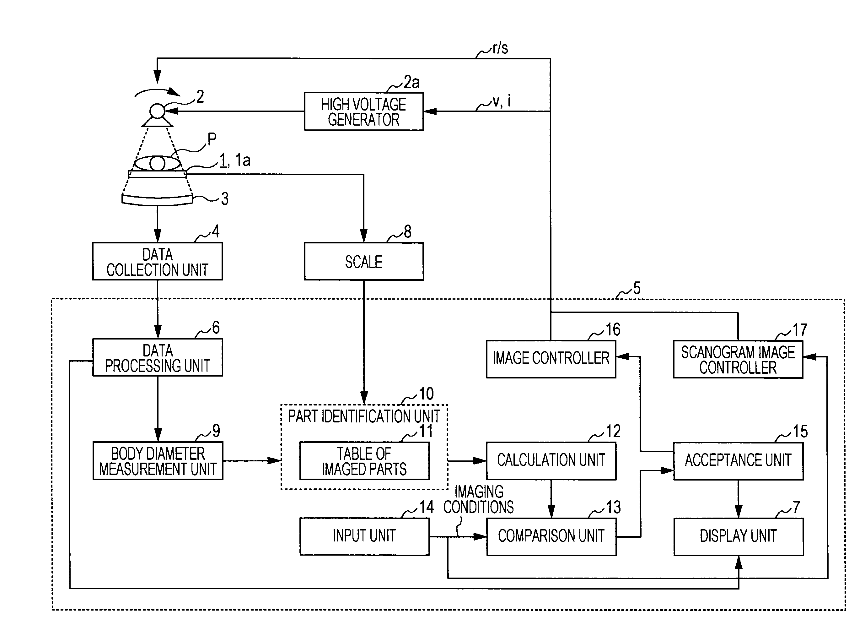
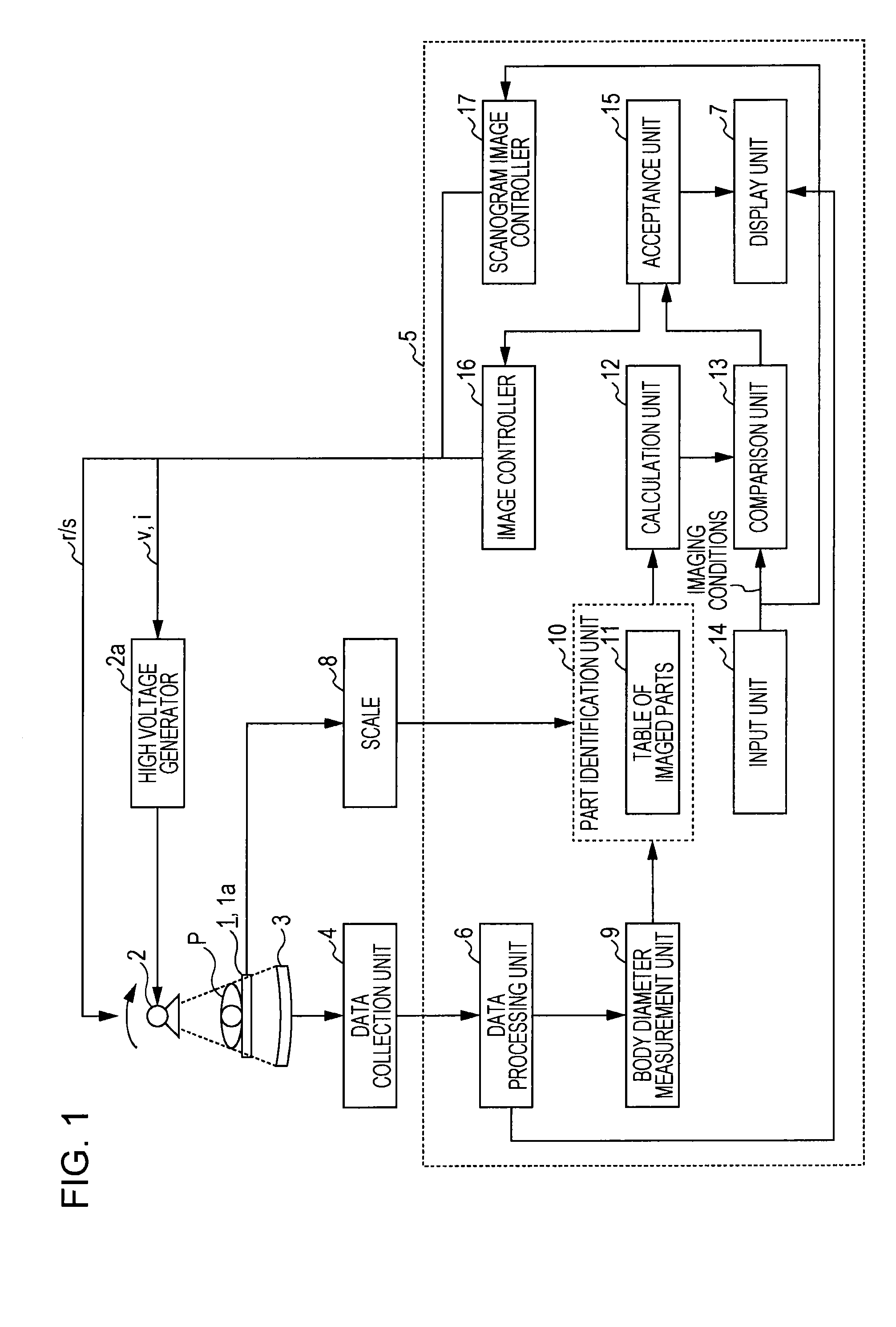
[0026]FIG. 1 is a block diagram indicating the composition of the X-ray CT system. As FIG. 1 indicates, the X-ray CT system comprises a couch 1, X-ray tubes 2, an X-ray detector 3, a data collection unit 4, and a console 5. The console 5 comprises a data processing unit 6 and a display unit 7.

[0027]The couch 1 comprises a couch top 1a on which the subject P is placed, a couch top driving means (illustration omitted) that enables adjustment of the imaging position by moving the couch top 1a towards the body axis of the subject and a helical scan, and a scale 8.

[0028]The X-ray tubes 2 and X-ray detector 3 are provided in a rotor (illustration omitted) such that they are placed facing each other with the couch top 1a on which subject P is placed between them. A high voltage generator 2a applies high voltage to the X-ray tubes 2 by receiving control information (described later) from the console ...


PUM


Abstract

An X-ray CT system is provided that determines input imaging conditions based on the attributes of subjects that were measured by the apparatus itself and can prevent excessive radiation exposure due to human error. The X-ray CT system comprises a measurement unit, a part identification unit, a calculation unit, a comparison unit, and a notification unit. The measurement unit measures the attributes of a subject placed on a couch. The part identification unit identifies the part of the subject being imaged based on the measured attributes of the subject. The calculation unit calculates a radiation dose based on the identified unit being imaged. The comparison unit makes a determination by comparing the calculated radiation dose and the radiation dose based on the imaging conditions input for examination. The notification unit notifies the results determined by the comparison unit.

Description

TECHNICAL FIELD[0001]The present invention relates to an X-ray CT system that reconstructs tomogram images of a subject based on X-rays transmitting the subject.BACKGROUND ART[0002]Acceptable radiation doses may differ according to the part to be imaged depending on the development risk of cancer and their hereditary impact in tissues exposed to radiation during X-ray imaging.[0003]The effective dose (Sv) indicating the acceptable radiation dose is represented by the following formula.[0004]Effective dose=total amount Σ all tissues (tissue weighting factor×equivalent dose)[0005]Here, the tissue weighting factor is a numerical value representing relatively the absorbed dose (Gy) which received in each tissue. Tissue weighting factors in the 2007 recommendations by the International Commission on Radiological Protection (ICRP) include 0.12 for the breasts, bone marrow (red), colon, and lungs; 0.08 for the stomach, gonads; 0.04 for the thyroid gland, esophagus, liver, and bladder; 0.01...

Claims


Application Information

Patent Timeline
Patent Type & Authority Applications(United States)
IPC IPC(8): A61B6/03A61B6/00
CPCA61B6/032A61B6/544A61B6/10
Inventor YANAGIDA, YUUJI
Owner TOSHIBA MEDICAL SYST CORP
Why Eureka
Try Eureka