Dual-modality scanning system for detecting breast cancer

a scanning system and breast cancer technology, applied in the field of dual-modality scanning system for detecting breast cancer, can solve the problems of not meeting the functional attributes identified above, not doing well in breasts, and not meeting the functional attributes of prior art, so as to improve the spatial resolution of this imaging modality

Inactive Publication Date: 2013-10-24
CAPERAY MEDICAL
View PDF13 Cites 19 Cited by
  • Summary
  • Abstract
  • Description
  • Claims
  • Application Information

AI Technical Summary

Benefits of technology

[0034]The processor preferably includes a tomosynthesis reconstruction algorithm that takes advantage of the multiple three-dimensional ultrasound images to enhance the spatial resolution of this imaging modality.

Problems solved by technology

Although digital X-rays significantly increase specificity in comparison with analogue film (Pisano et al., 2005), a major shortcoming of planar X-ray mammography is that it does not do well when the breasts are dense which is often the case for younger women who are less than 50 years of age (Nothacker et al., 2009).
Furthermore, none of the prior art fulfills all five of the functional attributes identified above.
However, this concept suffers from the shortcoming that the X-ray fan beam is scanned across the breast by a rotating X-ray tube with a fixed collimator slit, which means that tomosynthesis is not possible and the system can therefore only acquire 2D rather than 3D images.

Method used


Image

  • Dual-modality scanning system for detecting breast cancer
  • Dual-modality scanning system for detecting breast cancer
  • Dual-modality scanning system for detecting breast cancer

Examples


Embodiment Construction

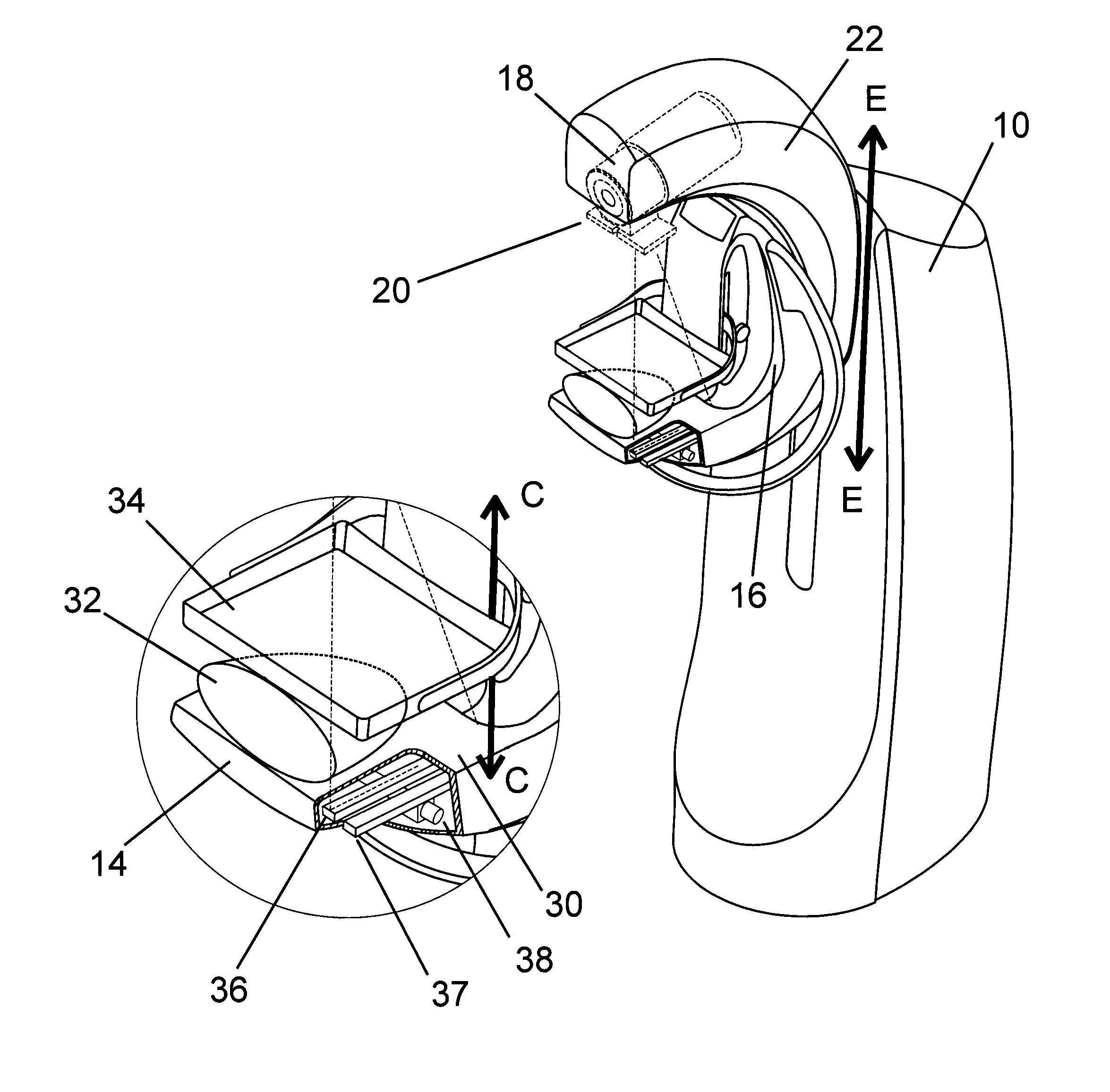
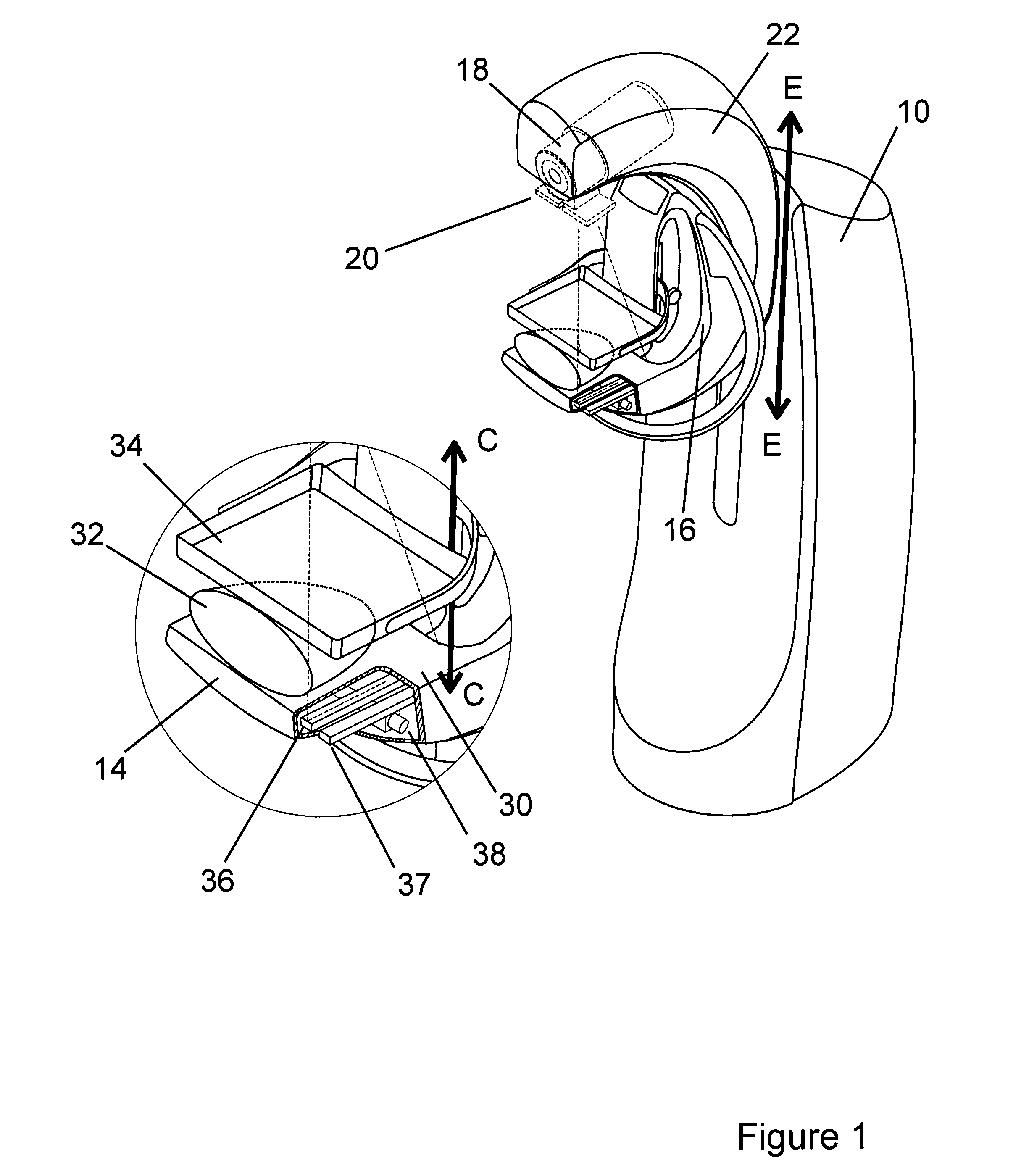
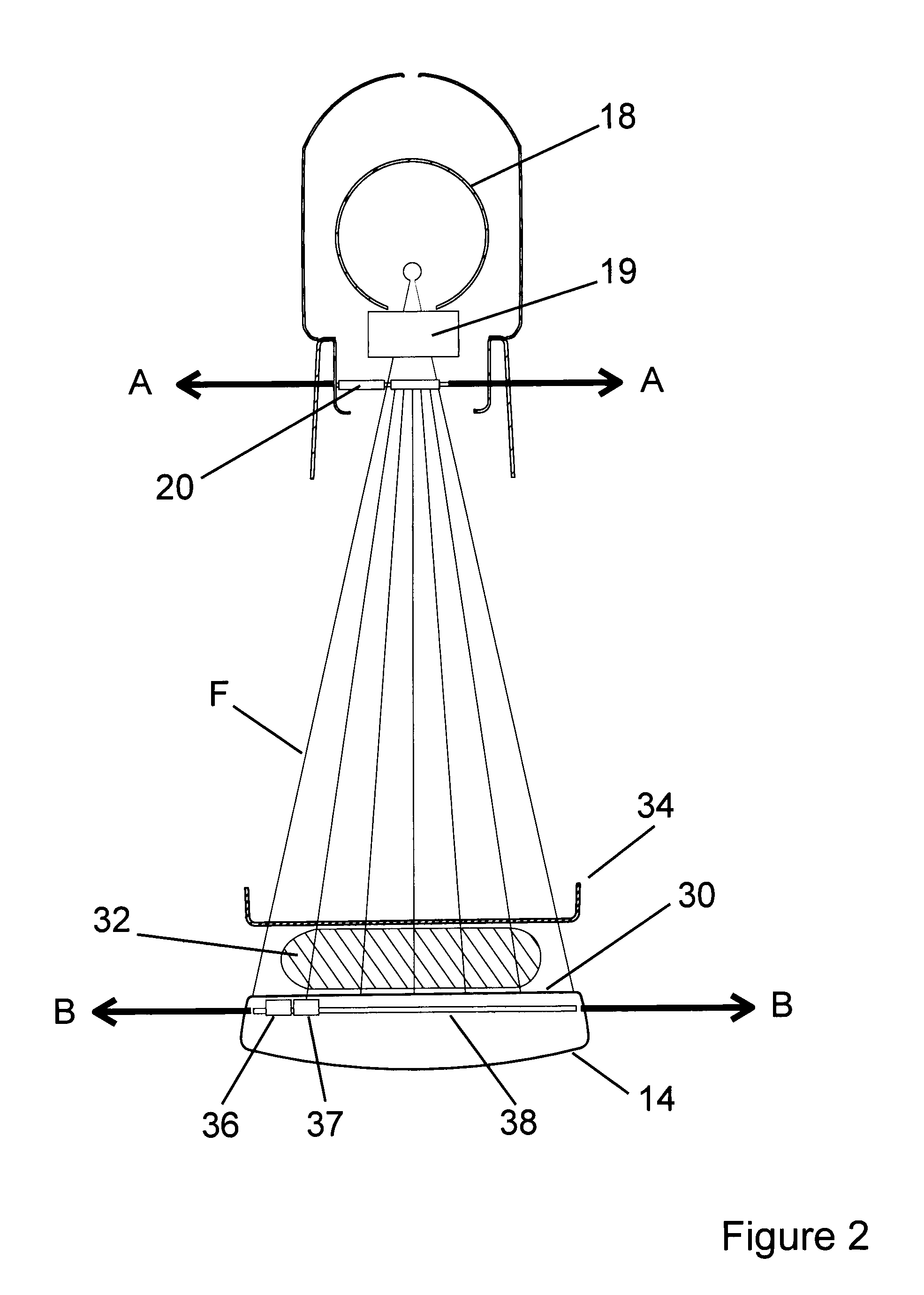
[0039]FIGS. 1 and 2 are pictorial illustrations of an example embodiment of the dual-modality scanning apparatus according to the present invention. The apparatus includes a support pillar 10 on which is mounted a C-arm. The C-arm includes a breast support platform 14, an upright member 16 mounted rotatable to the pillar 10, a further upright member 22 mounted rotatable to the upright member 16, and an X-ray source 18 with an associated beam-shaper 19 and pre-collimator 20, attached to the upright member 22, which extends parallel to the platform 14.

[0040]The entire C-arm can be moved up and down in the direction of the arrows E and rotated about its attachment point on the pillar 10 about an axis of rotation, as indicated by the arrows D in FIG. 3, by respective drives.

[0041]The breast support platform 14 defines a breast support surface 30 on which a breast 32 of a human subject can be placed. A breast compressor plate 34 with an associated clamp mechanism is positioned adjacent t...


PUM


Abstract

This invention relates to a scanning apparatus for imaging the breast that employs both X-ray and ultrasound technologies to enhance the diagnosis of cancer. There is provided a method for acquiring and co-registering the X-ray and ultrasound images in three-dimensional space, so that: the breast is in the same orientation and degree of compression when X-ray and ultrasound images are obtained; both sets of images are acquired simultaneously so as to minimize the time the woman's breast is held stationary; automated breast ultrasound images of the whole breast are acquired in a single scan; both image modalities are acquired in three dimensions; and radiation dose exposure to the woman is minimized. The method incorporates, in a single device, a system for acquiring full-field digital mammography and automated breast ultrasound images, and an algorithm for performing digital tomosynthesis reconstruction using these images.

Description

[0001]This invention relates to a scanning apparatus for imaging the breast that employs both X-ray and ultrasound technologies to enhance the diagnosis of cancer. In particular, the present invention provides a method for acquiring and co-registering the X-ray and ultrasound images in three-dimensional (3D) space.BACKGROUND TO THE INVENTION[0002]There is strong evidence to support the screening of women for breast cancer where the traditional imaging modality has been mammography which uses two-dimensional X-rays. The sensitivity (percentage of true positives) of X-ray mammography for the detection of breast cancer varies from 75% to 90%, while the specificity (percentage of true negatives) varies from 90% to 95% (Vaughan, 2011). Although digital X-rays significantly increase specificity in comparison with analogue film (Pisano et al., 2005), a major shortcoming of planar X-ray mammography is that it does not do well when the breasts are dense which is often the case for younger wo...

Claims


Application Information

Patent Timeline
Patent Type & Authority Applications(United States)
IPC IPC(8): A61B6/00
CPCA61B6/4417A61B6/0414A61B6/5247A61B8/0825A61B6/025A61B6/06A61B6/4078A61B6/502A61B8/403A61B8/4416A61B8/483A61B6/4441A61B8/5261
Inventor VAUGHAN, CHRISTOPHER LEONARDEVANS, MICHAEL DAVID
Owner CAPERAY MEDICAL
Why Eureka
Try Eureka