
Methods for detecting allosteric modulators of protein

a technology of allosteric modulators and protein, applied in the field of molecular detection and protein detection, can solve the problems of unintended clinical side effects, difficult competition with potential drugs, and the growth of the number of approved therapies targeting the most abundantly activated cancer-related genes

Inactive Publication Date: 2013-10-31
QUANTA THERAPEUTICS INC
View PDF2 Cites 15 Cited by
  • Summary
  • Abstract
  • Description
  • Claims
  • Application Information

AI Technical Summary

Benefits of technology

The patent describes a method for identifying and detecting agents that can change the structure of a target protein by using second harmonic generation technology. This method involves labeling the target protein with a second harmonic-active moiety and measuring the detectable signal generated by the moiety when the target protein binds to an agent. The method can be used to identify specific sites in the structure of the target protein that undergo a conformational change upon binding to an agent. The patent also describes the use of different interfaces, such as surfaces and amino acid residues, for labeling the target protein. Overall, the method provides a way to study the structure and function of target proteins in real-time using advanced technology.

Problems solved by technology

In that same time, however, the number of approved therapies which target the most abundantly activated cancer-related genes has grown only marginally.
An example of the latter case is found with proteins such as Ras which binds its ligand GTP with picomolar affinity making competition with potential drugs difficult.
Additionally, among other advantages (1), allosteric modulation of target proteins has the added benefit of not having to rely on inhibition or competition with the binding of the natural ligand to the protein, which can result in unintended clinical side effects.
However, it has been difficult to identify allosteric modulators using currently available conventional techniques.
For example, structural information obtained from X-ray crystallography or NMR methods is limited for drug discovery purposes due to low throughput, sensitivity, non-physiological conditions, size of the protein amenable to the technique, and many other factors.

Method used


Image

  • Methods for detecting allosteric modulators of protein
  • Methods for detecting allosteric modulators of protein

Examples


example 1

Identification of Allosteric Modulators of a Kinase by Addition of a Synthetic Ligand to the Kinase Active Site

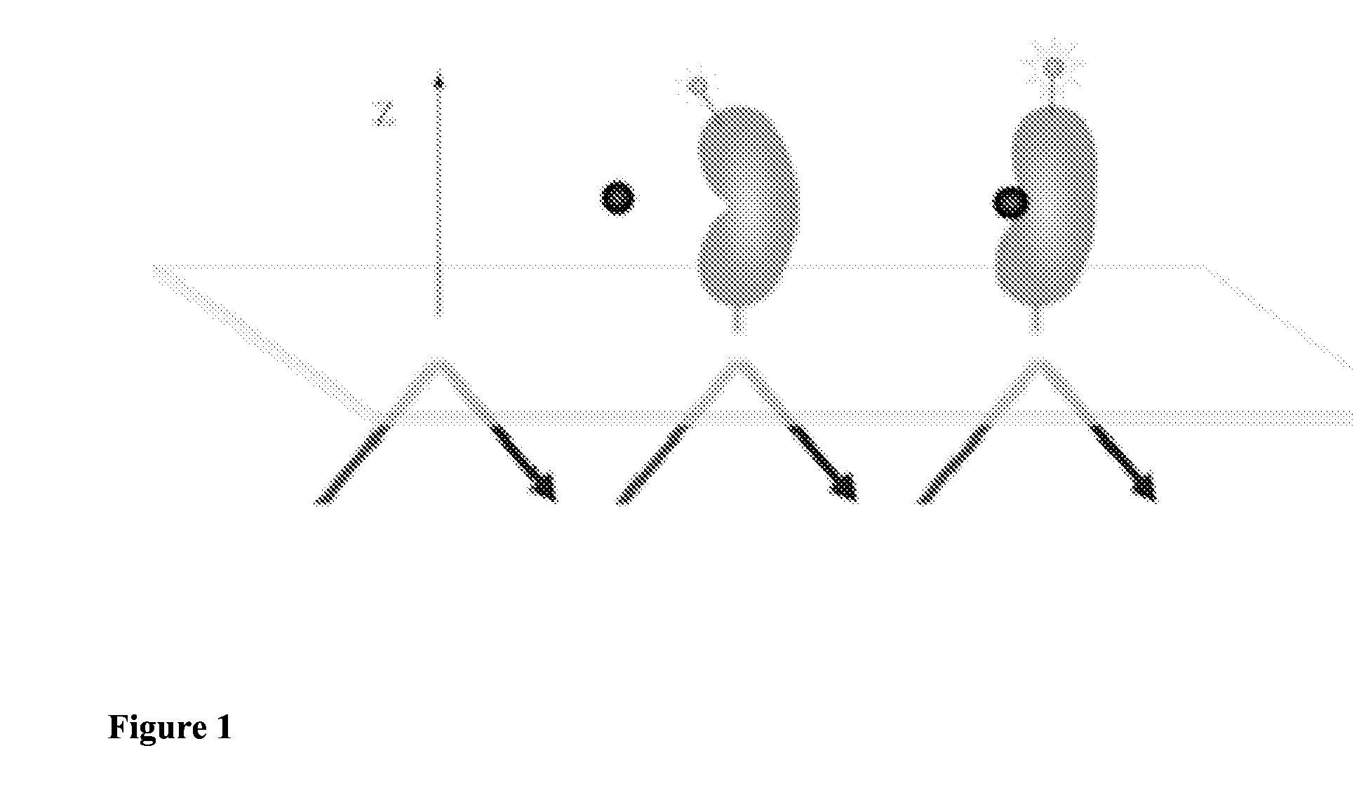
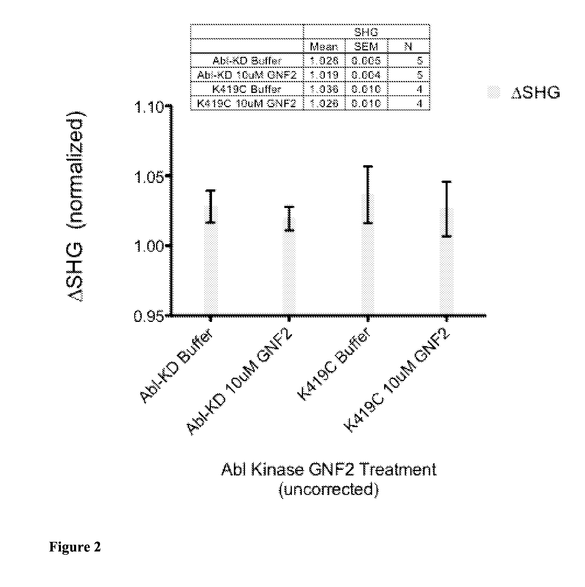
[0137]Kinases are a major class of drug targets with at least 30 distinct kinase targets currently in clinical trials. Most kinase drugs, known as type I inhibitors, bind to the kinase ATP binding site and work by mimicking and directly competing with ATP to stabilize the kinase activation loop into an active confirmation. Type II inhibitors, on the other hand, cause the activation loop of the kinase to shift into an inactive conformation by partially binding to the activation loop as well as other sites on the kinase. Type III kinase inhibitors, on the other hand, allosterically modulate kinase activity by binding to sites on the kinase other than the kinase activation loop. The identification of type III inhibitors is difficult, due to the fact that determination of the site of candidate inhibitor binding to a kinase typically requires X-ray crystallography to discern. In...

example 2

Labeling a Site Located in a Region of a Target Protein which Participates in a Partner Binding Event

[0146]Various mutations are made to insert cysteines in the region of H-Ras known to participate in PI3K effector binding. The native cysteine residue C118 can be optionally mutated to alanine or serine. The mutants are tested for an ability to be labeled by the SHG probe PyMPO-maleimide using mass spectrometry and protocols known to those skilled in the art. For example, the native residue tyrosine Y64 is known to participate in PI3K binding. Accordingly, recombinant 6×-His-tagged (N-terminal) H-Ras protein with a double mutation (C118A, Y64C) are prepared according to standard protocols (10). H-Ras is labeled using the cysteine-reactive SHG probe, PyMPO-maleimide in 0.1 M Tris pH 8.0, 20 mM NaCl, 0.5 mM TCEP, 5% glycerol and using a 12:1 dye:protein ratio for one hour at room temperature. Unreacted dye is purified away by gel filtration. Labeling of the cysteine at position 64 is c...

example 3

Labeling a Target in Two or More Different Ways, where at Least One of the Two or More Different Ways does not Involve Labeling a Native Residue

[0147]A slide with an oligo-PEG derivatization is prepared as follows: a slide-staining vessel is cleaned and dried in a vacuum oven at 75° C. / 20 inches Hg, and then allowed to cool to room temp. Enough SAT(PEG4) (N-Succinimidyl S-acetyl(thiotetraethylene glycol; Pierce) solution is added to the staining vessel to cover the entire slide (−50 mL). Ultrastick slides (amino-terminated silane derivatized slides; Thermo) are placed in a staining rack and submerged in the staining vessel. The slides are incubated in a hood at room temperature, stirring for 2-3 hrs. The slides are then removed from the SAT(PEG4) solution and transferred to a slide washing dish containing anhydrous chloroform. Sonicate the slides for 15 minutes by immersing washing dish to ⅔ its height in tap water. Transfer the slides to a second washing dish and rinse each with et...


PUM

PropertyMeasurementUnit
surface selective techniqueaaaaaaaaaa
structureaaaaaaaaaa
affinityaaaaaaaaaa

Abstract

The present invention discloses, inter alia, methods for labeling a target protein with an SHG-active probe for detection by second harmonic or sum-frequency generation in order to identify agents which bind to an allosteric site on the target protein thereby altering its structural conformation

Description

CROSS-REFERENCE TO RELATED APPLICATIONS[0001]This application claims priority to U.S. Provisional Patent Application No. 61 / 638,131, filed Apr. 25, 2012, the disclosure of which is incorporated by reference herein in its entirety.FIELD OF THE INVENTION[0002]The invention relates to the field of molecular detection and protein detection in particular. Specifically, provided herein are methods for identifying and detecting allosteric modulators of target proteins using second harmonic generation technology.BACKGROUND OF THE INVENTION[0003]The number of potential therapeutic targets in cancer biology has grown exponentially over the last twenty years with the advancement of the understanding of cancer genomics. In that same time, however, the number of approved therapies which target the most abundantly activated cancer-related genes has grown only marginally. Many cancer targets that are attractive from a biological perspective are considered intractable (“undruggable”) from a chemica...

Claims


Application Information

Patent Timeline
Patent Type & Authority Applications(United States)
IPC IPC(8): G01N33/68
CPCG01N33/68G01N33/573G01N2333/91205G01N2333/91215G01N2500/04G01N33/74G01N2333/726G01N21/6428G01N21/645G01N33/54366G01N2021/6463
Inventor SALAFSKY, JOSHUA S.
Owner QUANTA THERAPEUTICS INC
Learn More