

Novel Method for Diagnosing Lyme Disease Using a Cellular Immunological Test
a lyme disease and immunological test technology, applied in the field of lyme disease diagnosis, can solve the problems of poor sensitivity and specificity, inability to diagnose infected patients, and inability to reliably diagnose lyme disease based on humoral immunological tests based on antibody detection
- Summary
- Abstract
- Description
- Claims
- Application Information
AI Technical Summary
Benefits of technology
Problems solved by technology
Method used
Image

Examples
example 1
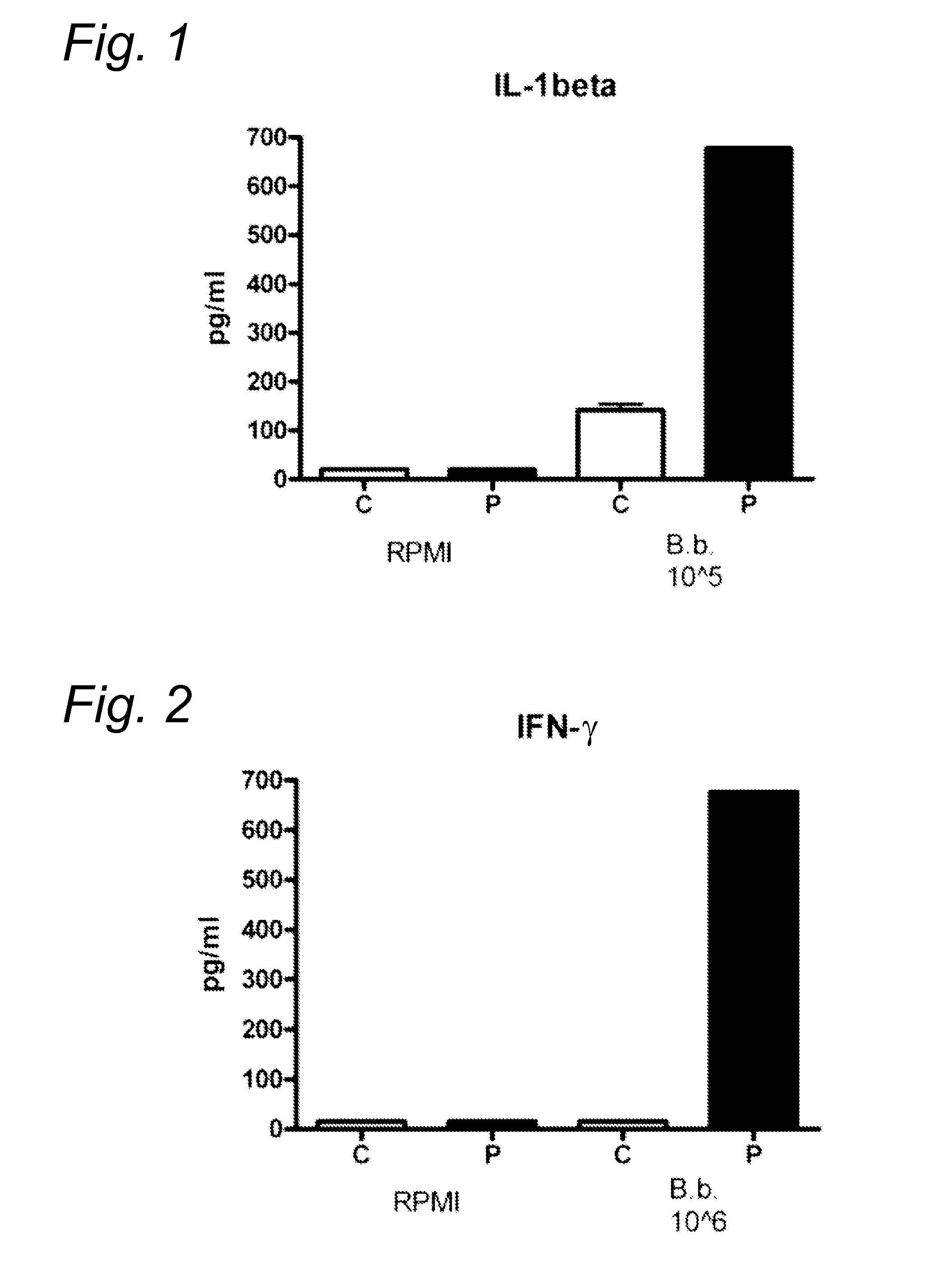
FIGS. 1, 2
[0081]Peripheral blood mononuclear cells (PBMC) were isolated from healthy volunteers (C) and patients with Lyme disease (P). 1.106 cells / ml were stimulated in RPMI 1640 for 24 h with the following strains of Borrelia: B. burgdorferi (ATCC 35210), B. garinii (ATCC 51383) and B. afzelii (ATCC 51567) that were first heat-inactivated by heating at 52° C. for 30 minutes (1.105 / ml). Thereafter IL-1β was determined by ELISA using antibody from R&D systems (IL-1F2). IL-1β was strongly detected in Lyme patients (see FIGS. 1, 2).
[0082]Blood (lithiumheparine) was taken from a subject suspected to have the Lyme disease. Blood was subsequently diluted in culture medium (1:5) and stimulated for 4 hours with 3 species of Borrelia common in Europe (see above). Subsequently, the supernatant was isolated via centrifugation and the presence of IL-1b was assessed by ELISA as described above.
example 3
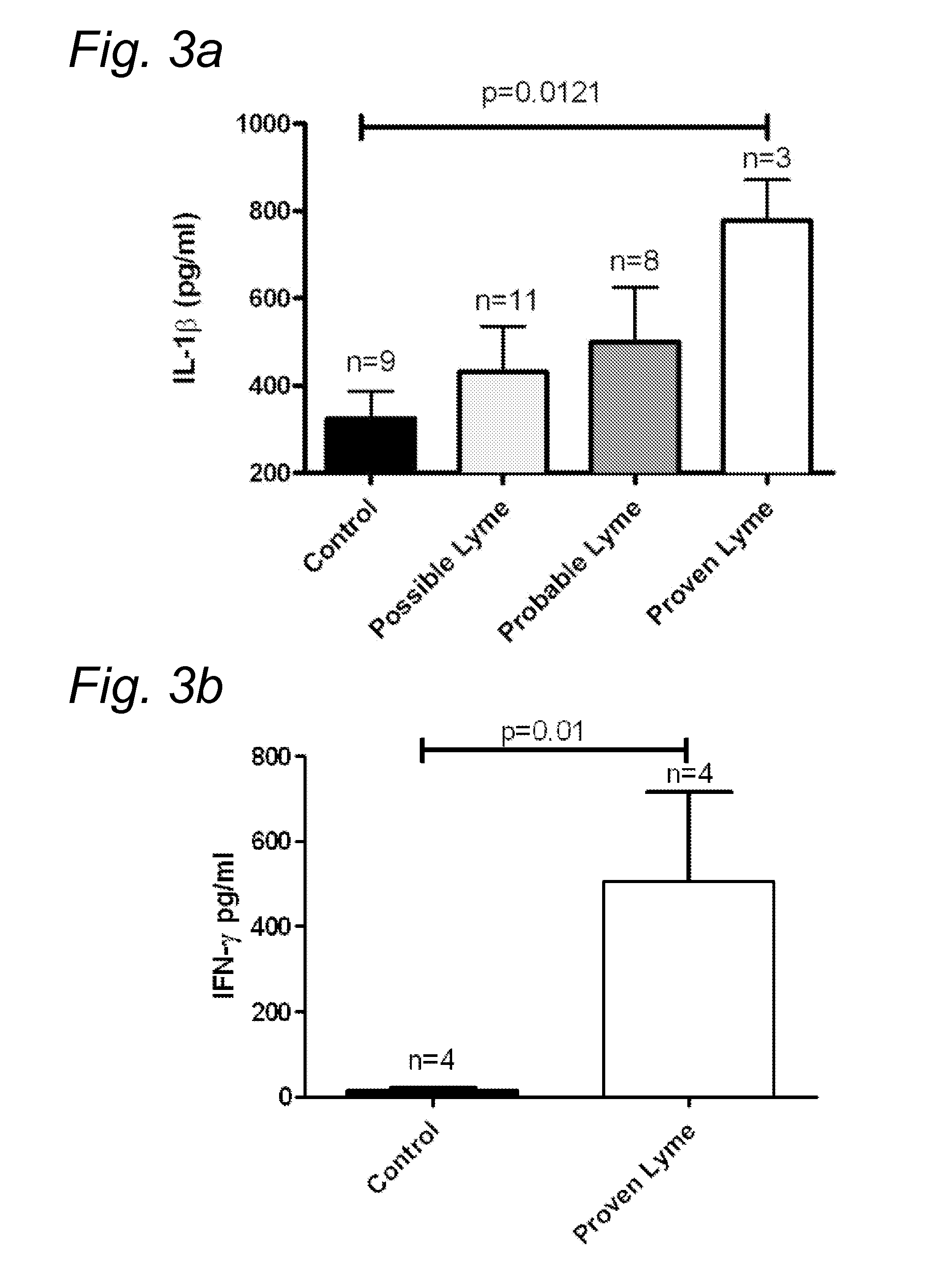
FIG. 3
[0083]Venous blood was drawn from the cubital vein of healthy volunteers and Lyme patients into 10 mL ethylenediaminetetraacetic acid (EDTA) tubes (Monoject). Peripheral blood mononuclear cells (PBMCs) were isolated according to standard protocols, with minor modifications. The PBMC fraction was obtained by density centrifugation of blood diluted 1:1 in PBS over Ficoll-Pague (Pharmacia Biotech). Cells were washed three times in PBS and resuspended in RPMI 1640 (Dutch modified) supplemented with 50 mg / L gentamycin, 2 mM L-glutamin, and 1 mM pyruvate. Cells were counted in a Coulter Counter Z® (Beckman Coulter), and adjusted to 5×10⁶ cells / mL. Mononuclear cells (5×10⁵) in a 100 μL volume were added to round-bottom 96-wells plates (Greiner) and incubated with either 100 μL of medium (negative control) or B. burgdorferi (ATCC 35210), at a dose of 1×10⁶ spirochetes per mL. The B. burgdorferi were first heat-inactivated by heating at 52° C. for 30 minutes. Concentrations of human I...
example 4
[0084]Blood samples were taken from patients and healthy individuals in EDTA vacutainer tubes (Becton and Dickinson, Leiden, The Netherlands). From these samples 200 μl were diluted 1:5 in culture medium (RPMI 1640) and incubated in 24 wells tissue culture plates (Costar, Badhoevedorp, The Netherlands). As a stimulus, 100 ng / ml of formaldehyde-inactivated (i.e. formalin fixated) B. burgdorferi (ATCC 35210), B. garinii (ATCC 51383) or B. afzelii (ATCC 51567) that were first heat-inactivated by heating at 52° C. for 30 minutes was added to these cultures. Formaldehyde-inactivated cells were prepared by transferring or incubating them with 4% formaldehyde for one hour. Subsequently cells were washed several times with PBS. No stimulus was added to the control cultures. The cultures were incubated at 37° C. and 5% CO2 for 48 hours. After this incubation period, the supernatants were harvested and centrifuged at 15000 g for 5 minutes, and thereafter stored at −20° C. until measurement of...
PUM
 Abstract
Description
Claims
Application Information