Theranosis of macrophage-associated diseases with ultrasmall superparamagnetic iron oxide nanoparticles (USPIO)

a superparamagnetic iron oxide and macrophage technology, applied in biocide, diagnostics, diagnostic recording/measuring, etc., can solve the problems of inefficient contrast agents at wavelengths used, inaccurate diagnosis and selective therapy of inflammatory diseases, and uspio nanoparticles of about 25 nm would be relatively inefficient contrast agents, etc., to achieve enhanced capillary permeability, facilitate extravasation, and suit the effect of siz

Inactive Publication Date: 2013-12-19
WOLF GERALD L +1
View PDF2 Cites 13 Cited by
  • Summary
  • Abstract
  • Description
  • Claims
  • Application Information

AI Technical Summary

Benefits of technology

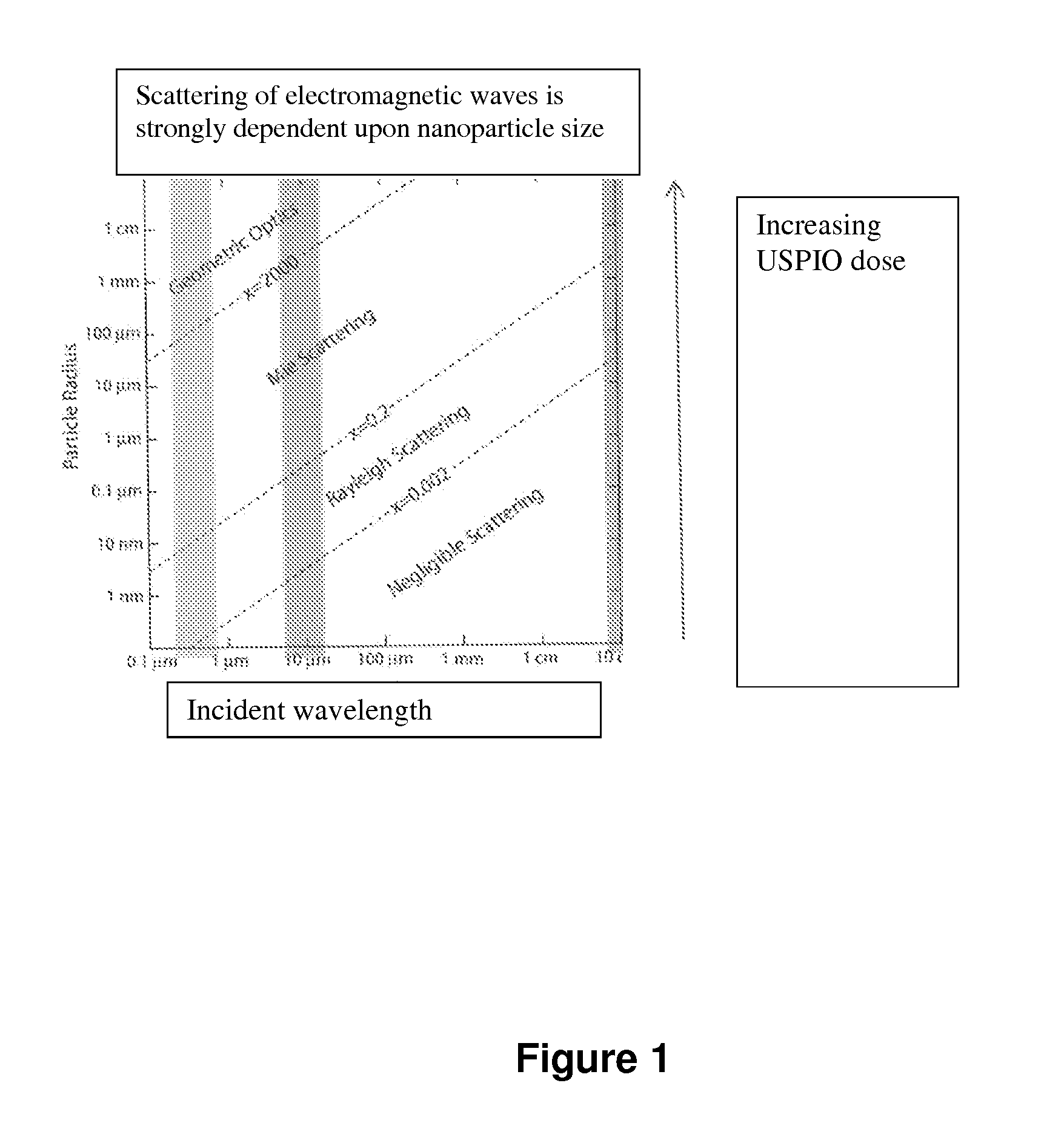
[0024]Fixed tissue macrophages and disease-associated activated macrophages readily ingest USPIOs from nearby body fluids. Useful USPIO formulations consist of a magnetizable iron oxide core and a biocompatible coating. For presentation to macrophages in macrophage-dependent or -associated diseases, the USPIO should have a suitable size to facilitate extravasation from the enhanced capillary permeability and interstitial diffusion to the disease location, where they are readily ingested and stored in lysosomes. This active process creates nanoparticle aggregates of much larger size, resulting in increased scattering and absorption of incident electromagnetic waves, including those of light (e.g., laser), radiofrequency (RF), microwave (MW), and ultrasound (US). As a result, the location and density of macrophages with the aggregated USPIO nanoparticles can be imaged, and the presence and severity of disease in that location effectively staged. Thus, the scattering and absorption from aggregated particles create different imaging mechanisms than that associated with the use of USPIO nanoparticles as MRI contrast agents.
[0025]Furthermore, these focal USPIO aggregates can be locally heated with hyperthermic devices, and the heat absorption will conform to the distribution of the activated macrophages, which closely mirrors the geometry of the tissue to be treated. During imaging or heating, the USPIO-loaded macrophages do not have to be (e.g., is not) subjected to any magnetic field, e.g., any static or time-varying magnetic field. In addition, the USPIO coat can be used to deliver useful payloads of bound drug to the diseased site, and suitable spacers and linkers can also provide for facilitated release due to, for example, the lysosomal pH or applied local heat.

Problems solved by technology

Accurate diagnosis and selective therapy for inflammatory diseases has not previously been achieved with a theranostic approach.
USPIO nanoparticles of about 25 nm would be relatively inefficient contrast agents at wavelengths used for ultrasonic imaging devices or for optical coherent tomography devices.

Method used


Image

  • Theranosis of macrophage-associated diseases with ultrasmall superparamagnetic iron oxide nanoparticles (USPIO)
  • Theranosis of macrophage-associated diseases with ultrasmall superparamagnetic iron oxide nanoparticles (USPIO)

Examples


example 1

Experiments Assessing Macrophage Loading

[0268]Extensive preclinical research on a representative USPIO, ferumoxtran-10, shows the broad range of macrophage loading feasible in mammals. Intravenous doses ranging from 2-400 mg Fe / kg in rats and dogs show no significant adverse effects. Bourrinet (supra). These doses of ferumoxtran-10 are cleared from the blood by body macrophages within 24 hours. Any of the tissues with fixed macrophages could be used to determine the detection characteristics of USPIOs when they are aggregated in macrophages, using ultrasound or optical coherence imaging devices.

[0269]One particularly useful method uses percutaneous administration into the subcutaneous tissue on the dorsum of the foot in rabbits or rats see Wolf (Acad. Radiol., 6(1):55-60, 1999, incorporated herein by reference). Percutaneous lymphography relies upon the access of nanoparticles through the clefts in the terminal lymphatics. These single celled vessels are tethered to the surrounding ...

example 2

Detection Sensitivity with Ultrasound (Macrophage-Enhanced Lymph Nodes: Method for Comparing the Imaging Detection and Characterization)

[0271]The rat or rabbit is injected with a challenge subcutaneous dose of USPIO, the injection site is gently massaged for 15 minutes, and then the animal is anesthetized for evaluation at an appropriate experimental time post injection. It is possible to use comparison substances injected in the same manner in the opposite hind paw.

[0272]The dose of UPSIO can be an experimental variable as well as the time after injection. These experiments allow the creation of loaded nodal macrophages where accumulation, retention, and metabolism can be evaluated. The experimental animals can be studied with any ultrasound device wherein ultrasound frequency, detection of backscatter, probe design, or other variable can be studied to determine detection parameters. Qualitative, semi-quantitative, or quantitative response can be obtained in reference to the in viv...

example 3

Detection Sensitivity with Optical Coherence Tomography

[0275]Using the methods above, OCT device variables and their detection sensitivity can be determined. The draining node in these animals is close to the surface, but surgical exposure can be used when experimentally appropriate.


PUM


Abstract

Macrophages sequester and aggregate ultrasmall superparamagnetic iron oxide nanoparticles (USPIO) in their lysosomes. The amount of USPIO loading of macrophages depends upon the route and dose of administration, and the pharmacokinetics of accumulation and removal. Both fixed macrophages and activated macrophages associated with inflammatory diseases and cancer phagocytize USPIOs, and the loaded macrophages can serve to identify the extent of a macrophage-dependent disease as well as to direct treatment options. Furthermore, the absorption of energy from incident electromagnetic waves by the aggregated nanoparticles can be used for conformal thermotherapy. The USPIOs can further be used to carry drugs to the same activated macrophages. The co-administered drugs can be bound to the USPIO by condition-dependent releasing links that are responsive to local pH or heating.

Description

REFERENCE TO RELATED APPLICATIONS[0001]This application claims the benefit of the filing date under 35 U.S.C. 119(e) of U.S. Provisional Application No. 61 / 690,005, filed on Jun. 18, 2012; U.S. Provisional Application No. 61 / 690,006, filed on Jun. 18, 2012; U.S. Provisional Application No. 61 / 742,382, filed on Aug. 9, 2012; U.S. Provisional Application No. 61 / 743,428, filed on Sep. 4, 2012; U.S. Provisional Application No. 61 / 797,757, filed on Dec. 14, 2012; and U.S. Provisional Application No. 61 / 779,123, filed on Mar. 13, 2013. The entire content of each of these applications, including drawings, are incorporated herein by reference.BACKGROUND OF THE INVENTION[0002]Medical imaging is the technique and process for creating images of the live human body or parts thereof for clinical purposes, most notably for diagnostic uses or for medical science research. Over the years, numerous medical imaging devices and technologies have been developed based on distinct scientific principles t...

Claims


Application Information

Patent Timeline
Patent Type & Authority Applications(United States)
IPC IPC(8): A61K49/18
CPCA61K49/1818A61K41/0052A61K47/6923
Inventor WOLF, GERALD L.SCHMIDT, KARL F.
Owner WOLF GERALD L
Why Eureka
Try Eureka